William Henry Bates, 1841 - 1930
by Brian Stevenson
last updated December, 2025
W.H. Bates was a physician and surgeon in Brooklyn, New York. Consistent with his occupation, known microscope slides by Bates are of human and other animal tissues (Figure 1). He advertised to exchange histological slides with other microscopists in 1881-82, and known slides probably date from that time (Figure 2).
A biographer noted that Bates “was one of the first to recognize the fact that bacteriology must be known in connection with surgical work. On account of his researches along that line he was made a fellow in the Royal Microscopical Society of London - the first Brooklyn physician to receive that honor”. Bates was also a member of the New York Microscopical Society, the Medical Microscopical Society of Brooklyn, and the Department of Microscopy of the Brooklyn Institute of Arts and Sciences
.

Figure 1.
Microscope slides by William H. Bates, probably dating from around 1881-81, when he advertised to exchange histological and pathological slides (see Figure 2). Bates stained and sectioned a variety of tissues from humans and other animals, including unusual material such as the penis of a bat (Figure 3).
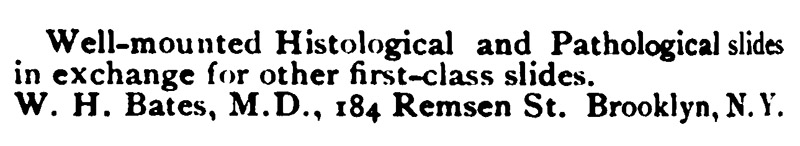
Figure 2.
Bates published exchange offers in multiple issues of “The American Monthly Microscopical Journal” during 1881 through early 1882. I have not found any exchange requests from after 1882.
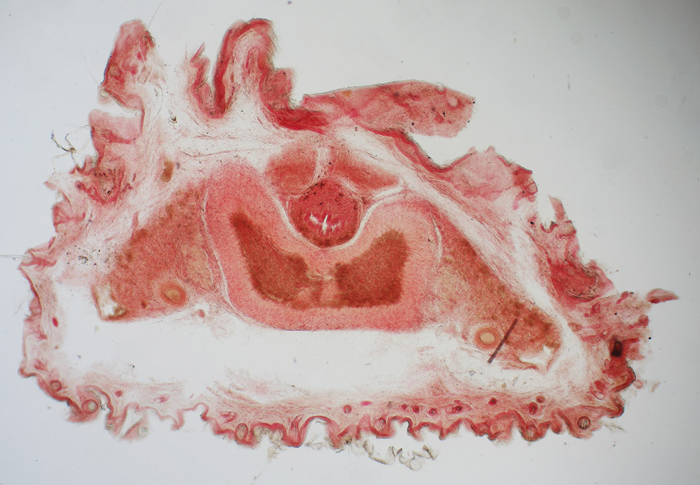
Figure 3.
“Transverse section of bat’s penis”, prepared ca. 1881 by W.H. Bates (see Figure 1). Imaged with a 3.5x objective lens and C-mounted digital SLR camera on a Leitz Ortholux II microscope.
William Henry Bates was born on December 25, 1841, in Westhampton, Long Island, New York. He was the fourth child, and fourth son, of Edmund Ogden and Charlotte (nee Bishop) Bates. Father Edmund was a minister in the Methodist Episcopal church. The family moved several times, presumably associated with Edmund’s ministry. By 1855, they were living in Brooklyn.
After education in Brooklyn private schools, William attended New York University. He obtained his M.D. degree in 1862, then worked in a hospital for about a year.
The US Civil War had broken out in 1861. William Bates joined the US Navy in 1863 as a surgeon. According to Ross (1902), Bates was, “assigned to duty on gunboats on the Potomac River. Later he was transferred to the South Atlantic squadron, engaged in blockading the port of Charleston, and there remained until the fall of the city (February, 1865). He was afterward on the ‘Benton’ on the Mississippi river, and was present at the time of the surrender of Kirby Smith (May, 1865)”. The USS Benton was an ironclad gunboat.
Upon the end of the war, Bates resigned his commission and opened a medical practice in Brooklyn. In addition, he served as demonstrator of anatomy in the Long Island College Hospital from 1867 until 1874 (Figure 4), attending surgeon of the Brooklyn Hospital from 1888 until 1897, consulting surgeon in the Kings County Hospital from 1893 through at least 1902, on the staff of the “psychopathic department” of Kings County Hospital from ca. 1900 onward, and consulting physician at Adrendale Sanitorium from 1897 onward (Figure 5). Arendale was “a private sanitarium for the care and treatment of cases of neurasthenia, drug and alcoholic habituation … established to meet the demand felt by the medical profession for a strictly ethical institution where patients of the better class could receive the benefit of the best care and treatment and experience of men well known in this line of work”.
He married Mary Amanda Libby in 1875. The couple had three children, all girls.
Bates attended meetings of the New York Microscopical Society as early as 1880. He became a member in 1882. As noted above, he published requests to exchange microscope slides in 1881 and early 1882 (Figure 2). He was elected Fellow of the Royal Microscopical Society on February 13, 1884. He was also a member of the Department of Microscopy of the Brooklyn Institute of Arts and Sciences.
An example of Bates’ use of the microscope in medical practice was published in 1886. A report to the Brooklyn Pathological Society described the case of a man who “had a growth on the right lower lid, projecting two-thirds of an inch, with an ulcerated surface. It had first been noticed, as a warty excrescence, a year or two before, and it had lately begun to grow rapidly. Microscopical examination, by Dr. W.H. Bates, showed it to be clearly an epithelioma”.
Bates was instrumental in the formation of the Medical Microscopical Society of the City of Brooklyn in 1887, and served as its first president. The group was “composed of medical practitioners, for the advancement of medical microscopy. The society holds its meetings on the first Wednesday of each month in the year, excepting July and August. Its membership is limited to twenty-five, twenty of the members being active working microscopists, and five being associate members and not necessarily active workers with the microscope. Papers are read monthly on medical microscopical topics, and are illustrated with microscopical preparations”. Bates made a presentation at the group’s first meeting, "Remarks on Bacterium Lactis from human milk, illustrated with slides and cultures”.
Bates had a longstanding interest in mental health and alcoholism. He coordinated a Prize on Alcoholic Pathological Microscopy in 1889, which was broadly advertised throughout the world. Announcements stated, “A prize of 100 dollars has been offered on behalf of the American Association for the Cure of Inebriates, by Dr. L.D. Mason, one of the Vice-Presidents, for the best original essay on ‘The Pathological Lesions of Chronic Alcoholism, capable of Microscopic Demonstration’. Along with the essay, the competitors have to send carefully prepared microscopic slides, with accurate drawings or micro-photographs of these. Conclusions which have been founded on experiments with animals will be admissible. The object of the essay will be to demonstrate the facts as to these two questions: (1) Are there pathological lesions due to chronic alcoholism? (2) Are these lesions peculiar or not to chronic alcoholism? An authentic alcoholic history must accompany each microscopic preparation, complications such as specific disease being excluded. The essays, with slides, drawings, or microphotographs, are to be forwarded to Dr. W.H. Bates, Chairman of the Prize Committee, 175, Remsen Street, Brooklyn, New York, not later than October 1st, 1890. After adjudication the successful competitor will be asked to read and demonstrate, either personally or by proxy, his essay, at a meeting of the Medical Microscopical Society of Brooklyn. The essay, after publication in the Journal of Inebriety, will be the property of the author”. The prize was awarded to Dr. Pierre Francois Spaink, Baarn, Netherlands, whose essay was published in 1891.
Bates volunteered for military duty during World War I, but was turned down on account of his age. He died from throat cancer on October 19, 1930, at the age of 88.
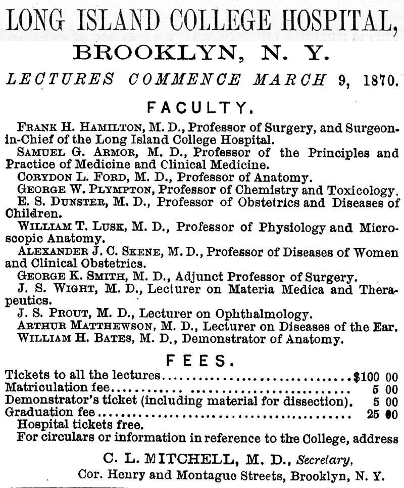
Figure 4.
William H. Bates served as a demonstrator of anatomy at Long Island College Hospital from 1867-1874. Advertisement from “The Physican and Pharmacist”, 1870.
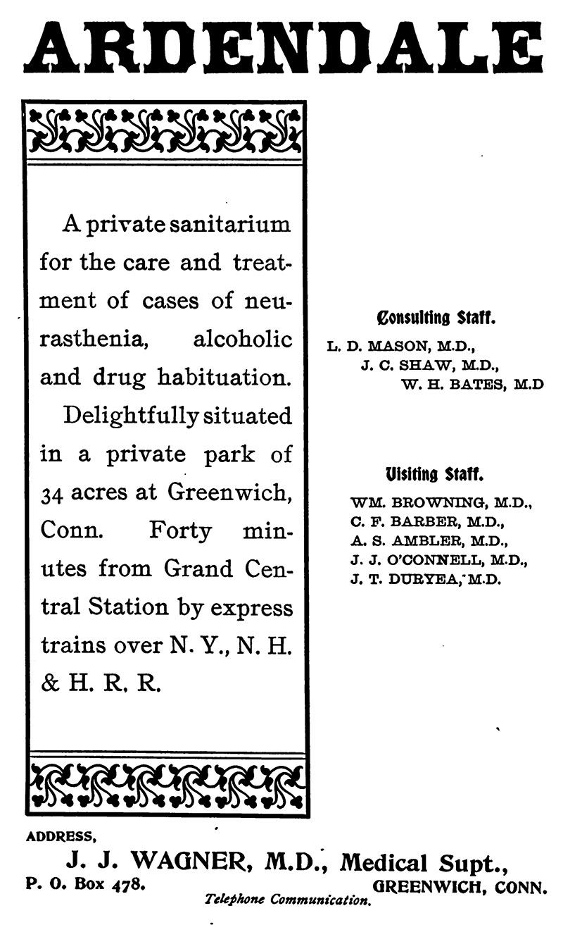
Figure 5.
William H. Bates served on the staff of Ardendale sanitorium, “a strictly ethical institution where patients of the better class could receive the benefit of the best care and treatment and experience of men well known in this line of work”. Advertisement from “The Quarterly Journal of Inebriety”, 1902.
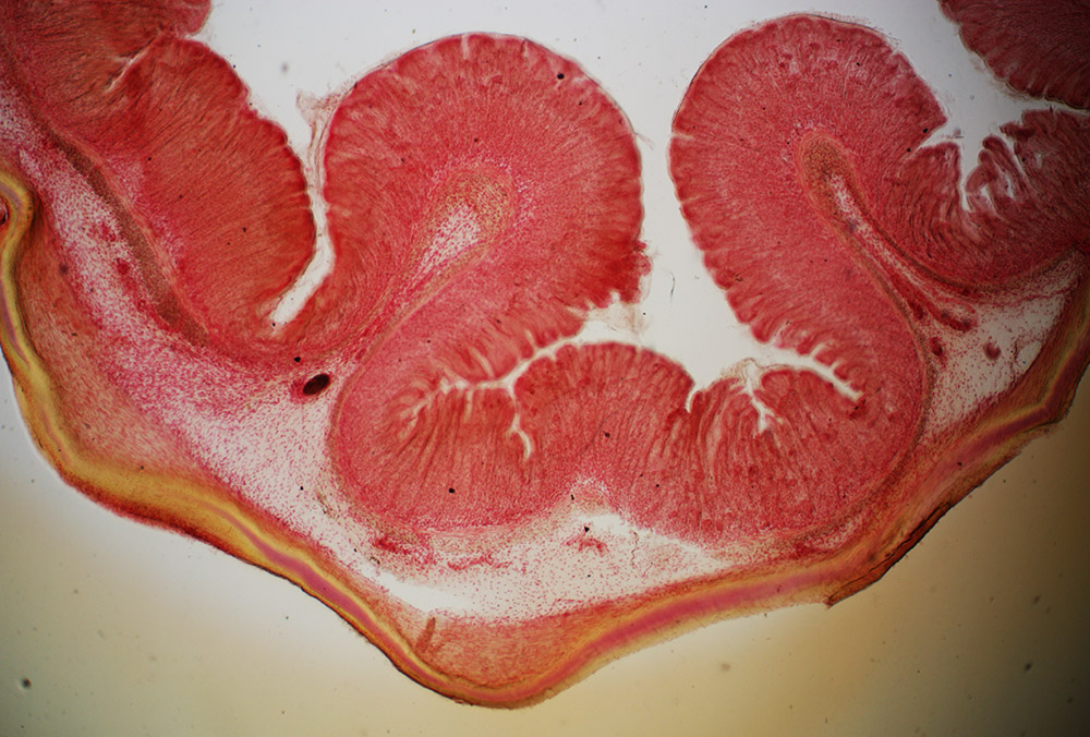
Figure 6.
“Epididymis of rabbit”, prepared ca. 1881 by W.H. Bates (see Figure 1). Imaged with a 3.5x objective lens and C-mounted digital SLR camera on a Leitz Ortholux II microscope.
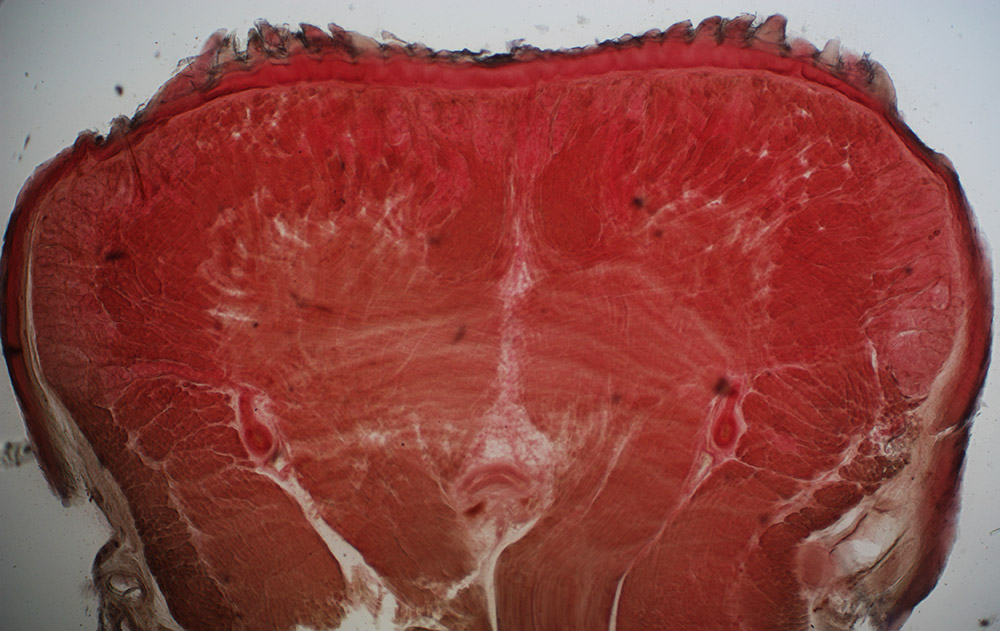
Figure 7.
“Section of bat’s tongue”, prepared ca. 1881 by W.H. Bates (see Figure 1). Imaged with a 3.5x objective lens and C-mounted digital SLR camera on a Leitz Ortholux II microscope.
Resources
The American Monthly Microscopical Journal (1875) On mounting and staining pollen, page 206
The American Monthly Microscopical Journal (1881) Exchange offers from William H. Bates, pages 180, 200, and 240
The American Monthly Microscopical Journal (1882) Exchange offersfrom William H. Bates, page 20
The Bates Bulletin (1931) Dr. William Henry Bates, April issue, page 75
British Medical Journal (1888) Prize on alcoholic pathological microscopy, page 1301
Brooklyn Medical Journal (1887) The Medical Microscopical Society of the City of Brooklyn, pages 75-76
Brooklyn Medical Journal (1897) Ardendale, page 183
Journal of the New-York Microscopical Society (1887) Medical Microscopical Society of Brooklyn, page 97
Journal of the New-York Microscopical Society (1889) Members, “1882, Bates, William H., M.D., 175 Remsen Street, Brooklyn, N.Y.”
Journal of the Royal Microscopical Society (1884) Annual meeting of 13th February, 1884, pages 327-329
The Naturalists' Directory (1890) “Bates, H. Wm., M.D., F.R.M.S., 116 Schermerhorn St., Brooklyn, N. Y. Gen. Biol., Histol. of Vert., Infusoria. C. Ex.”, page 12
New York Times (1930) Dr. W.H. Bates dies; a Civil War veteran, October 21, page 25
The Physican and Pharmacist (1870) Advertisement for Long Island College Hospital, February issue, page 5
Quarterly Compendium of Medical Science (1886) Epithelioma of the eyelid removed by applications of benzol, page 122
The Quarterly Journal of Inebriety (1891) Report of the Mason Prize Essay, Vol. 13, pages 44-52
The Quarterly Journal of Inebriety (1902) Advertisement for Ardendale, advertising section
Ross, Peter (1902) William H. Bates, M.D., A History of Long Island: From Its Earliest Settlement to the Present Time, Vol. 2, pages 253-254
Transactions of the Medical Society of the State of New York (1897) Medical Society of the County of Kings, page 515
US census and other records, accessed through ancestry.com
Year Book of the Brooklyn Institute of Arts and Sciences (1889) Department of Microscopy, page 55