Microscopist “Complex Monogram, B”:
probably the Reverend John Bramhall, 1809 – 1889
by Brian Stevenson and Richard Courtiour
last updated Deecmber, 2021
Many Victorian makers and owners
marked their microscope slides with identifying monograms, which ranged in
style from simple letters-in-a-row to complex overlays of intertwined letters
and curlicues, serifs, etc. Extreme examples of the latter are shown in Figure
1. Yet even monograms that initially appear to be undecipherable may be
interpreted through a few basic “rules”. A key to monograms is that the
predominant letter is the first letter of the owner’s last name. The
initial of the last name is generally placed in the center of the monogram.
Mirror images are often used to create symmetry.
The monograms marked “B1a” and “B1b”
in Figure 1 are clearly comprised of the letter “B”. Evidence provided below
suggests that the stem of the “B” is not merely a curlicued affectation, but is
the letter “J”. Monograms “B1a” and “B1b” are found on slides with the same
labels and other shared characteristics, and were undoubtedly marks of the same
person (Figure 2).
Monogram “B2” also contains a central
“B”, with a “J” alongside, suggesting that the owner of this monogram also had
the initials J.B. (Figure 1). Evidence is presented in this essay that monogram
“B2” was used by John Bramhall.
The overall similarities between
monogram “B2” and “B1a/b” – the same letters, the mirror imaging, the
curlicues, and the same shield device – suggest that they are variations used
by the same person. In addition, there are similarities between the microscope
slides labeled with monograms “B2” and “B1a/b”, such as the use of paired
labels (which may have been custom-made, as these patterns have been identified
only on “B1” and “B2” slides) and the dating of almost all slides (few
microscopists did this). We consider it likely that microscope slides labeled
by “B1” were also property of John Bramhall.
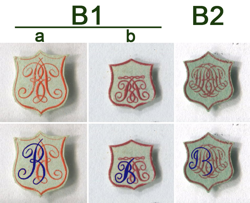
Figure 1.
Monograms and interpretations. Labels with monograms
“B1a” and “B1b” are found on microscope slides that have identical label
styles, and are undoubtedly from the same person. The stylistic similarities
between the three monograms suggest that they were all used by the same person.
All three monograms contain a recognizable letter “B”, which is centrally
located and most likely indicates the last name. The letter “B” is ordinarily drawn
with symmetry, with a similar loop on top and bottom. Instead, the bottom loop
of “B” in these monograms is separate from the vertical stem, suggesting a
separate significance to the shape of the stem. The stem has a curl at the
bottom, thereby resembling a “J”.
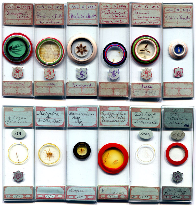
Figure 2.
Examples of English microscope slides with the
labels and monograms of microscopists “B1” (top row) and “B2” (lower row).
Slides without monograms are not uncommon. Although there are few apparent
similarities between the handwriting on “B1” and “B2” labels, there are often
enormous differences in writing styles on slides with the same monogram.
The slides in this figure show several such examples. Additional handwriting
variations are highlighted in Figure 3. While both “B1” and “B2” put dates on
slides, the listed years are not necessarily accurate (see Figure 4). This indicates
that labels on some slides were applied quite some time after their production.
Thus, the two label and monogram styles may represent two periods of one
microscopist’s life.
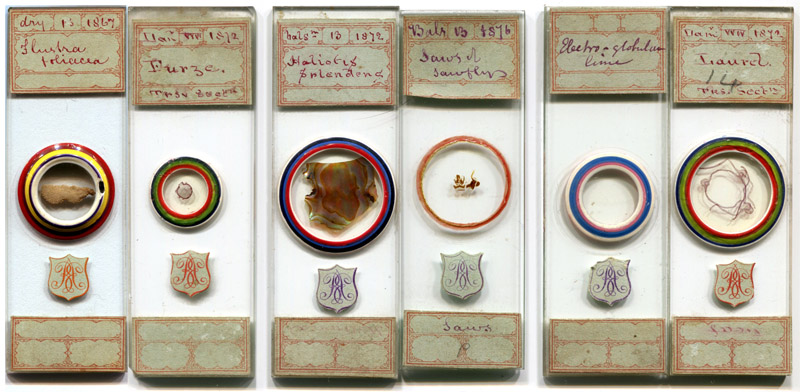
Figure 3.
Examples of slides with monogram
“B1”, yet their labels have very different handwriting styles. Compare the
letter “F” in the left pair, letter “S” in the central pair, and “L” in the
right pair. The variations within “B1” slides indicates that handwriting
differences between “B1” and “B2” slides need not signify that they were owned
by different people.
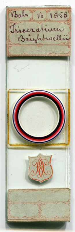
Figure 4.
A strew of Triceratium brightwellii diatoms, labeled
by “B1”. The label is dated 1858, although this diatom was not given this
species name until 1860. This indicates that the label was added after the
actual date of production had been forgotten. It is, therefore, possible that
other dates on “B1” slides are not accurate and/or “B1” labels were attached many years after the slides were produced. This slide is also of particular interest in that it has a square cover slip but circular ringing. This ringing does not fulfill the normal, structural purpose of binding a cover slip to the slide, but is instead purely ornamental. It is possible that the colorful ringing was added to a pre-existing slide.
Connecting “B” with John Bramhall
A large microscope slide collection
of parasitic arthropods was recently located, which included several dozen
slides with “B2” monograms and labels. Among these was a “B2” slide of a tick
(“Ixodes”) from an Indian bullock
(Figure 5). Also written on the slide is the word “Pygidia”. A second slide in this collection, with a different type
of label, is of a tick from a tiger, also labeled “Pygidia”. Pygidia (singular pygidium) are sensory organs of fleas,
which were frequently used in Victorian times as test objects, to evaluate the
resolving power of microscope lenses. However, ticks do not possess pygidia,
nor do they have analogous organs. We were able to locate only two references
in which organs of ticks were called pygidia, both of which were written in
1877 by Reverend John Bramhall of Terrington St. John, Norfolk.
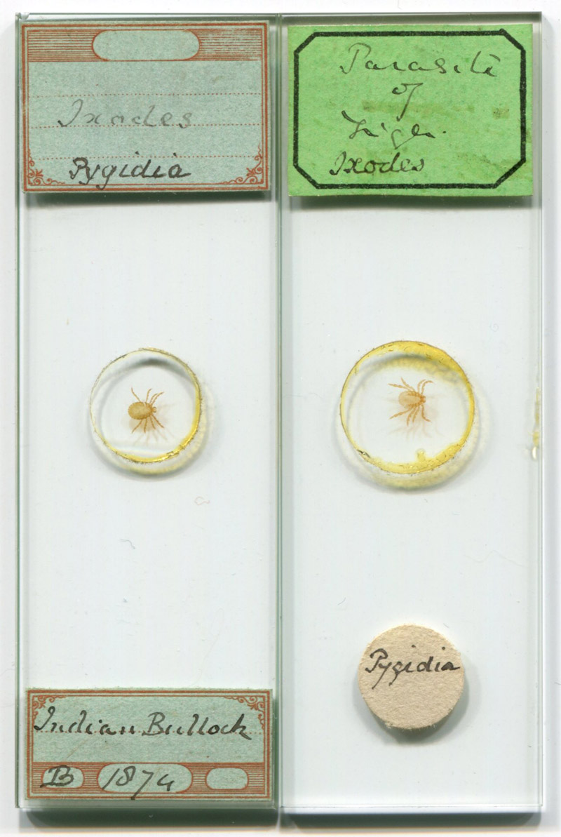
Figure 5.
Two microscope slides from the same extensive
collection, holding ticks from an Indian bullock and a tiger. Both slides
emphasize the pygidia. The slide on the left has labels by microscopist “B2”.
During Victorian times, ticks were often called “ixodes”. That name is
currently a genus name, as in the Lyme disease vectors Ixodes scapularis and
Ixodes ricinus, and all ticks fall into the Order Ixodida.
The first known use of “pygidia” in reference to ticks was
published by Reverend John Bramhall in Hardwicke’s
Science-Gossip in January, 1877. Notably, Bramhall specifically described
pygidia of ticks from Indian bullocks and tigers:
“A word about the ‘pygidium’ - That old well-known ‘test’, the pygidium
of the flea, is one of the first objects a young microscopist desires to
possess, and a very curious apparatus it is. I shall be thankful to any one
learned in such matters who will tell me what is supposed to be its use to its
proprietor. I cannot find it mentioned in any work on insects in my possession.
But the flea is not the only possessor of a pygidium, though it certainly is A1
in that line; nor is it always single, or to be looked for in the same
position. Generally it is to be found in
pairs at the extremity of the abdomen; but not always, for in the Ixodes of the tiger and Indian bullock we find two on the underside of the
abdomen, nearer the upper than the lower end. The Chrysopa perta and vulgaris have pygidia in the usual locality; and, I believe, several other
insects have the same, but I cannot recall their names. Perhaps the most
uncommon pygidia are those of the Agrion
pulehellum, a very interesting insect in many points. Like all
dragon-flies, it is a voracious feeder, and devours all it can catch in the
insect line daily. It possesses a powerful set of jaws for breaking up its
prey, and gastric teeth, well suited for ‘grinding the bones to make its
bread,’ like the giant of our nursery days, save that he preferred Englishmen
to English insects. The ovipositor has a formidable set of jaws, something like
those of the Sirex, and its
pygidia are large and mammiform, quite at the extremity of the abdomen. Its
wings are also worth studying. In short, I know no insect possessing more
points of interest, and strongly recommend it to the notice of those who take a
pleasure in such things. If asked where it is to be had, I may say that it is
not in any list of objects that I have seen. My specimens of Agrion pulehellum and Chrysopa are by Mr. Enoch (sic), of 30, Russell Road, Seven Sisters' Road,
N., who has, I believe, a good supply of both. But, if the want be made known,
others possibly may be found able to supply them – John Bramhall”.
Two months later, this was followed
by the only other known publication on tick pygidia:
“The
Pygidium - Allow me to correct an error. I stated in my paper on the Pygidium (p. 15), that I had found a
pair on the Ixodes of the tiger and Indian bullock. Further
examination with a higher power convinces me that these are not Pygidia but spiracles – John Bramhall.”
The aforementioned slide collection
contains several other specimens that were described by Bramhall (Figure 6). He
wrote to the Quekett Microscopical Club in March, 1877, in reference to an
article from C.F. George on a bat-associated tick named Argas fischerii (now Carios vespertilionis):
“I was much interested in Mr. George's paper and illustrations of the
'Blyborough Tick,' and have no doubt about its connection with the Bat. In 1875
I found, in a seat in my church, one of these creatures in company with two
genuine Bat bugs. Another tick was brought to me, having been taken from the
shawl of a lady who sat in the same seat; indeed, this led to my examination of
the seat, which for some time had been constantly covered with the excrements
of bats. In order to put an end to this nuisance, our churchwardens took up a
portion of the lead on the roof above the seat, and, in a very small space,
took out from between the lead and boards of roof no less than 287 bats of
various kinds - large, small, and eared, I believe. Unfortunately I was from
home at the time, and all the ' vermin' were destroyed - a fact which greatly
vexed me when I heard of it, as no doubt I should have obtained a large
quantity of both ticks and bugs. The shape of the creature, together with the
markings, and form, and locality of the spiracles, make me sure it is the same
as those found by Mr. George at Blyborough. I marked mine 'Ixodes of Bat,' but shall alter it to Argas Fischerii, as that seems to be
its true appellation. Some years ago a friend gave me a small 'parasite of
bat,' which I take to be a young Argas. I believe he took it off the bat. It has only six legs, and the shape of
the body is rather different to the adult insect. I expect the fourth pair of
legs would be produced from the shoulders, and so fill up the circularity of
the body. This one is mounted, after soaking in turpentine only, and shows the
caeca perfectly. I rather spoiled those I found by too much liq. pot., as all
the contents of the body are gone; still, it is a beautifully marked skin, and
shows the spiracles. The flea of the bat is worth mounting, and so is the bug,
which is a delicate form of our detested enemy the B. flat. I can see very
little difference between the pigeon bug and the B. flat, though I am told
there is a difference. What is the best work on Ticks?”
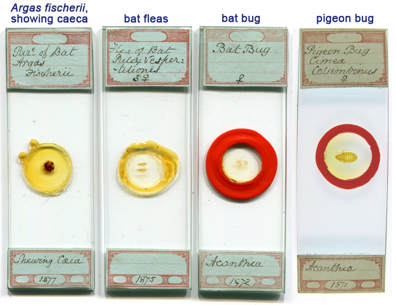
Figure 6.
Slides by “B2” with specimens described by John
Bramhall in 1877. He noted that he had previously labeled a slide “as Ixodes of Bat’, but shall alter it to Argas Fischerii, as that seems to be
its true appellation”. Bramhall described that he had mounted an Argas
fischerii which “shows the caeca perfectly”. He also wrote of mounting bat
fleas, bat bugs and pigeon bugs. Bat bugs and pigeon bugs are relatives of the
human bedbug.
In summary: John Bramhall is the
only person known to have discussed pygidia of ticks/ixodes. He wrote of bat
ticks, fleas and bugs, and of pigeon bugs. All of those specimens were
described by him in the context of microscopy. Microscope slides by mounter
“B2” were identified that match Bramhall’s descriptions of those parasitic
arthropods. The monogram of “B2” is comprised of the letters “J” and “B”, with
“B” central and dominant, indicating it to be the initial of the last name.
Based upon this evidence, we hypothesize that mounter “B2” was John Bramhall.
Due to the similarities between “B1” and “B2”, we think it is likely that
Bramhall was also “B1”.
John Bramhall
John Bramhall was born December 5,
1809, in Westminster (now part of London), the child of Thomas and Sophia
Bramhall. John attended Exeter College at Oxford University, graduating in 1834
with a B.A. in Literature and Humanities. He was made an Anglican deacon in
1837, and a priest in 1838. Bramhall apparently served as a minister in
Avening, Gloucestershire, being recorded as living in that town on the 1841
census, in the household of his aunt, Frances Clutterbuck. Bramhall was
appointed Vicar of Terrington St. John, Norfolk, in 1843, and remained at that
post until the early 1880s. He also served as Justice of the Peace from 1852,
and Rural Dean from 1865.
Bramhall married Clara Gilchrist in August, 1841, in
Middlesex (London). According to census records, they did not have any children.
The first record of Bramhall’s
interest in microscopy is a request for diatom samples, published in Hardwicke’s Science-Gossip, in 1868, “Wanted in exchange, or by purchase,
Eupodiscus Rogersii, Glyphodiscus stellatus, Brightwellia elaborata, B.
Johnsoni, B. coronata, and Heterodictyon splendidum. - Address, Rev. J.
Bramhall, St. John's Vicarage, King's Lynn”. He posted another request for
diatoms in 1870, “Wanted, in exchange or
otherwise, Valves of Aulaeodiscus
Kittoni, with more than four rays, or processes, and A. Comberi, with more than three
ducts”. A third request, from 1876, “Wanted
- Slides of Stauroneis spicula and Schizonema cruciger, in
exchange for well mounted Slides of Diatoms from Subpeat, Troy.
New Hampshire, U.S.A.”.
His other microscopy exchange offers
involved attempts to trade lenses. In 1872, “For exchange or otherwise, an excellent English 1/8-in.object glass of
120 degrees angular aperture, in perfect condition. - Apply to the Rev. J. Bramhall, St. John's Vicarage, near Lynn, Norfolk”.
He appears to have advertised the same lens in 1875, “For exchange or otherwise, a first class 1/8 Object Glass, by Thomas
Ross, especially selected for the late owner”. Another lens was offered in
1876, “For exchange or otherwise, a 4-in
object-glass by Ross, and an Achromatic Condenser, in perfect order”.
Bramhall
created a stir in microscope circles in 1876, with the publicizing of his
invention that came to be called the “Bramhall Oblique Illuminator”. This
simple device consisted of a platform on which to place a microscope slide,
with an inset mirror placed approximately 1/8 inch (2-3 mm) below the surface
(Figure 7). In use, light focused by a bull’s-eye condenser would be shone at a
sharp angle, reflecting off the mirror, then off or through the specimen on the
slide. This was especially useful for oblique lighting of diatoms, as a means
to emphasize their structures.
The famed diatomist Frederick
Kitton raved about Bramhall’s Oblique Illuminator to the Royal Microscopical
Society, “I have much pleasure in
calling the attention of those interested in the resolution of the striae on
the finely lined forms of Diatomaceae to a simple form of apparatus invented by
the Rev. J. Bramhall, of Lynn, which after a careful trial I am able to say
is superior to anything I have hitherto had the opportunity of trying. This
illuminator in its simplest form consists of a disk of silvered glass about one
inch in diameter, mounted in a wood or brass fitting similar to a selenite
stage; the disk should be sunk in it about one eighth of an inch. Mr. Bramhall informs me that with sunlight it resolves striae far
better than any other mode of illumination. My own experience has only been
with the ordinary micro lamp; this requires to be elevated from three to four
inches above the stage, and the light, after passing through the large
bull's-eye condenser, should impinge on a smaller one placed close to the
stage, the height of the lamp from the surface of the condenser of course regulating
the obliquity of the reflected ray. The performance of the reflector is
improved if, instead of being constructed of silvered glass, a disk of speculum
metal is substituted, and in place of the supplementary stage the reflector
should be mounted on an adapter fitting into the sub-stage of the microscope;
by this means the distance from the lower surface of the slide can be
regulated. This illuminator can also be used in place of the ‘Spot Lens’ with
the lower powers, but a short tube to slide on the objective is necessary to
prevent the reflexion from the upper surface slide passing into the objective.
I find that the tube attached to the Lieberkuhn (the reflecting portion being
detached) answers very well. Careful adjustment of the objective is necessary,
and the object should be perfectly parallel with the stage of the microscope”. Kitton
also wrote in Hardwicke’s Science-Gossip,
“The utility of a more or less oblique pencil of light has
long been recognized by those who use the higher power of the microscope. The
instruments contrived for obtaining this have been various, and many of them
very complicated and expensive, and, so far as my own experience goes, none of
them have produced a more effective oblique ray than can be obtained by a mirror
on a separate stand, or the lamp placed at an angle with the axis of the
microscope. The Rev. J. Bramhall, of Lynn, has called my attention to the
following arrangement, and asks if it is new, which, so far as I am aware, it
is .. The objectives with which the illuminator was tested were a 1/8 of
Baker's, made many years ago, but a very good glass, and a ‘Beneche No. 7’, using the following objects as
tests:—Navicula rhomboides, N.
cuspidata. N. peregrina, N. rostellum, Synedra robusta, Nitzschia sigmoidea, N.
sigma (the finely-marked variety called by Möller N. curvula), and Lepidocyrtis curvicollis. The 1/8
resolved the transverse striae of N.
rhomboides, which it had not done before; the longitudinal striae on N. cuspidata shown very sharp and
distinct, as were also the transverse striae on costae of N. peregrina and S. robusta. The beautiful curved
striae on N. rostellum were
better shown than I have ever before seen them. N. sigmoidea - The striae resolved with as much ease as those on Pleurosigma angulatum with a 1/4. N. sigma as difficult as N. rhomboides. The ‘Beneche’ resolved
N. sigmoidea, N. rostellum. N.
peregrina, and S. robusta. The
podura scale was shown well, but not better than by a less oblique ray. The
mirror would probably be more effective if mounted so as to slide up or down in
the substage tube; but this, of course, would be more expensive. It is
desirable that the cover should not be attached to the slide with the usual
black varnish, but with ‘dammar’, or some similar transparent cement”. Kitton also noted that the C.
Baker optical works was manufacturing and selling Bramhall’s Illuminators
(Figures 8 and 9).
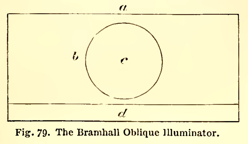
Figure 7.
The diagram of a Bramhall’s Oblique Illuminator that accompanied Frederick
Kitton’s 1876 ‘Hardwicke’s Science-Gossip’ article. Kitton wrote, “The
subjoined diagram will enable any one possessing the slightest mechanical
ability to manufacture one of these illuminators without difficulty, a is a
piece of wood 3 1/4 inches in length, 1 1/2 inch in breadth, and 3/16 of an
inch thick; the central perforation b is 3/4 of an inch in diameter, in which
is placed a silvered disk of glass or metal, c, the face of which should be not less than 1/8 of an inch
below the upper surface of the wood, d, a ledge for the slide to rest against. We will now describe the method
of using it, premising that a light of considerable obliquity is required. The
wooden stage, with its mirror, is placed on the stage of the microscope, and
the lamp elevated some 3 or 4 inches above it, and a beam of light condensed upon
the mirror by means of a ‘bull's-eye’. I need scarcely observe that the
obliquity of the reflected beam depends upon the angle at which the ray
impinges on the mirror”.
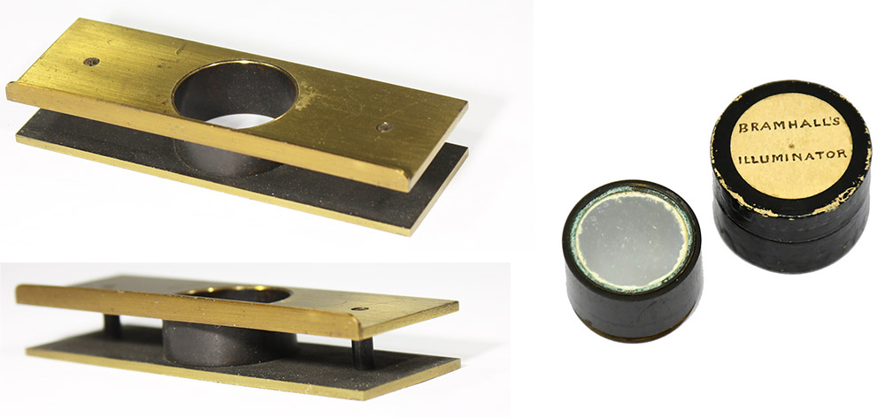
Figure 8.
Components of a commercially-produced Bramhall’s Oblique Illuminator, probably manufactured ca. 1878 by Baker (see Figure 9). (Left) Brass base, the size of a standard microscope slide (1 x 3 inches), and 7/16 inch (1.1 cm) tall, with a central cylinder to accept the mirror, and a lip along one edge to hold a microscope slide in place. (Right) A mirror insert and its case. Images adapted for nonprofit, educational purposes from an internet sale site.
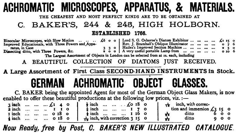
Figure 9.
An 1878 advertisement from Baker. At that time, they were selling Bramhall’s Oblique Illuminators for 7 shillings 6 pence.
Bramhall subsequently wrote to Hardwicke’s Science-Gossip, “Mr. Kitton, one of our first authorities on
all that concerns diatoms, has been pleased to express his approbation of this
invention, and to name it after its inventor. I can confirm all that he has
said of its resolving power. Whatever an object-glass is capable of doing, I
believe it will enable it to do. In addition to the tests named by Mr. Kitton,
I may add, that with a Siebert's No. 7 immersion, and the A eyepiece, I have by
the aid of the Illuminator resolved the following, which I consider the most
difficult tests in their class. Pleurosigma
Macrum, Navicula Crassinervis, Frustulia Saxonica, and Amphipleura Pellucida. The only test
that has so far beaten me is Stauroneis
Spicula, and of that I have ‘glimpsed’ the markings. Before I found out
this method of illuminating, I could never really resolve any of the more
difficult tests, not even Navicula
Rhomboides, in spite of achromatic condenser, spot lens, prisms, and
stops of various kinds, but by its aid, that old difficulty N. Rhomboides, dry or in balsam, is
as easy as P. Angulatum was of
old. The illuminator is made in two forms. The one, represented in Mr. Kitton's
paper, does for such microscopes as have no sub-stage; the other, and the best,
fits the sub-stage, rising and falling by aid of the rackwork. I use a silvered
glass mirror of ¾ inch in diameter, and a polished metal disc of about an inch,
which fit the same holder. On the whole, I prefer the metal; the chief merits
of the invention are its simplicity, efficiency, and cheapness. I use only
sunlight or clear daylight, but believe it will work equally well with the
lamp. Nothing can be more easy than its use. Throw the light on to the object more or less
obliquely, as that particular object requires, and regulate the reflector
beneath, as experience alone can determine. I hope my brother microscopists
will have as much success in the use of this illuminator as I have had, and I
am sure they will be satisfied. - John
Bramhall, St. John's Vicarage, near Lynn”.
William K.
Bridgman further modified Bramhall’s device, creating a finely-adjustable
substage mechanism that afforded precise control of the mirror for directing
oblique light (Figure 10). He wrote in the Journal
of the Quekett Microscopical Club, in 1876, “Having for several years past been endeavouring to obtain a controlling
command over the obliquity of an illuminating pencil of light by means of a
metallic speculum, placed between the condenser and the object, I was
altogether unsuccessful until the advent of the ‘Bramhall’ reflector, which, by
suggesting the substitution of a small bull's-eye lens in the place of the
ordinary hemispherical condenser, gave a new turn to the direction of the
investigation, which has resulted in a complete success. The Bramhall
arrangement consists of a reflecting surface placed beneath and within a very
short distance of the object; whilst the light, being thrown down as for an
opaque substance, first passes through the glass slip, and is then reflected
upwards, so as to produce a transparent illumination of great obliquity. There
are, however, several very important drawbacks in addition to the difficulty of
adjustment, and the very limited range of action, which restricts its use to a
comparatively small class of objects, and of these, to only a small proportion
of such as are mounted in the ordinary way; yet, all who have once seen the
marvellous sharpness and force with which the most difficult diatom tests can be displayed, must
be quite ready to admit the principle to
be very far in advance of all other modes of illumination, making it highly
desirable that it should form the basis of some other special arrangement, that
would not only be equally effective, but be easily managed, and free from its
other objections, at the same time extending alike superiority of effect to all other classes of objects, and
equally applicable under all the common conditions of ordinary mountings -
properties that are now claimed for the present arrangement”.
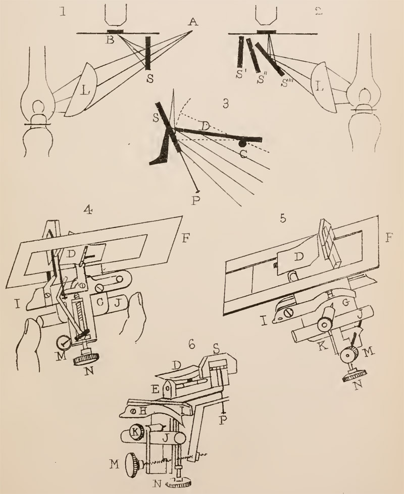
Figure 10.
Diagrams that accompanied W.K. Bridgman’s variation
on the Bramhall’s Illuminator, as described in the Journal of the Quekett
Microscopical Club, 1876. Bridgman’s device held a mirror below the microscope’s
stage, and permitted fine adjustment of the mirror to precisely control the
angle at which oblique light may be shone upon an object.
Controversy quickly arose as others claimed prior invention of the Bramhall Illuminator. Since Baker was marketing Bramhall’s device, substantial money was at stake.
The Royal Microscopical Society’s Monthly Microscopical Journal reported,
in 1876, “Dr. R.H. Ward has sent us the
following note, contributed by him to the December number of the 'American Naturalist', and as it bears upon a question as
to priority of invention, it will doubtless interest our readers: ‘At the
Indianapolis meeting of the American Association for the Advancement of
Science, in August, 1871, P.H. Van der Weyde, of New York, described a contrivance,
believed to be new, for oblique illumination of transparent objects. It was
designed chiefly to facilitate the resolution of lined or dotted objects, and
consisted of a plane mirror lying beneath the object-slide and parallel to it,
from which mirror light, condensed upon it from above by means of a bull's-eye
condenser, would be reflected back at the same angle through the object and
into the objective. These illuminators were shown in successful operation at
the meeting, working best with moderately high powers, and were freely
distributed among the members present. They were briefly described in the
'American Naturalist' for September, 1871, being there estimated as 'a little
expedient of great practical convenience’. Ever since that time the present
writer, among others, has used them habitually, shown them freely, and not
unfrequently given them away. The mirror may be either of silvered glass or of
polished metal. In some cases the object-slide may lie directly upon it while
it rests upon the stage; but frequently the object-slide is best elevated
slightly above it. The mirror is most conveniently made of the size of a slide
(3 x 1), and furnished with glass strips at the ends to support the slide at
any required height; but it may be made smaller, say one inch square or round,
and sunken in a brass or wooden stageplate, or for stands having a sub-stage of
any kind it may be made of suitable size and supported from the sub-stage and
adjusted for height in the same manner as the achromatic condenser. It has the
advantage of great ease of manipulation and applicability to any stand, and the
drawback of being liable to be interfered with by the presence on the slide of
such obstructions as paper covers or opaque cells or rings of varnish. Within a
few months past it has been brought forward by Rev. John Bramhall,
of Lynn, England; its previous use and publication having either escaped
the notice or slipped from the memory of himself as well as of the
distinguished microscopist who has indorsed it and proposed to name it after
him".
Kitton then responded, “The
oblique illuminator, which I proposed to name after my friend the Rev. J. Bramhall, was, I know, a new thing, so far as he is
concerned. On discovering the effective performance of a reflector parallel to
the slide, he at once wrote to me to ask my opinion of its value, and also to
ask whether it was new. I made a rough trial, and found, with every
disadvantage, it resolved striae with ease, that had formerly taken me some
time to bring out; and as I was not aware that this method of illumination had
previously been described, I wrote a short description of it for
'Science-Gossip' and this Journal, calling it the ‘Bramhall Illuminator’. On June 30, 1876, I received a letter from the Secretary
of the Microscopical Department of the Providence
Franklin Society (Mr. John Peirce), in which he tells me that ‘Mr. Norman
Mason, of Providence, R.I., accidentally discovered some time ago identically
the same thing as you describe as 'Bramhall's Oblique Illuminator’. He was
endeavouring to find a piece of plate glass of uniform thickness. He used
pieces of broken mirrors, strewed with Lycopodium, for the purpose of
focussing, and was astonished at the illumination. He afterwards made use of
this illumination for diatoms, and found, as you state, that the distance of
the mirror from the object made quite a difference. Yours respectfully, John
Peirce’. On July 10, 1876, I received a letter from my friend Professor H.L.
Smith, of Hobart College, N.Y., from which I quote the following remarks: ‘I
have just noticed your account of Mr. Bramhall's oblique light illuminator. It
is an old dodge over here. I have one, made over ten years ago; a piece of
looking-glass plate with a ledge’. It thus appears that this method of
illumination has had several discoverers. It seems to have been known in New
York five or six years before Mr. Van der Weyde of that city described it in
August 1871, as probably a new contrivance; Mr. Mason, of Providence,
discovered it ‘some time ago’ (this may mean six or seven years); and lastly,
Mr. Bramhall ‘discovers’ the same thing about
twelve months ago. But leaving the discoverer out of the question, it is the
best and easiest plan for the resolution of striae I am acquainted with”.
Bramhall wrote in 1877, “Allow me to say, in answer to the concluding
paragraph of Dr. Ward's letter on Mr. Van der Weyde's claim to the invention,
which goes by the name of the Bramhall Oblique
Illuminator, that, so far as I am concerned, it is original, as I had never
heard of Mr. Van der Weyde, or of that other American microscopist who claims
to have used the same form more than ten years ago. I believe I may say the
same on behalf of Mr. Kitton”.
The editor
of The American Journal of Microscopy appears to have put an end to the
controversy with this 1877 editorial, “The Bramhall Illuminator – This little piece of apparatus, which
was fully described in a former number, is receiving great favor in England,
many of the microscopists there declaring that by its means they are enabled to
resolve objects which were previously entirely beyond their reach. It seems,
however, to have been known to several American microscopists, some of whom now
lay claim to the honor of inventing it, while one microscopist of note, who had
employed it some ten years ago, considers it of very little value. It seems to
us, however, that if there is any great merit in the device, the only person to
whom the microscopic public owe thanks for it, is Mr. Bramhall. The other inventors have taken no pains to give it to their fellows, or
to have it manufactured and placed in market. Mr. Bramhall promptly handed it to a well-known microscope maker, and rendered it
accessible, in its best condition, to every one. He then published a
description, with figures, in a journal of large circulation, and so told
almost every one interested, just where they might get it. The simultaneous
invention of a particular device by several different persons is nothing
unusual. Neither is it uncommon for an invention which in its earlier form was
rude, inefficient or inconvenient, to be brought forward at a subsequent period
and be generally accepted. In one sense there is nothing new under the sun; in
a higher and better sense, every day sees something new. The invention of the
nose-piece is generally attributed to Mr. Brookes; it is called by his name,
and he gets the credit of having invented and introduced it, and justly so. But
we had in our possession, a short time ago, a London microscope, made about the
close of the last century, which possessed a very elaborately constructed
nose-piece! We have now before us a book which was published nearly two hundred
years ago, in which there is an engraving and description of a binocular
microscope. In the same work there are figures of telescopes, or, rather, opera
glasses, in which the distance of the two tubes from each other can be
regulated to suit the distance at which the eyes of the observer are placed
apart - just as we find in the modern binocular microscope. But all this does
not detract one iota from the credit of the men who have given us the modern
binocular. In order to have the credit of an invention, the inventor must not
only present it in a usable form, but he must so publish it that other workers
can avail themselves of its advantages. This Mr. Bramhall has done, and this
the others left undone”.
As Francis
Darwin said in another context, “In
science, the credit goes to the man who convinces the world, not to whom the
idea first occurs”.
John
Bramhall was elected to membership of the Quekett Microscopical Club on May 25,
1877. He remained on the rolls through 1880.
Bramhall also published several brief
hints on methods for preparing microscope slides and, in 1870, a letter on the
pseudoscorpion, Chelifer, and on pheasants,
“Chelifers - Allow me to say a word on
behalf of the much-maligned chelifer, which has been described by a writer in
your periodical as of a most quarrelsome and sanguinary disposition. I have
kept numbers of Chelifer cancroides together
for several months; and though I have constantly watched them, I never saw a
quarrel or even an attempt at one; on the contrary, they seemed to be on the
very best terms, and very polite and well behaved. I wish also to mention a
curious fact in natural history which has come under my notice, and of which I
believe there have been many instances recorded. A neighbour of mine had a pair
of silver pheasants, male and female; last year the male bird died, and since
then the female has assumed the complete
plumage of the male, and appears, I presume from the greater amount of
plumage, to have increased in size. This bird is now to be seen at Levell's
Hall, Terrington St. Clement, near Lynn.— John
Bramhall, St. John's Vicarage.”
In addition to microscopy, Bramhall
had strong interests in apple growing and archery. He appears to have been a
member of the British Pomological Society, and may have developed a new apple
variety. He was noted to be an expert target archer, and was deeply involved
with promotion of that sport.
Bramhall evidently gave up his microscopy hobby in 1885. In May of that year, he advertised to sell his Smith and Beck binocular microscope, associated apparatus, and three cases holding about 2000 slides (Figure 11). John was retiring from his Norfolk ministry during that time. His wife, Clara, died the following year, when they were living in Eastbourne, Sussex. John died on November 1, 1889, at home in Eastbourne. His estate was left to his half-brother William and one “May Kingsford of Sunbury-on-Thames”, of unknown connection.
.
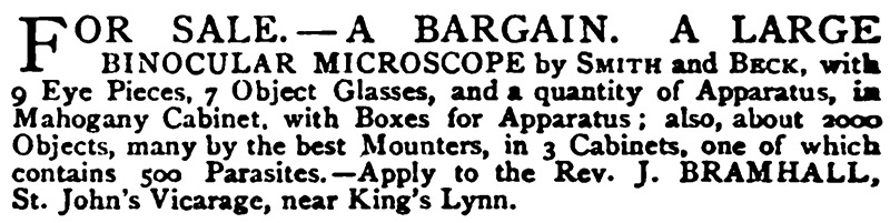
Figure 11.
May, 1885 advertisement from "Hardwicke's Science-Gossip".
Acknowledgements
Many thanks to Howard Lynk, Tom Schwan and Maureen Carter for their helpful comments.
Resources
The American Journal of Microscopy (1876) A new oblique light illuminator, Vol. 1, page 96
The American Journal of Microscopy (1877) The Bramhall illuminator, Vol. 2, page 48
Baptism record of John Bramhall
(1810) St. James Piccadilly parish records, accessed through ancestry.co.uk
Bracegirdle, Brian (1998) Microscopical Mounts and Mounters,
Quekett Microscopical Club, London, pages 182 and 188, and plates 39-O and 42-K
Bramhall, John (1870) Chelifers, Hardwicke’s Science-Gossip, Vol. 6, page 120
Bramhall, John (1876) The Bramhall
oblique illuminator, Hardwicke’s
Science-Gossip, Vol. 12, page 159
Bramhall, John (1876) The Bramhall
oblique illuminator, Hardwicke’s
Science-Gossip, Vol. 12, pages 231-232
Bramhall, John (1877) A word about
the “pygidium”, Hardwicke’s
Science-Gossip, Vol. 13, page 16
Bramhall, John (1877) How to resolve
test diatoms without any special apparatus, Hardwicke’s
Science-Gossip, Vol. 13, page 16
Bramhall, John (1877) The pygidium, Hardwicke’s Science-Gossip, Vol. 13,
page 66
Bramhall, John (1877) Letter to the
editor, Monthly Microscopical Journal,
Vol. 17, pages 106-107
Bramhall, John (1878) To clean old
slides, Hardwicke’s Science-Gossip,
Vol. 14, page 15
Bramhall, John (1878) Removing
surplus balsam, Hardwicke’s
Science-Gossip, Vol. 14, page 15
Bridgman, W.K. (1877) On a new
universal reflecting illuminator, Journal of the Quekett Microscopical Club, Vol. 4, page 214
Crockford’s Clerical Directory (1870) “Bramhall,
Joh, Terrington St. John, King’s Lynn, Norfolk. - Ex. Coll. Ox. B.A. (4th el. Lit. Hum.) 1832; Deac. 1837. Pr. 1838 by Bp of G. and B. V. of Terrington St. John, Dio. Nor.
1843. (Patron, the Crown; Tithe
206£ 4s 11d; Glebe 5 acres; V.’s Inc. 220£; Pop. 793.) J.P. 1852; Rural Dean
1865”, page 88
England census, birth, marriage and
death records, accessed through ancestry.co.uk
The Florist, Fruitist, and Garden Miscellany (1859) Report on the May 5 meeting
of the British Pomological Society, “Rev. J. Bramhall, of St. John's near Lynn, again
sent specimens of Clissold's Seedling, or Lodgemore Nonpareil, a seedling which
he submitted to the society at the meeting of May 6th last year. A very high
opinion was then expressed regarding it, and which, this year, was fully
sustained. It is a most valuable dessert Apple at this late season”,
Vol. 12, page 245
Hardwicke’s Science-Gossip (1868) Exchange offer from John Bramhall, Vol. 4, page 72
Hardwicke’s Science-Gossip (1870) Exchange offer from John Bramhall, Vol. 6, page 120
Hardwicke’s Science-Gossip (1872) Exchange offer from John Bramhall, Vol. 8, page 168
Hardwicke’s Science-Gossip (1875) Exchange offer from John Bramhall, Vol. 11, page 168
Hardwicke’s Science-Gossip (1875) Exchange offers from John Bramhall, Vol. 12, pages
24 and 216
Hardwicke’s Science-Gossip (1878) Exchange offer from John Bramhall, Vol. 14, page 48
Hardwicke’s Science-Gossip (1878) Advertisement from C. Baker, Vol. 14, January issue,
advertising section
Hardwicke’s Science-Gossip (1885) Sales offer from John Bramhall, Vol. 21, page liv
Journal of the Quekett Microscopical Club (1876) Exhibits at the October 27
Ordinary Meeting: Navicula cuspidata
(with Bramhall illuminator) by Mr. Roper, Vol. 4, page 237
Journal of the Quekett Microscopical Club (1877) Notes from the March 23
Ordinary Meeting, Vol. 4, page 292
Journal of the Quekett Microscopical Club (1877) Election of John Bramhall to
Membership, May 25
Kitton, Frederick (1876) The
“Bramhall” reflector, or new oblique light illuminator, Monthly Microscopical Journal, page 102
Kitton, Frederick (1876) A new
oblique light illuminator, Hardwicke’s
Science-Gossip, Vol. 12, page 136
Kitton, Frederick (1876) Markings on
Surirella gemma, Hardwicke’s Science-Gossip, Vol. 12, page 160
Kitton, Frederick (1877) The oblique
illuminator, Monthly Microscopical
Journal, Vol. 17, pages 106-107
Longman, C.J. and W. Wolrond (1894)
John Bramhall, Archery, Longmans,
Green & Co., London, pages 300-302
Marriage record of John Bramhall and
Clara Elizabeth Gilchrist (1841) Sudbury on Thames St. Mary parish records,
accessed through ancestry.co.uk
The Monthly Microscopical Journal (1877) Van der Weyde’s oblique illuminator, Vol. 17, page
49
Probate of
John Bramhall (1889) Accessed through ancestry.co.uk
West, T.
(1860). Remarks on some Diatomaceae new or imperfectly described and a new
desmid. Transactions of the Royal
Microscopical Society, Series 2, Vol. 8, pages 147-153