George Howe Curtis, 1842 - 1901
by Brian Stevenson
last updated July, 2024
American G.H. Curtis was an expert on the diatoms near his homes in Ohio and Kansas. He wrote authoritative descriptions of fossil diatoms in the Cincinnati Group of mineral layers, and of freshwater diatoms in central Kansas. Curtis donated slides to the Smithsonian Institute, as well as his local scientific societies. Slides that are encountered nowadays probably came from his exchanges with fellow diatomists or from breakup of Curtis’ collection.
Known slides by Curtis bear labels with his address in Kingsville, Ohio, which is located near Lake Erie, between Cleveland and the Pennsylvania border (Figure 1). Yet, at least one of those was prepared in 1891, when he lived near Cincinnati. This suggests that Curtis purchased custom-made labels when he lived in Kingsville, but continued to use them after he left that town (which would have been before 1889)
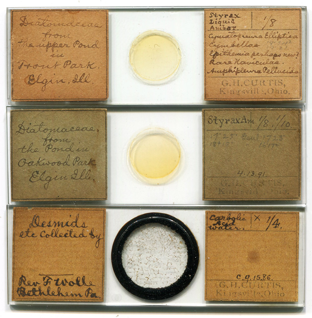
Figure 1.
Examples of microscope slides that were prepared by George H. Curtis. The top two are gatherings of freshwater diatoms, and the bottom is a mix of desmids that he obtained from Reverend Francis Wolle of Bethlehem, Pennsylvania. Curtis wrote lists of interesting species on the right-hand labels. All of these slides bear labels with his address as Kingsville, Ohio, even though one is dated April 13, 1891, when Curtis lived at the other end of Ohio, near Cincinnati.
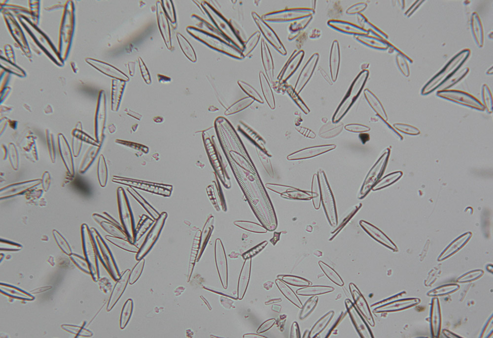
Figure 2.
A mix of diatoms from “the upper pond in Trout Park, Elgin, Illinois”, mounted in Styrax liquid ambar (see Figure 1). Curtis listed some notable genera and species on the slide: “Cymatopleura eliptica, Cymbellae, Epithemia perhaps new?, rare Naviculae, Amphipleura pellucida”. Imaged with a 25x objective lens and C-mounted digital SLR camera on a Leitz Ortholux II microscope.
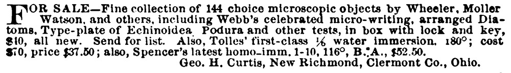
Figure 3.
An 1899 advertisement from George H. Curtis, when he lived near Cincinnati, Ohio. From “The Microscope”.
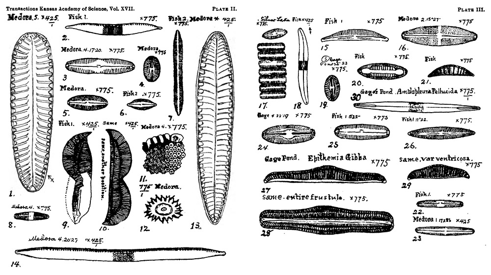
Figure 4.
Some diatoms of Reno County, Kansas, drawn by George H. Curtis. From his 1900 paper on that subject.
George Howe Curtis was born during December, 1839, in New York (possibly in the city of Ithaca). He was the eldest of eight children of Orren and Emily Curtis. The 1850 national census listed Orren’s occupation as “tin smith”. During the 1850s, the family moved to Ashtabula County, Ohio. The town of Kingsville, where our microscopist and other members of his family lived, is located in that county. Orren died in 1858. The 1860 census listed George’s occupation as “telegraph operator”, and he was probably the main breadwinner of the family. George’s whereabouts for the next 29 years are uncertain. As noted above, a microscope slide dated 1891 has a label with a Kingsville address, suggesting that he lived there a relatively short time before that date. The 1900 US census listed Curtis's employment as "manager", without further details. He was also listed as being a “widower”, but no information is known about his wife. The probate of his will stated that he had no next of kin except siblings and nieces/nephews.
The earliest record of George Curtis’ scientific interests is a January, 1885 donation of “ten microscopic slides of hairs and scales” to the Cincinnati Society of Natural History. In July of that year, he donated another two slides of unknown subjects. A diatom slide was donated in August, 1886.
A person with initials “G. H. C.” wrote an article on preparing glycerin mounts for The Microscopical Bulletin in 1888. It was later reprinted in The Journal of the Royal Microscopical Society.
It is possible that the above-described donations by Curtis to the Cincinnati Society of Natural History occurred before he move to that area, and while he lived in Kingsville. But, he was certainly in the Cincinnati area by 1889. That year’s annual report of the Smithsonian Institute acknowledged a donation of “mounted slides showing the Diatomaceae of the Cincinnati group” from “Curtis, George H. (Cincinnati, Ohio)”. He also advertised to sell microscope components and professionally-prepared slides in October, 1889, giving an address of New Richmond, Ohio, which is on the outskirts of Cincinnati (Figure 3).
In 1890, Curtis published to articles on microscopical techniques, “On the cleaning and mounting of the diatomaceae” and “How to mount objects in motion for examination by polarized light” (the latter was reprinted in The Journal of the Royal Microscopical Society). Both are reproduced at the end of this essay, since that may be of benefit to modern slide-makers.
Curtis probably moved to Kansas during the early 1890s. He was almost certainly there by the spring of 1893, based on his donation of “Guinea corn and milo maize, from Kansas” to the Cincinnati Society of Natural History in April of that year.
His interest in diatoms grew considerably, probably because the species of the American plains had never before been described. In 1899 and 1900, Curtis published three papers in The Transactions of the Kansas Academy of Science. They were republished in 1901 in The American Monthly Microscopical Journal. He noted, “Although the Diatomacea of the eastern United States have been pretty thoroughly investigated by several competent observers, I believe that little is known as to what forms exist in the West. With the exception of Thomas and Chase's catalogue of the Diatomacea of Lake Michigan, which embraces 34 genera and 214 species, and my own catalogue of the Cincinnati forms, 42 genera and 972 species, there are no investigations of western or central-western forms known to me. Under these circumstances, perhaps a short paper upon Kansas forms may possess sufficient interest to justify reading it”.
George H. Curtis died on April 5, 1901, in Painesville, Ohio. His brother Frank, lived in that town. The short time between his Kansas diatom papers and his death in Ohio suggests that George had fallen ill and moved in with his brother for care.
__________________________________
George H. Curtis, “On the cleaning and mounting of the diatomaceae”, from The Microscopical Bulletin and Science News, 1890:
“I usually collect in phials of 2 drams as I find a large quality of material unnecessary. After settling, put a dram of the sediment into an 11⁄2 oz. test tube, pour on about twice as much nitric acid and boil for 15 minutes, then add bichromate of potash, size of a pea, crushed fine, boil again for a minute; then pour all into a tumbler of cold rain water, stir up and let settle for half an hour, carefully decant and add fresh water, repeating until the acid is all washed out. Transfer the sediment to clean test tube and add 1⁄2 ounce rain water and a piece of yellow ‘palm soap’, size of a pea, broken smaller, boil for two or three minutes, then turn into a tumbler of rain water preferably hot, as a better solvent of soap, let settle for half an hour and repeat until the soap is entirely washed out. Return the white sediment to a clean test tube which half fill with rain water and thoroughly shake up to wash off adherent fine sand and clean the Diatoms. Let stand 10 minutes, and half that time if you only want the larger forms. Pour all the water except the last half dram into another test tube and save it for the small forms.
This is the process of Prof. H. L. Smith, substantially, and I now come to mine:
Prepare a little handle, say 3 inches long and 1/8 inch in diameter, make a hole in one end with a needle, in which insert a fine bristle, or white horse hair, taper that end of the handle small and leave the bristle half an inch long.
After settling 10 minutes take up a small quantity of sediment from bottom of test tube with a pipette and lay at the end furthest from you of a clean glass slide of the usual size. A drop or two of water with the sand and sediment will be sufficient, as you only want enough to float the Diatoms. Now spread it out a little on the plate with the end of the pipette, and standing near a window, with the carpet, or something dark for a background, slightly incline the glass sideways, and all the Diatoms will run down toward the right, or long side of the plate, leaving the sand lying where you placed it. Now coax the Diatoms along down the edge of the plate towards you, inclining it just sufficiently to flow them and leave other matters not wanted behind; and pushing back any little specks of dirt or stuff with the bristle. Diatoms are very thin and small and only barely enough water should be used to float them and leave the impurities which require more depth lying stranded behind. If the stream inclines to stop, breathing on the plate will start it, or a little track can be made with the bristle. In this way they can be led along to the corner toward you. Lay under the microscope and examine, follow the course of the stream back as some large ones may be stranded above which can be brought down to the corner with the bristle. If not sufficiently clean wipe all dirt off the plate and lead the Diatoms to another corner, leaving the dirt behind as you go.
Have ready a cover as clean as possible, especially from greasiness, set the corner of the plate on it, scrape off the Diatoms and spread them out over it with the bristle. If they do not spread evenly and stay where placed, your cover is not clean. Set aside to dry, and mount just as soon as dry enough, as many absorb unsightly bubbles of air if not mounted immediately. Mount in ‘sweet gum’, or styrax liquidambar dissolved in a mixture of benzine and alcohol. Put a small drop in centre of slide, turn the cover over it and hold over a kerosene lamp chimney until it boils up and the bubbles get large, then adjust cover to centre and set aside to cool.
By this process, sand, the thing usually complained of, is no trouble at all. The difficulties are: some Diatoms grow in a kind of jelly, apparently siliceous, which resists the action of acid and leaves a kind of white, fluffy, or jelly like substance, behind after treatment, of about the same sp. gr. as the smaller Diatoms, which washes and settles with them. This may usually be eliminated by subsidence and manipulation with the bristle. Sometimes agitation in the test-tube with a little aqua ammonia for 5 or 10 minutes will help it, washing it out afterwards. Levigated earth is troublesome in some gatherings. In such cases probably not enough soap was used and it may be necessary to soap again, or it may perhaps be got rid of by shortening up on the time allowed for subsidence in the different washings, so as to give just time enough for the Diatoms to settle, but not this fine stuff.
Perfectly fresh gatherings do not clean up as well as those that stand a week or two before cleaning.
As a rule the longer you boil in nitric acid the less trouble you will have afterwards.
Probably the best and quickest way to combine both small and large forms in one mount would be to treat the sediment from 1st and 2d tubes on the glass slide separately and mix on the cover with the bristle. Do not press the cover down too hard in mounting, as many large forms are broken by it.
I have mounted slides in this way as good as any I have seen, and it is little trouble after you get accustomed to it.
*In a note accompanying the article, Mr. Curtis says: In reply to your question, I would say that sand by this process gives me no trouble at all and I easily make slides by it that are perfectly clean. I have carefully sought to make the article as short as possible, but I do not see how it can be cut down any shorter without omitting important details. Neither do I think the remarks at the end can be profitably omitted, but I leave it to your judgment. I should have been glad to extend them so as to say a word on quality of soap, and more than I have on the important matter of subsidence, etc., but realizing that you perhaps had not space I have cut it short as I could”.”
__________________________________
George H. Curtis, “How to mount objects in motion for examination by polarized light”, from The Microscopical Bulletin and Science News, 1890:
“None of the manuals I have consulted give directions for preparing slides of objects in motion for polariscope. Rubber cells are best and they should be about 1/16 of an inch deep and preferably for a 3/4 cover. The medium I use is Canada balsam thinned with a not quite equal bulk of spirits turpentine. Stir well together, and when dissolved filter through cotton. Cement the cells to slide with something not acted on by turpentine, say shellac, or sealing wax in alcohol. Le Page's liquid glue I think would answer, but I have not tried it. The fragments may be quartz, agate, sand or anything not soluble in turpentine, which polarizes well. One of the best is transparent gypsum or sulphate of lime. It is unnecessary to cement the cover on; set aside for a couple of days and the balsam will get dry enough to hold it. Should you wish to ring them with Brunswick black, size first with a coat or two of liquid glue made thin enough to flow, or the black will probably run in and spoil the slide”.
Resources
Curtis, George H. (1890) On the cleaning and mounting of the diatomaceae, The Microscopical Bulletin, page 12
Curtis, George H. (1890) How to mount objects in motion for examination by polarized light, The Microscopical Bulletin, page 2
Curtis, George H. (1899) Some diatomaceae of Kansas, Transactions of the Kansas Academy of Science, pages 67-78
Curtis, George H. (1899) Diatomaceae of Gage’s Pond, Topeka, and of Silver Lake, Transactions of the Kansas Academy of Science, pages 67-70
Curtis, George H. (1900) Diatomaceae of Reno County, Kansas, Transactions of the Kansas Academy of Science, pages 70-75
Curtis, George H. (1900) The food of fish in central Kansas, Transactions of the Kansas Academy of Science, pages 75-78
Curtis, George H. (1901) Diatomaceae of Gage’s Pond, Topeka, and of Silver Lake, Curtis, The American Monthly Microscopical Journal, pages 263-267
Curtis, George H. (1901) Diatoms, the food of fish in Kansas, The American Monthly Microscopical Journal, pages 275-280
Familysearch.org (accessed July, 2024) George Howe Curtis, https://www.familysearch.org/tree/person/details/GC7L-5LZ
Find-a-Grave (accessed July, 2024) Frank B. Curtis, https://www.findagrave.com/memorial/79174012/frank-b-curtis
Find-a-Grave (accessed July, 2024) George H. Curtis, https://www.findagrave.com/memorial/79174512/george-h-curtis
Find-a-Grave (accessed July, 2024) Orren Curtis, https://www.findagrave.com/memorial/54257387/orrin-curtis
G.H.C. (1888) Glycerin mounts, Microscopical Bulletin, page 42
Journal of the Cincinnati Society of Natural History (1885) Minutes of the January 6 meeting, page 1
Journal of the Cincinnati Society of Natural History (1885) Minutes of the July 7 meeting, page 163
Journal of the Cincinnati Society of Natural History (1886) Minutes of the August 3 meeting, pages 133-134
Journal of the Cincinnati Society of Natural History (1893) Minutes of the April 4 meeting, page 5
Journal of the Royal Microscopical Society (1888) Glycerin mounts, page 309
Journal of the Royal Microscopical Society (1890) How to mount objects in motion for examination by polarized light, page 414
The Microscope (1889) Advertisements from George H. Curtis, October issue and later
Probate of the will of George Howe Curtis (1901) accessed through ancestry.com
Report Upon the Condition and Progress of the U.S. National Museum (1889) “Curtis, George H. (Cincinnati, Ohio”. Mounted slides showing Diatomaceae of the Cincinnati group”, page 78
Transactions of the Kansas Academy of Science (1901) “The committee on necrology reported the death during the past year of Geo. H. Curtis, of McPherson, an active member”, page 12
US census and other records, accessed through ancestry.com