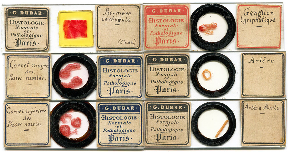
Figure 1. Microscope slides of human anatomical specimens by Georges Dubar. They probably all date from his time as a histologist at Charité Hospital, 1883 - ca.1899. Magnified details of several of these specimens are shown below as Figure 3-6.
Georges Carlos Henri Dubar, 1855 - ca. 1900
by Brian Stevenson
last updated January, 2020
From 1883 until about 1900, Georges Dubar worked as a histologist/pathologist in the surgery department of Charité Hospital, Paris. In addition, he taught courses in “de technique microscopique et de diagnostic histologique et pathologique” (“microscopic technique and histological and pathological diagnosis”). Evidently, he sold microscope slides to his students, and probably to all medical students at the hospital (Figure 1). I have not found any advertisements from Dubar for the sale of his slides, so they were probably purchased directly by people who knew him. That probably accounts for their relative scarcity. Dubar’s known slides are generally of high quality, both in preparation of the anatomical specimens and finish of the slides, with attractively printed labels and clear handwriting.
Figure 1.
Microscope slides of human anatomical specimens by Georges Dubar. They probably all date from his time as a histologist at Charité Hospital, 1883 - ca.1899. Magnified details of several of these specimens are shown below as Figure 3-6.
Georges Dubar was born in Lille, France, on April 11, 1855. His 1888 MD thesis gives some details of his early career (Figure 2). Dubar was appointed prosector in the Lille medical school in 1875. From 1876 until 1878, he served as “Aide d’Anatomy” to the medical college, as well as performed an internship at the Lille Hospital. In 1879, he moved to Paris for a three-year externship. Afterward, Dubar worked as an aid in the pathology laboratory of the Faculté de Mèdecine de Paris, and, in 1883, “M. Dubar (Georges-Carlos-Henri), bachelier ès lettres et ès sciences, est nommé aide du laboratoire des cliniques de la Faculté de médecine de Paris, à l'hôpital de la Charité, en remplacement de M. Bergeron, démissionnaire” (“M. Dubar (Georges-Carlos-Henri), Bachelor of Arts and Sciences, was appointed assistant to the laboratory of the clinics of the Faculty of Medicine of Paris, at the Charité hospital”). After completing his Doctor of Medicine training in 1888, Dubar worked as second in command of the Charité surgical laboratory, for as long as he appears in records.
A brother, Louis, followed a similar career track, and records of the two Doctors Dubar are often confusing. Louis was evidently older, joining Société Anatomique de Paris in 1881, whereas Georges joined in 1892. Louis moved back to Lille before 1890, becoming Professor of Surgery at the Faculté de Médecine de Lille.
Evidence of Georges’ extramural teaching comes from an 1889 post in La France Médicale, “M. le Dr. Dubar, préparateur au laboratoire de la clinique chirurgicale de la Charité, recommencera son cours de technique microscopique et de diagnostic histologique et pathologique le mardi 7 mai à 4 heures. - On s'inscrit de midi à 2 heures, rue Antoine-Dubois [près la Faculté de médecine]” (“Dr. Dubar, preparer at the laboratory of the Charité surgical clinic, will resume his course in microscopic technique and histological and pathological diagnosis on Tuesday May 7 at 4 am. - We register from noon to 2 am, rue Antoine-Dubois [near the Faculty of Medicine]”). This was soon after he completed his M.D. degree, suggesting that Dubar took a break from teaching while he wrote his thesis.
Confirmation that the Dr. Dubar who taught anatomical microscopy comes from his 1891 voter registration card, which places George Carlos Dubar, born April 11, 1855 in Lille, at 4 Rue de Antoine Dubois. In 1892, he joined the Société Anatomique de Paris, giving the same address.
Dubar took a special interest in researching cancers. His thesis was entitled “Essai sur la Sarcomatose Ostéoïde” (Figure 2). He presented on “Sarcome du Creux Poplité” in 1892, which included “préparations microscopiques et dos dessins relatifs à la tumeur” (“microscopic preparations and drawings relating to the tumor”).
An 1886 report in La Lancette Français included “La pièce a été examinée par M. le docteur G. Dubar, aide de clinique, au laboratoire d'histologie de la Charité. Les coupes faites en différents sens ont démontré que nous avions affaire à un sarcome développé dans le muscle grand dorsal, avec de nombreux noyaux de calcification, mais nulle part de traces d'ossification” (“The piece was examined by Dr. G. Dubar, clinical assistant, at the Charité histology laboratory. The cuts made in different directions demonstrated that we were dealing with a sarcoma developed in the dorsal muscle, with numerous nuclei of calcification, but nowhere traces of ossification”).
Other presentations also described Dubar’s microscopical studies. His 1883 examination of liver histology noted “Le foie, conserve dans le liquide de Millier, a été durci par la gomme et l'alcool. Les coupes sont colorées par le pioro-carminatc neutre d'ammoniaque et conservées dans la glycérine. Les lobules (grossissement 20 d.) de ce foie sont séparés les uns des autres par des espaces-portos normaux; il n'existe pas la moindre trace de cirrhose. Une coupe, à laquelle on a fait subir l'action des vapeurs d'acide osmique, vue au même grossissement, montre, dans toute son étendue, les lobules hépatiques colorés en jaune très sombre, surtout à leur périphérie” (“The liver, preserved in Millier's liquid, has been hardened by gum and alcohol. The sections are stained with neutral ammonia pioro-carminate and stored in glycerin. The lobules (magnification 20x) Of this liver are separated from each other by normal port spaces; there is not the slightest trace of cirrhosis. A section, which was subjected to the action of osmic acid vapors, seen at the same magnification, shows, in all its extent, the liver lobules colored in very dark yellow, especially at their periphery”).
An 1888 report by a Dr. Ozenne included “Notre ami le Dr. G. Dubar a eu l'obligeance de faire l'examen microscopique du segment nerveux réséqué; nous le remercions de la note suivante qu'il nous a remise et qui nous montre que l'on était en présence d'une transformation fibreuse, résultat d'une névrite interstitielle” (“Our friend Dr. G. Dubar was kind enough to do the microscopic examination of the resected nerve segment; we thank him for the following note he gave us and which shows us that we were in the presence of a fibrous transformation, the result of interstitial neuritis”).
Georges Dubar is recorded as being an adjunct member of the Société Anatomique de Paris from 1892 through 1899. He is not listed in later editions. His brother, Louis, who worked in Lille, was recorded as being a member well into the twentieth century. I have not found any records of Georges after 1899. He may have been the “Henri Dubar” who died on November 19, 1901 at Hospital Invalides, although that man was reported to be 51 years old, whereas Georges Dubar would have been 56.

Figure 2.
The cover page of Georges Dubar’s 1888 medical school thesis. It provides his date and place of birth, as well as relevant employment history up to that time.
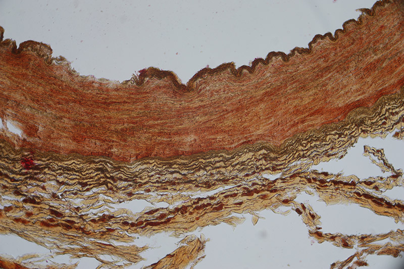
Figure 3.
Detail of a cross-section of a human artery, stained with Verhoef iron hematoxylin for elastica, and counterstained with Van Gieson picrofuchsin for connective tissue (see Figure 1). Photographed with a 10x objective lens and C-mounted digital SLR camera.
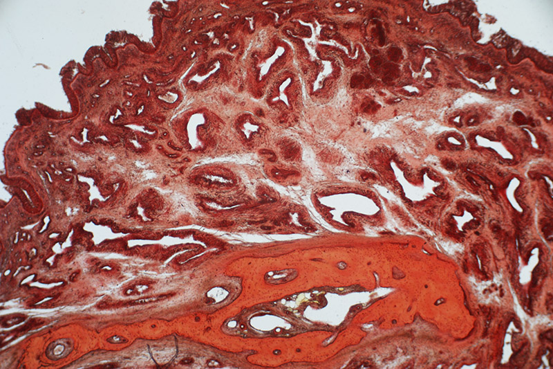
Figure 4.
Detail of a medial turbinate from a human nasal cavity (see Figure 1). This is a common surgical specimen, frequently removed from patients with chronic nasal obstruction due to sinusitis or a deviated nasal septum. Photographed with a 3.5x objective lens and C-mounted digital SLR camera.
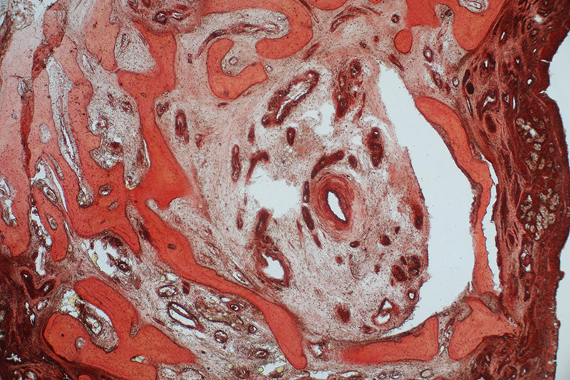
Figure 5.
Detail of an inferior turbinate from a human nasal cavity (see Figure 1). Photographed with a 3.5x objective lens and C-mounted digital SLR camera.
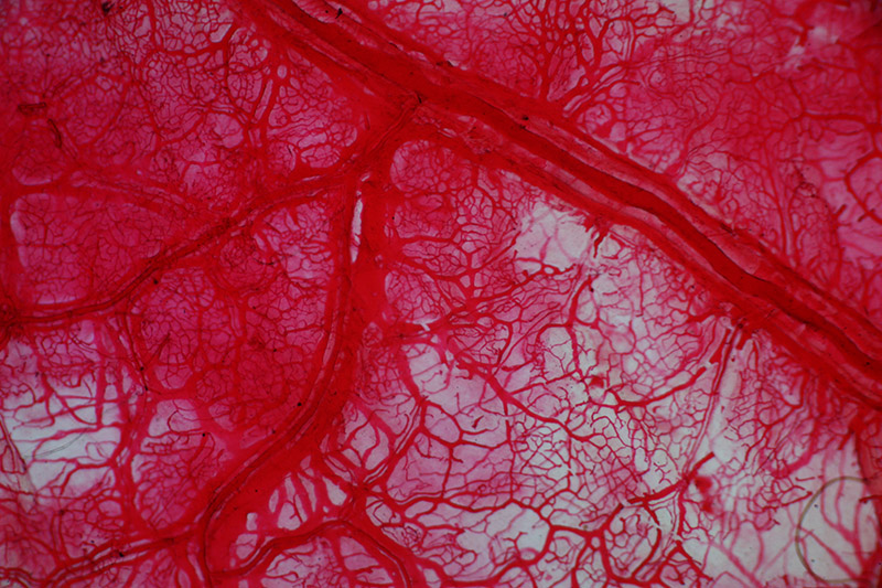
Figure 6.
Detail of spread of pia mater membrane from a dog (see Figure 1). Photographed with a 3.5x objective lens and C-mounted digital SLR camera.
Acknowledgements
Thank you to Jeffrey Silverman for generously providing insights on Dubar’s histological preparations.
Resources
Bulletin Administratif (1884) “M. Dubar est nommé pour trois ans, à partir du 1er août 1884, aide du laboratoire de clinique chirurgicale à la Faculté de médecine de Paris, à l'hôpital de là Charité”, Vol. 36, page 48
Bulletin Bibliographique (1883) Faculté de médecine de Paris, Vol. 32, page 471
Bulletins de la Société Anatomique de Paris (1885) “Members: 1881. Dubar, rue Monge, 68, Paris” (Georges’ brother, Louis), page 11
Bulletins de la Société Anatomique de Paris (1894) “Members adjoints: 1892. Dubar, rue Antoine-Dubois, 4, Paris”, page 9
Bulletins de la Société Anatomique de Paris (1899) “Members adjoints: 1892. Dubar, rue Antoine-Dubois, 4, Paris”, page 5
Death record of Henri Dubar (1901) accessed through ancestry.com
Dubar, Georges (1888) Thèse pour le Doctorat en Médecine: Essai sur la Sarcomatose Ostéoïde, accessed from https://openlibrary.org/authors/OL7449366A/Georges_Dubar
La France Médicale (1889), Vol. 1, page 600
Gazette médicale de Paris (1891) “Laboratoire de Cliniques: Clinique chirugicale (Charité). - MM. Cazin, chef ; Dubar, aide”, Vol. 62, page 587
Lancette Française (1874) “Élevès de 1re année … 13 Dubar (Georges Carlos Henri)”, Vol. 47, page 959
Lancette Française (1886) Vol. 59, page 857
Le Mercredi Médical (1892) Sarcome du creux poplité, page 431
Ozenne, Dr. (1888) Traitement des néuralgies sous-orbitaires par la résection du nerf, Journal de Médecine de Paris, Vol. 14, pages 746-751
Paris voter record of Georges Dubar (1891) accessed through ancestry.com
Le Progrès Médical (1883) Examen histologique du foie, pratiqué par M. Georges Dubar, Vol. 11, page 527