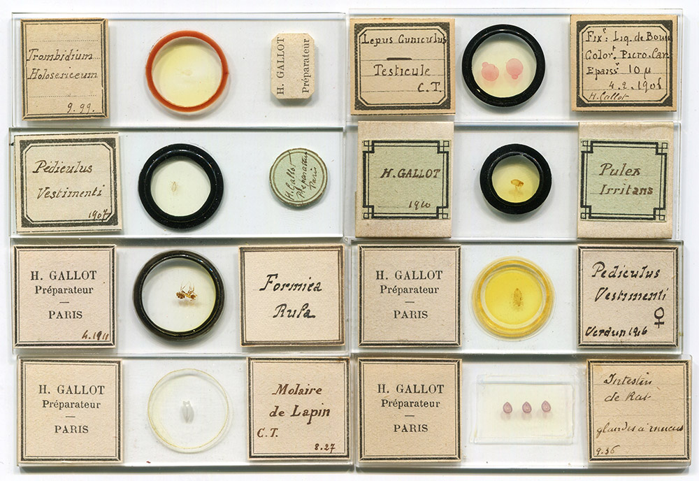
Figure 1. Microscope slides by Henri Gallot, dated between 1899 (upper left) and 1936 (lower right). His earlier slides tend to have off-the-shelf labels, while later works used custom-printed labels with Gallot’s name on them.
Henri Gallot, ca. 1880 – after 1936
by Brian Stevenson
last updated April, 2025
Henri Gallot was a surgeon-dentist in Paris, and an expert amateur microscope slide-maker. He was a member of the Société Francaise de Microscopie, serving as the Society’s President in 1936. His slides show considerable skill with staining and sectioning of histological specimens, as well as expertise with preparing insects. Gallot often wrote the date of preparation on his slide labels, with a known range of between 1899 and 1936.
Figure 1.
Microscope slides by Henri Gallot, dated between 1899 (upper left) and 1936 (lower right). His earlier slides tend to have off-the-shelf labels, while later works used custom-printed labels with Gallot’s name on them.

Figure 2.
Trombidium holosericeum (red velvet mite), prepared by Henri Gallot in 1899 (see Figure 1). Imaged with a C-mounted digital SLR camera and 3.4x objective lens on a Leitz Ortholux II microscope.
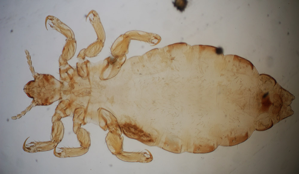
Figure 3.
Human body louse (Pediculus vestimenti) from Verdun, prepared in 1916 by H. Gallot. The Battle of Verdun took place from February through December, 1916. Body lice were a serious danger during World War I, due to the “trench fever” diseases that these insects spread among troops and displaced civilians. Imaged with a C-mounted digital SLR camera and 3.4x objective lens on a Leitz Ortholux II microscope.
Henri Gallot first appears in Paris directories in 1903, as a dentist working at 51 Rue Bonaparte. He was not listed in the 1902 or earlier editions, indicating that he opened the business in 1903. A reasonable guess is that he would have completed his training by his early twenties, suggesting a birth date of around 1880. Gallot’s earliest known microscope slide is dated September, 1899 (Figure 1), which was likely prepared during the time of his education.
The 1909 Annuaire du Commerce Didot-Bottin indicates a change in location (Figure 4). It lists the dentist H. Gallot at 51 Rue Bonaparte, and a new entry for Dr. Gallot who treated “diseases of the mouth and teeth” at 3 Rue de Choiseul. Additional information described below show that the Dr. Gallot at Rue de Choiseul was our microscopist Henri Gallot. This was the last entry for Gallot of Rue Bonaparte. I conclude that both entries are for our microscopist. Later publications indicate an address change from 3 to 1 Rue de Choiseul during the early 1930s.
Henri Gallot became a member of the Société Francaise de Microscopie. In 1931, he served on the Society’s Council. A Society publication of that year described Gallot as “chirurgien-dentiste, biologiste micrographe” (“surgeon-dentist, biologist microscopist”). The 1932 list of members gave his address as 1 Rue de Choiseul. He was elected President of the Société Francaise de Microscopie in 1936 (Figure 5).
I have not located any information about Gallot after 1936.
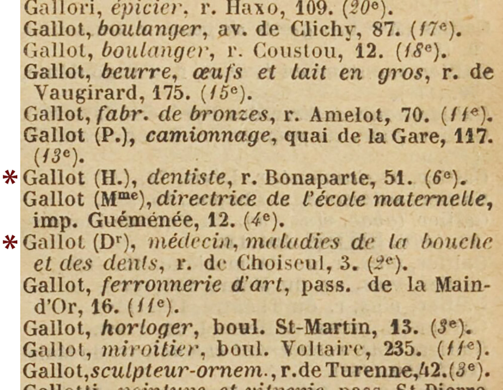
Figure 4
Excerpt from the 1909 “Annuaire du Commerce Didot-Bottin”. Evidence discussed above indicate that both the “dentiste” at Rue Bonaparte and the “médicin, maladies de la bouche es des dents” at Rue de Choiseul were our microscopist Henri Gallot. The double entry was due to his move from Rue Bonaparte to Rue de Choiseul.

Figure 5.
Header for the “Bulletin de l’Société Francaise de Microscopie” report of the February 24, 1936 meeting, stating that Henri Gallot was the Society’s President.

Figure 6.
Section through a rabbit's tooth, prepared by Henri Gallot (see Figure 1). Imaged with a C-mounted digital SLR camera and 3.4x objective lens on a Leitz Ortholux II microscope, with standard transmitted light (left) and between crossed polarizing filters (right).
Resources
Annuaire-Almanach du Commerce (1893 – 1908) accessed through https://gallica.bnf.fr
Annuaire du Commerce Didot-Bottin (1909 – 1926) accessed through https://gallica.bnf.fr
Annuaire du Commerce Didot-Bottin (1935) "Gallot"
Bulletin de l’Société Francaise de Microscopie (1932) Members, page 3
Bulletin de l’Société Francaise de Microscopie (1936) pages 4 and 7
Revue d'Optique Théorique et Instrumentale (1931) La Société Française de Microscopie, page 435