William Gatrell, 1864 - 1902
by Brian Stevenson
last updated March, 2022
During his brief career, William Gatrell was very highly regarded as a preparer of diatom microscope slides. Edmund Spitta, in the 1899 book Photo-micrography, stated “We know of no mounter of diatoms in the United Kingdom that can surpass Mr. Firth, of Clifton Park Avenue, Belfast, and few that can equal him, save Mr. Gatrell, of Barnes, whose work is of the most excellent quality” (pages 129-130), “Mr. Gatrell has sent us some amphipleura pellucida mounted in realgar
and other diatoms in quinidine and piperidine which are of the highest order of merit, especially the amphipleura pellucida, which of late have been so difficult to obtain” (page 130), and “Amphipleura pellucida ... None that the writer has ever seen can compare with those prepared in realgar by Dr. Van
Heurck, excepting those mounted by Mr. Gatrell” (page 140).
Gatrell used
pale yellow-brown labels on his slides, with handwritten descriptions of the
specimens. His name was printed,
along with his location in Barnes, Surrey. Examples of labeling variants are shown in Figure 1.
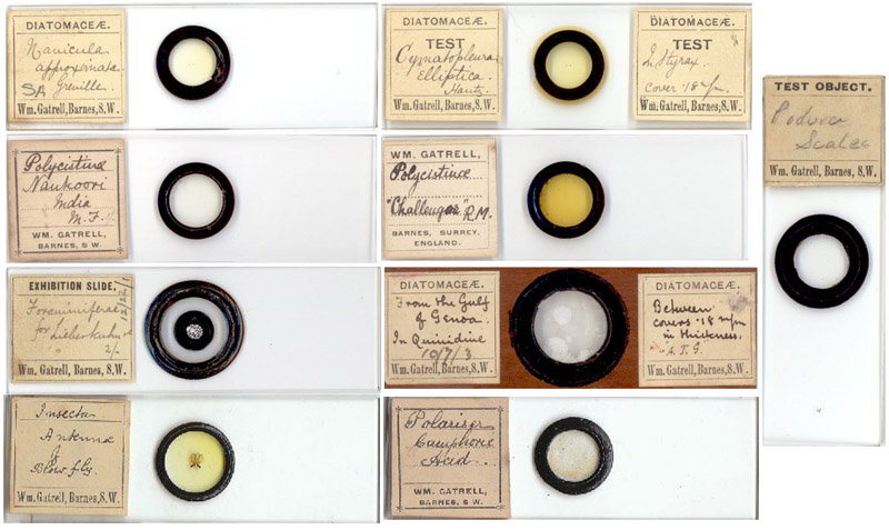
Figure 1.
Nine slides by William Gatrell. Three are of diatoms (the 2 glass slides have arranged single-species diatoms, the wooden slide holds a strew of mixed species), two of polycystina (including a sample from the H.M.S. Challenger expedition), an arrangement of foraminifera, a non-diatomaceous test slide (Podura scales), an insect slide (blowfly antennae), and a chemical slide for the polariscope (camphoric acid). The wooden slide holds a strew of diatoms between two very thin glass slips, and the quinidine mountant has crystallized over the years, adding a new and interesting dimension to views through the microscope.
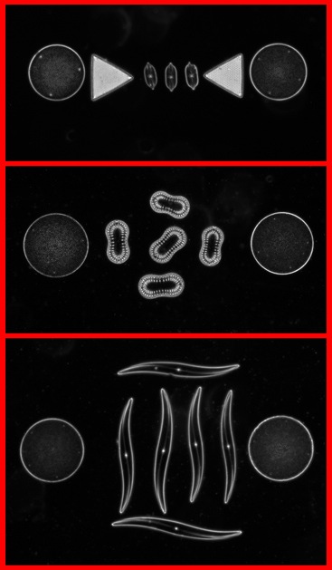
Figure 2. Three single-species diatom slides by William Gatrell (see Figure 1). In all the examples of single-species slides I have seen, he mounted a half dozen or so specimens between at least one pair of larger diatoms. The larger diatoms are visible even without a microscope, and serve as guides for centering the specimens prior to viewing.Top to bottom, the specimens were labeled by Gatrell as Navicula approximata Greville, Surirella lata Wm. S., and Rhoicosigma robusta v. niflexa H. Pen. Photographed using an Olympus BX51 microscope, 4x objective lens, phase contrast illumination and a Retiga 2000R Fast 1394 with QCapture Pro software.
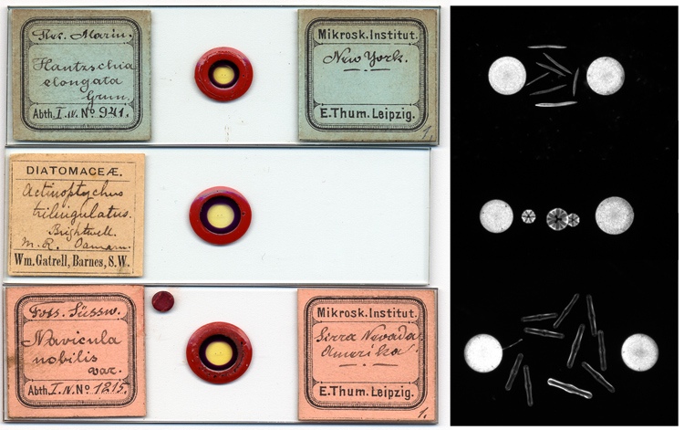
Figure 3. Gatrell’s style of mounting diatom specimens between larger circular diatoms was also used by Eduard Thum (1847-1926). Gatrell may have adapted his technique from copying earlier mounters such as Thum.Note that the ringing style is identical in all three slides, and is distinct from the usual Gatrell black ringing shown in Fig. 1. Did Gatrell re-label a Thum mount and sell it as his own? At the right are darkfield photomicrographs of the specimens in the three slides.
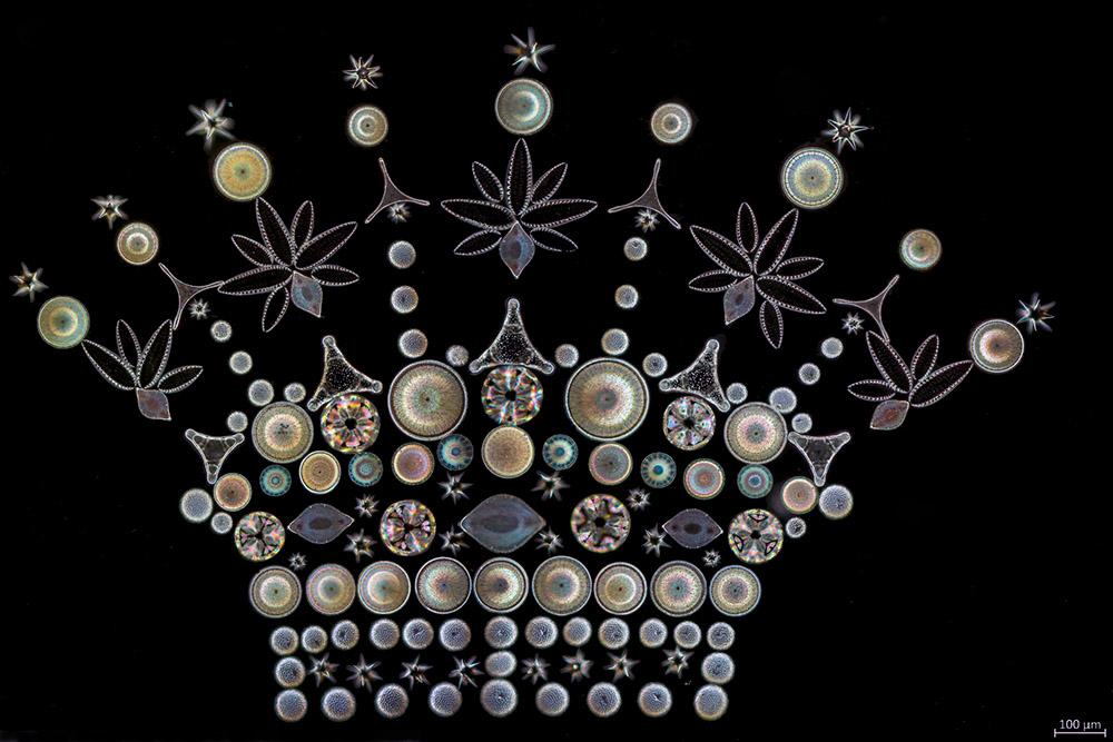
Figure 4.
An arrangement of diatoms by William Gatrell. Photographed using top lighting. Image generously provided by Ian Jones.
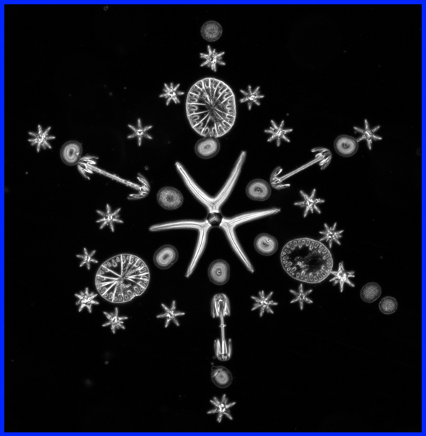
Figure 5.
The label is printed “Exhibition Slide”, with handwritten “Rosette. Sponge Spicules. 37 forms.” Photographed using phase contrast illumination.
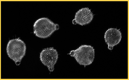
Figure 6.
A polycystina slide by William Gatrell, labeled Anthrocystis ventricola Ehr. Photographed using dark field illumination.
William Herbert Gatrell was born in Southampton, Hampshire,
at the start of 1864. His parents,
Maurice and Ellen, were both originally from the Isle of Wight. The 1861 English census lists Maurice
as being a millwright. Almost all
of the Gatrell’s neighbors worked at jobs related to ships, so it is probable
that Maurice was involved in ship building and/or repair. In 1871, Maurice was recorded as being
an engineer in the Merchant Marine, and the family consisted of 9 year-old
Agnes, 7 year-old William and 2 year-old Josephine. Intriguingly, Josephine was born in the United States,
suggesting that the family lived overseas during the late 1860s.
In April, 1879, 15 year-old William began training in
dentistry with Frederick Sleep at his practice in Great Russell St.,
London. With the exception of
father Maurice (who may have been out at sea), the whole family lived in St.
John Hackney, London at the time of the 1881 census. William sat for his exams in September of that year, and was
certified in dentistry on 30 Sept., 1881.
William
married Caroline Lillian Stay during early 1887, in Hackney. In 1891, he was employed as an
operative dentist and lived in Christchurch, Hampshire with Caroline, his 16 year-old sister Francis, a fellow dentist, and two domestic servants.
The Gatrells
moved back to the London area during early or mid 1897. The 1901 census (conducted at the end
of March) lists their son Maurice as being 3 years old and born in Sandown,
Hampshire. However, Brian
Bracegirdle’s Microscopical Mounts and
Mounters notes that Gatrell advertised from 7 White Hart Lane, Barnes,
Surrey during 1897. As noted
above, Spitta raved about William Gatrell’s diatom mounts in 1899. Dr. Gatrell appears to have put a lot
of effort into his microscopy business during that time, as the 1901 census
recorded him as being a “microscopic optician” who worked on his own
account. This business clearly
included more than just selling prepared slides: the box of blank card slides
imprinted with William Gatrell’s name, shown in Figure 8, was undoubtedly for
sale to customers interested in making their own mounts.
William
Gatrell’s acclaimed career as a commercial producer of microscope slides lasted
only 5 years, perhaps a little longer if he began during his stay in
Hampshire. Gatrell died at home on
14 August, 1902, at the age of 38. The listed cause of death was “heart failure, due to disease of the
heart and other organs (natural).” His death record further indicates his occupation as “dentist”,
suggesting that he had also continued with that trade. Two years after his death, he was still
listed as being a “diatoms expert, especially mounting” in the Naturalists'
Universal Directory.
Caroline
remarried in 1904, to Samuel Wells, in Portsmouth, and later moved back to
Sandown, Isle of Wight. Younger
son, Felix Gatrell, was killed in 1917 during World War I, and is buried in
Deir El Belah War Cemetery, Palestine, just east of the Egyptian border.
It is probable
that William Gatrell became familiar with the microscope during his dental
training, where histological training would have been stressed. In addition, dental journals of the
late 1800s contain frequent references to lectures and slide exhibitions on
microscopic analyses of tooth development, connective tissues, dental caries,
comparative tooth morphologies, etc.
We can only
speculate on why Gatrell decided to specialize in mounting diatoms. He may
have been directed by a very strong connection between dentistry and diatoms:
the abrasiveness of diatomaceous earth made it a major component of dental
powders and pastes. The August,
1881 issue of The
American Monthly Microscopical Journal contained the following letter from “C.W.G.” of Palisades, New York, “On examining some of the powder known as ‘Vegetable
Sozodont Tooth Powder’, I found it to be composed chiefly of diatomaceous
material, the forms being in both perfect and fragmentary states. The other constituent seemed to be some
flavoring material, which gives the powder its peculiar taste. Various forms of
Navicula and Pinnularia occur in great abundance. Also small and delicate forms. When mounted in Balsam, the powder makes an interesting
object.” C.W.G.’s letter was
quoted later that year in The Journal of
the Royal Microscopical Society. During 1882, Thomas Partridge wrote “On Diatoms” in The Journal of the Postal Microscopical Society, stating “The well-known ‘Vegetable Sozodont
Tooth-powder’ is composed of diatomaceous material, and makes a good
microscopic slide”. In 1897,
Edward Nelson observed in The American Monthly Microscopical Journal that “One of the best diatoms to work on with the
higher powers is the large N. rhomboides, found in ‘Sozodont’ tooth-powder.” Professor
Albert Mann reported in a 1902 issue of Harper's
Monthly Magazine that “from a certain
brand of tooth-powder” he had “secured seventy-six species of Diatoms”. Lewis Wright, in his A Popular Handbook to the Microscope,
noted that the “the tooth-powder sold as
‘Sozodont’ will furnish for sixpence a large quantity (of diatoms), being a
diatomaceous earth, but it varies a great deal; some year or two ago there were
many fine forms in it, but the last sample I obtained contained only very
common forms.”
An 1891 issue of The
Journal of the British Dental Association noted an exhibition by J. Howard
Mummery of “a slide showing specimen of
tooth powder, consisting of various species of diatoms”, along with
histological slides “illustrating the
incorporation of connective tissue with the dentine” and “of caries showing
micro-organisms in the dentinal tubes.” That same exhibition included a display of tooth
developmental slides by John J. Andrew, a noted dentist, preparer of microscope
slides and colleague of W.A. Firth in the Belfast Naturalists’ Field Club. Dr. Andrew later donated “a very fine collection of diatoms” of “almost 100 professionally mounted slides of
world wide range” to that club.
In addition, diatom structures were being studied by dental
anatomists. Dr. Otto Walkoff of Braunschweig compared the structure of Triceratum diatoms with tooth enamel
structures in his “Contributions Relating to the More Minute Structure of the
Enamel and to the Development of the Dentine”, published in The Journal of the British Dental
Association during 1898.
Whatever inspired William Gatrell to mount diatoms, he certainly left behind a legacy of skillfully constructed beauties.
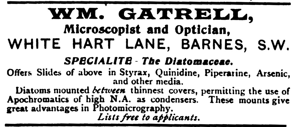
Figure 7. Gatrell was primarily known as a mounter of diatoms. This advertisement from an 1901 issue of Science-Gossip indicates that he prepared diatom slides using a variety of mounting media.
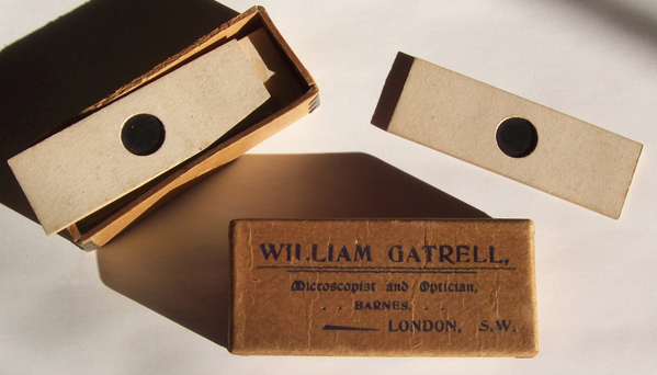
Figure 8. A cardboard box with William Gatrell’s name and address, holding a half dozen unused card slide blanks. Apparently offered by him for sale to customers who wanted to make their own deep mount slides.
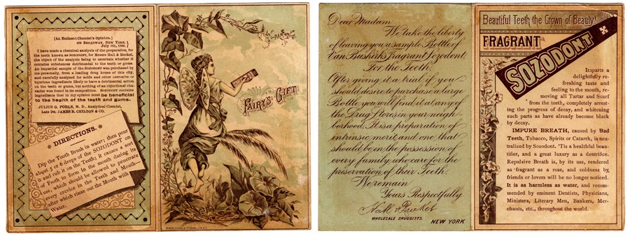
Figure 9.Advertisements for Sozodont tooth powder/paste, from ca. 1866.
Acknowledgements
Many thanks to Ian Jones for providing a photograph of a Gatrell slide from his collection, Peter Paisley for information on J.J. Andrew and Victorian dentistry, and to Peter Hodds for help in locating
Gatrell’s slides.
Resources
Bracegirdle, Brian (1998) Microscopical Mounts and Mounters, Quekett Microscopical Club, London.
Commonwealth War Graves Commission. Felix Gatrell. http://www.cwgc.org/search/casualty_details.aspx?casualty=645607
“C.W.G.” (1881) letter, The American Monthly Microscopical Journal, August, page 158
TheDentists Register (1882) page 207 and 1884, page 225
England vital statistics, accessed through ancestry.co.uk
The Irish Naturalists' Journal (1965) Page 349
Journal of the British Dental Association (1891) vol.12, page 595
Journal of the Royal Microscopical Society (1881) series 2, vol. 1, pp 786-787
Harper's Monthly Magazine, page 228
The Medical Times and Gazette (1881) Sept. 24, page 400 “Medical News”
The Naturalists'
Universal Directory (1904) 19th edition, S.E. Cassino, Salem, Mass, USA, 1905. page 291
Nelson, Edward M. (1897) Tests for microscope objectives, The American Monthly Microscopical Journal, vol. 18, March, pages 80-83
Partridge, Thomas (1882) On diatoms, Journal of the Postal Microscopical Society, 1882, pages 22-25
Samworth, Michael (1998) Sponge spicules, Micscape Magazine August issue. http://www.microscopy-uk.org.uk/mag/artaug98/spiccy.html
Science Gossip (1901) Advertisiment, new series, Vol. 7, p. 352
Spitta, Edmund Johnson (1899) Photo-micrography, Scientific Press, London
Walkoff, Otto (1898) Contributions relating to the more minute structure of the enamel and to the development of the dentine, Journal of the British
Dental Association, vol. 19, pages 688-696
Wright, Lewis (1895) A Popular Handbook to the Microscope,
The Religious Tract Society, London, page 234