An amateur microscopist, Joseph H. Hardy undertook to prepare slides of thin-sectioned echinoderm spines. His known slides are of very high quality, both macroscopically (Figure 1) and microscopically (Figure 3-7). The spines of sea urchins and other echinoderms are difficult to prepare in cross-section, as they are hard enough to require the same sort of hand-grinding necessary to prepare thin-sections of minerals, yet so fragile that a misstep can destroy their fine structure. Exactly why a railway clerk such as Hardy decide to specialize in echinoderms is not known, but by the turn of the 20th Century, he was an internationally recognized expert.
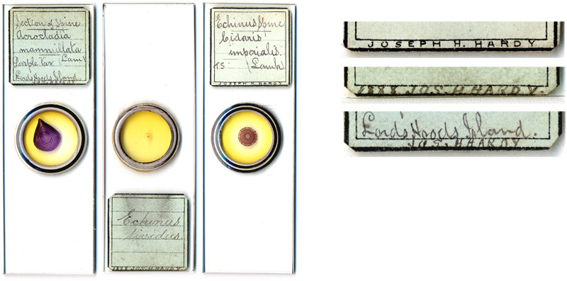
Figure 1.
Examples of microscope slides prepared by Joseph
Hardy. All three are of thin-sectioned echinoderm spines. To the right are
magnified views of Hardy’s name on these labels: the top one is machine
printed, and the lower two were printed by hand. The middle slide, a spine of
Echinus lividus, was dated 1888. Magnified views of the specimens are shown in
Figures 3 - 7.
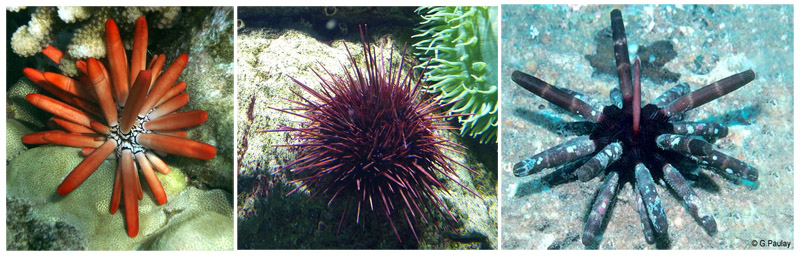
Figure 2.
Living examples of the species whose prepared spines
are illustrated in Figures 1 and 3-7. Left to right:
Acrocladia mammillata (now Heterocentrotus mamillatus),
Echinus lividus (now Paracentrotus lividus), and
Cidaris imperialis (now Phyllacanthus imperialis). Images used for nonprofit
educational purposes, in accordance with Creative Commons. Sources are listed
in the ‘Resources’ section, below.
John Quekett recognized both the beauty of sectioned echinoderm slides and the difficulty of their preparation, “The spines of the Echini, after having been cut transversely with a saw, and then ground down and mounted in Canada balsam, form some of the most beautiful objects for a microscope of low power; considerable difficulty will, however, be found in getting a section perfect and at the same time very thin”. The method for preparing echinoderm spine sections described by Cross and Cole, contemporaries of Hardy, are as follows:
“(1) With a fine saw make transverse . .sections.
(2) Take a hone (water of Ayr stone), moisten it with water, and rub one side of the section upon it until it is quite flat and smooth.
(3) Wash in water, and set aside until quite dry.
(4) Take some dried Canada balsam, place a piece on a square glass, and warm gently over a lamp until the balsam melts; allow it to cool a little, and then press the smooth side of the section into it, and set aside until cold.
(5) With a fine file rub the section down as thinly as possible.
(6) Take the hone again, and grind the section down until thin enough, using plenty of water.
(7) Place it, glass and all, in methylated spirit until the section comes away from the glass, then wash well in (clove oil).
(8) (mount in Canada balsam in the usual way)”.
In these modern times, we often forget that the Victorians did not have electric power tools. All cutting and grinding would have been accomplished by hand.
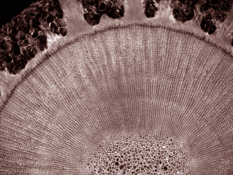
Figure 3.
Section of a Cidaris imperialis spine, prepared by Joseph Hardy
and photographed using darkfield illumination, with a Olympus BX51 microscope
eqipped with an Olympus PlanN 4x 0.10 NA objective lens and Q-Imaging Retiga
2000R Fast 1394 black/white digital camera. Pseudocolored with Photoshop.
Joseph Hawarth Hardy was born during the summer of 1842, in the Ecclesall Bierlow district of Sheffield, Yorkshire. While most records list Hardy’s middle name as “Haworth”, he signed it “Hawarth” on the 1911 census. He was the second child, and eldest son, of John and Sarah Hardy. The 1861 census recorded 11 sons and daughters in the Hardy family. Joseph’s father was a clerk for the railway, an occupation also followed by Joseph.
Joseph married Anne Richardson in 1868. The 1871 census located the couple and their first child living with Anne’s widowed mother on Stockport Road in the Manchester suburb of Rusholme. In 1881, Joseph, Anne, their then 5 children and Anne’s mother lived on the opposite side of Manchester, in Moss Side. The Hardy family stayed at that address through at least 1891.
One of the slides illustrated in Figure 1, above, is dated 1888. That is the earliest record I located of Hardy’s involvement with microscopy and echinoderms. The quality of this preparation, shown in detail in Figure 4, implies that Hardy already had considerable experience with preparing echinoderm spines by 1888. He displayed sections of at least fourteen different species’ spines at meetings of the Manchester Microscopical Society in 1889.
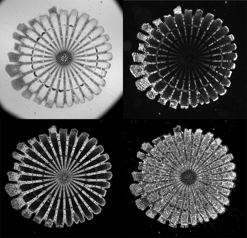
Figure 4.
Section of Echinus lividis spine, prepared by Joseph
Hardy and photographed using four different types of illumination. Clockwise
from upper left, brightfield, darkfield, phase contrast 1 and phase contrast 3.
Equipment as described in the legend of Figure 3, without pseudocoloring.
Hardy’s microscopical specialties included more that echinoderms. The February 27, 1892, issue of Chemist and Druggist reported, “Some seventy gentlemen interested in pharmaceutical microscopy, including about thirty old students, assembled on Tuesday night in the Manchester College of Pharmacy to witness an interesting exhibition of microscopes and of botanical and pharmaceutical slides. Amongst those present were many members of the Manchester Pharmaceutical Association and of the Microscopical Society. Eleven microscopes were shown by students, two by the Messrs. Turner, and four by friends—viz. starches of physostigma, arum, &c. by Mr. J. Beaton, M.A..; mosses, by Mr. H. G. Willis, M.A.; and sections of spines of echinoderms and sections of flowers, by Mr. Joseph H. Hardy. The Manchester College of Pharmacy exhibit gave a general view of botanical microscopy, including cells, leaves, stems, pollen, cryptogams, and drugs, such as cinchona, rhubarb, ipecacuanha. Other students showed in the laboratories the preparation and estimation of spirit of nitrous ether and estimation of hydrocyanic acid, myrrh, &c.”
In 1897, Hardy donated “Sixteen Micro-photographs of sections of Echinoderm Spines” to the Free Liverpool Library.
Joseph Hardy was a member of the Manchester Microscopical Society for many years, at times serving as a Society officer. His name does not appear in the 1905 or later lists of members.
By 1901, Hardy and family had moved back across town to Stockport Road, near where they had lived three decades previously. The 1911 census reported that Joseph had by then retired from the railway. He died on February 9, 1917.
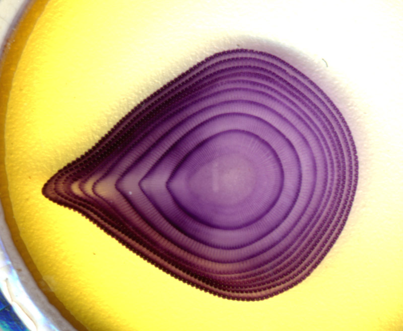
Figure 5.
Close-up flatbed scan of an Acrocladia mammillata spine, prepared by Joseph
Hardy.
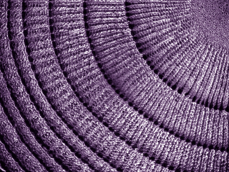
Figure 6.
Magnified view of the Acrocladia mammillata spine, using darkfield illumination.Equipment as described in the legend of Figure 3,
with pseudocoloring.
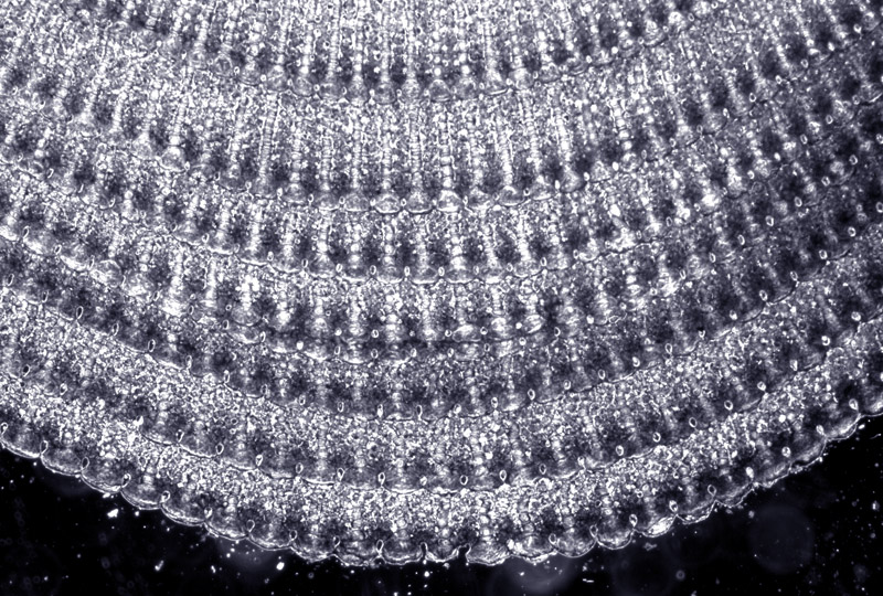
Figure 7.
Another magnified view of the Acrocladia mammillata spine, using phase contrast
illumination. Equipment
as described in the legend of Figure 3, with pseudocoloring.
Resources
Annual Report and Transactions, Manchester Microscopical Society (1889) [snippet view obtained from Google.com]
Annual Report and Transactions, Manchester Microscopical Society (1893) [snippet view obtained from Google.com]
Annual Report and Transactions, Manchester Microscopical Society (1895) [snippet view obtained from Google.com]
Annual Report and Transactions, Manchester Microscopical Society (1896) [snippet view obtained from Google.com]
Annual Report and Transactions, Manchester Microscopical Society (1900) [snippet view obtained from Google.com]
Annual Report and Transactions, Manchester Microscopical Society (1905) List of members (Hardy was not included)
Annual Report and Transactions, Manchester Microscopical Society (1906) List of properties, page 127
Chemist and Druggist (1892) Pharmacy and the microscope, Vol. 40, page 296
Cross, M.I. and M.J. Cole (1895) Modern Microscopy: A Handbook for Beginners : Combining I. The Microscope, and Instructions for its Use and II. Microscopic Objects: How Prepared and Mounted, 2nd edition, 1895, Balliére, Tindall and Cox, London, pages 154-155
Davis, George E. (1889) Practical Microscopy, New & Revised Edition, W.H. Allen & Co, London, 281-285
England census, birth, marriage and death records, accessed through ancestry.co.uk
Life Lore (1889) Manchester Microscopical, Vol. 1, pages 214 and 240
Probate record of Joseph H. Hardy (1917) accessed through ancestry.co.uk
Quekett, John T. (1855) A Practical Treatise on the Use of the Microscope: Including the Different Methods of Preparing and Examining Animal, Vegetable, and Mineral Structures, 3rd Edition, published by H. Bailliere, pages 355-356
Report of the (Free) Public Library of Liverpool (1897) page 14
Zoologisches Adressbuch (1901) “Hardy (Joseph H.). 11 Stockton Road, Chorlton-cum-Hardy near Manchester. Echinod.” Vol. 2, R. Friedlander and Son, Berlin, page 158
Information and photographs of echinoderms:
Acrocladia mammillata (Heterocentrotus mamillatus),
WoRMS, http://www.marinespecies.org/aphia.php?p=taxdetails&id=569228
http://en.wikipedia.org/wiki/File:Heterocentrotus_mammillatus_in_situ_from_Hawaii.JPG
Cidaris imperialis (Phyllacanthus imperialis)
WoRMS, http://www.marinespecies.org/aphia.php?p=taxdetails&id=179611
http://www.marinespecies.org/photogallery.php?album=694&pic=38435
Echinus lividus (Paracentrotus lividus)
WoRMS, http://www.marinespecies.org/aphia.php?p=taxdetails&id=124316
http://en.wikipedia.org/wiki/File:Ocean%C3%A1rio_de_Lisboa_%2810%29_-_Mar_2010.jpg