Alexander Hett’s microscope slides were highly regarded in his day. His preparations were of human and other animal tissues, generally injected with bright red pigment, in fluid-filled cells. He is known to have been a commercial slide preparer by the late 1840s, and was still active in the trade at the time of the 1862 London Exposition. Today, Hett’s slides are sought after by collectors, and often command high prices.
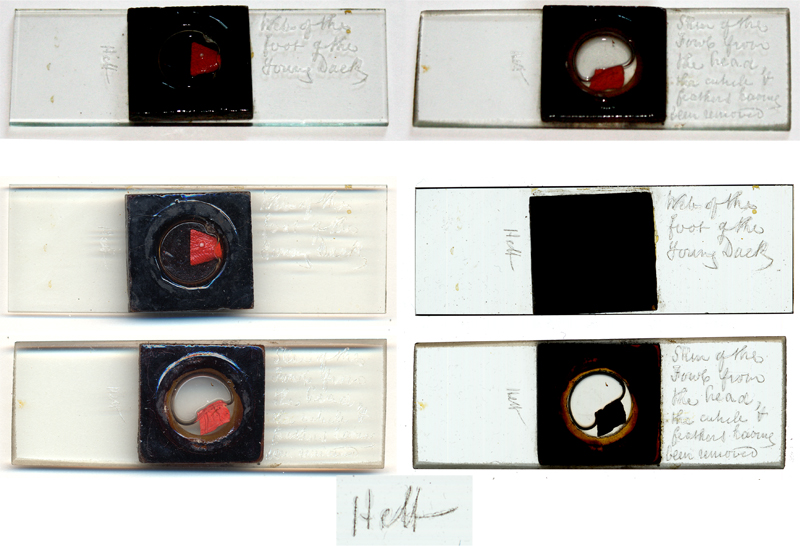
Figure 1. Two examples of
typical microscope slides prepared by Alexander Hett, photographed using incident light (top), and scanned with a flatbed scanner using both
reflective light (lower left) and transmitted light (lower right). Each slide holds a thick
section of animal tissue, with vascular structures injected with a bright red
pigment, probably vermilion, and held in a deep, fluid-filled cell made of dark
glass. Upon the slide, Hett etched his name and a description of the specimen.
A magnified view of Hett’s signature is shown below the slides.
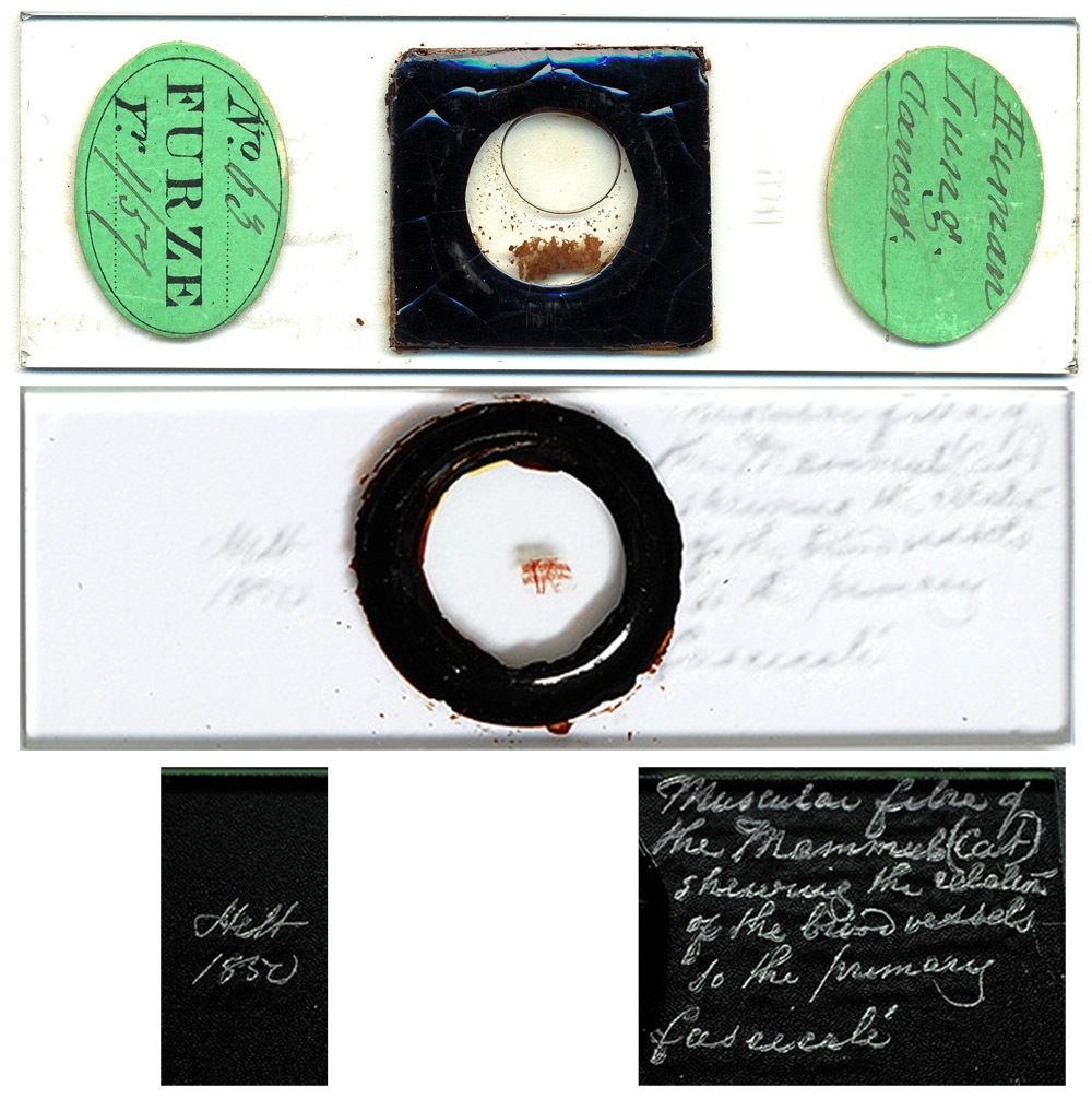
Figure 2. Two unusual Hett
slides. The upper slide used a thin (approximately 1mm thick) cell, and the
specimen was not injected with dye. This slide was once owned by fellow
Microscopical Society of London member, John Furze,
who attached his own, characteristic labels. The lower slide is an injected
preparation of cat muscle, mounted in what appears to be balsam (not a fluid
mount). Also rather unique, Hett dated this slide, 1850.
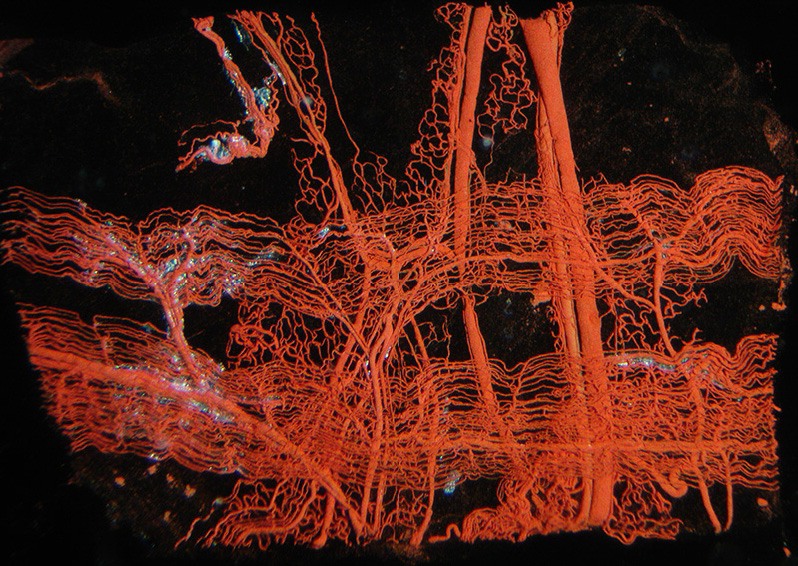
Figure 3. Detail of a Hett
slide of cat muscle fibers (Fig. 2). Courtesy of David Walker.
Census and other records suggest that Alexander Hett was the child christened February 9, 1807 at St. Peter’s, Leeds, Yorkshire, son of William Hett.
In 1828, The Lancet published a disparaging report of a surgery performed by Bransby Cooper at Guy’s Hospital, London. Alexander Hett was among the 174 “Dressers and Pupils” of the Borough Hospitals who rose to Cooper’s defense in a letter that appeared shortly thereafter in The Lancet. Bransby Cooper sued The Lancet’s editor, Thomas Wakley, for libel, and won a significant judgment. Ironically, Hett wrote another letter to The Lancet, in 1829, complaining bitterly about the behavior of Cooper.
Hett became a Fellow of the Royal College of Surgeons of London in 1829. Afterwards, he rarely stayed in the same location for long. He moved to Downham Market, Norfolk by 1833. His younger brother, Edward, was a solicitor in that town. On June 11, 1833, Alexander Hett married Lydie Claudine Francoise Mourcet, “daughter of the late Baron Mourcet”, in Paris, France. The Hett’s first child, Amelia, was born ca. 1836 in Downham Market. Their second, Julia, was born in 1838 in Islington, London. Their 1840 address was Gower St., Bedford Square, St. Giles in the Fields, Middlesex. By 1848, and through at least 1852, Hett was at 24 Bridge St., Southwark. Hett’s patent announcement of 1853 placed him in Stoke Newington, Middlesex. In 1855, he was at 8 Eastbourne Terrace, Paddington. During 1861, the family lived on a farm in Surrey. In 1862 and ca. 1865, Hett’s address was 4 Albion Grove, Islington. Later in 1865, he again lived in Stoke Newington. He died in 1870, in Lewisham, Kent.
Alexander Hett advertised his microscope slides of injected tissues in John Quekett’s 1848, first edition of A Practical Treatise on the Microscope (Figure 4). Since he was by then accomplished at the art, it is reasonable to assume that Hett began slide-making some years prior to 1848. Hett also advertised in Quekett’s second and third (final) editions. Advertisements in the 1855 edition indicate a business relationship between Hett and the microscope manufacturing firm of Smith and Beck (Figure 4). Selling through Smith and Beck would have greatly increased the number of Hett’s customers. An 1849 pamphlet “On the Structure of the Sudoriparous Glands” (sweat glands), by George Rainey, recommended Hett as the source for “microscopic preparations illustrative” of these glands. Dr. Boon Hayes, writing in 1853 on “histological anatomy and microscopical manipulation”, and Lionel Smith Beale, in the 1865 How to Work the Microscope, also recommended Hett’s preparations.
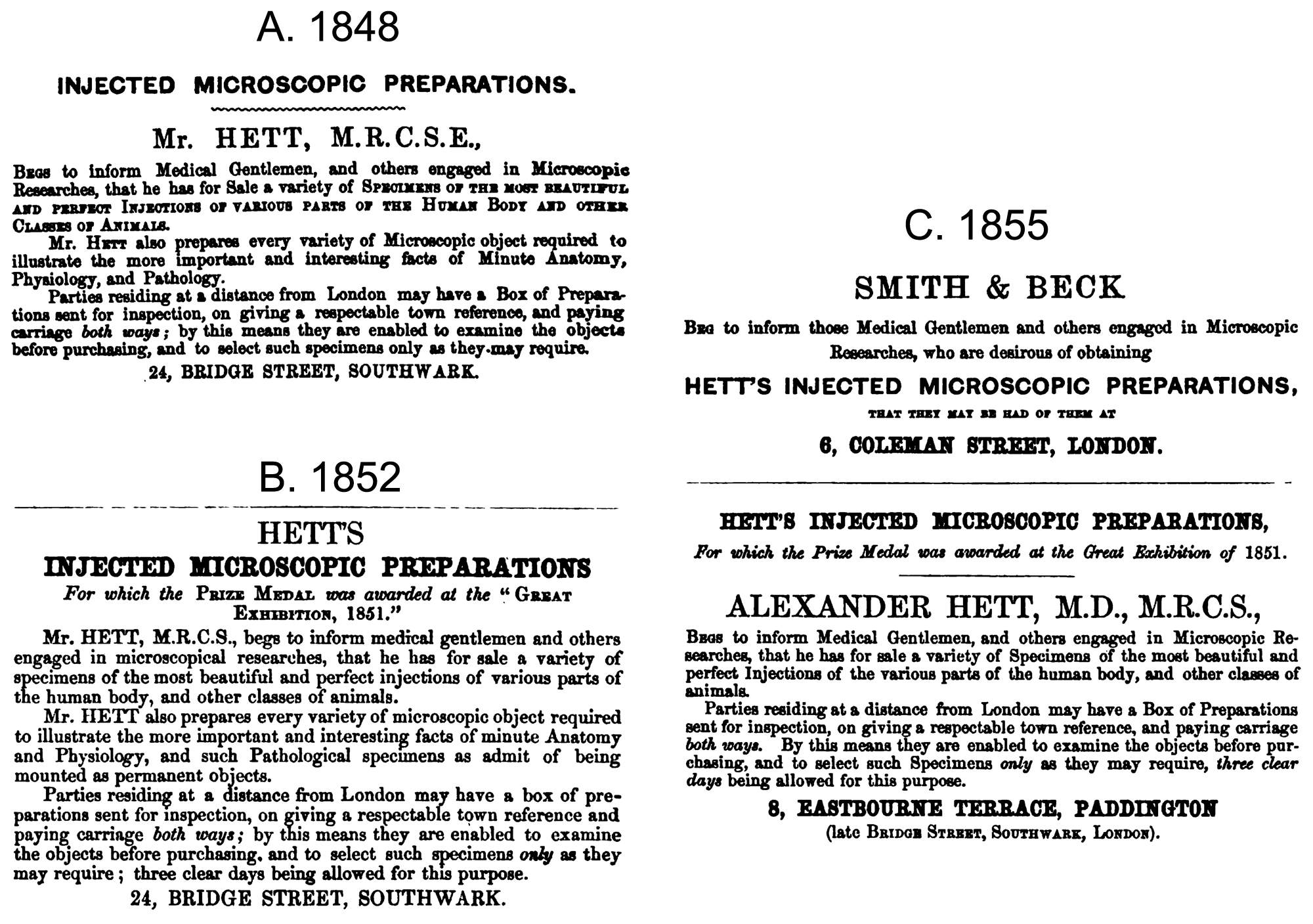
Figure 4. Paid
advertisements that appeared in the three editions of John Quekett’s A
Practical Treatise on the Microscope. Hett’s 1855 advertisement was immediately preceded by an ad from microscope makers Smith and Beck. Hett published a very similar advertisement in the English translation of Adolphe Hannover’s 1853 “On The Construction and Use of the Microscope”, with an address of Vicarage Lane, Chigwell, Essex, which indicates that Hett moved from Southwark between 1852 and 1853.
On May 16, 1850, Alexander Hett was elected to be a member of the Microscopical Society of London (which later became the Royal Microscopical Society)
Hett made several donations of specimens to the Royal College of Surgeons Museum, noted by John Quekett in the 1850 Descriptive and Illustrated Catalogue of the Histological Series, Volume 1. These include “A portion of adipose tissue attached to the skin of the neck of a Rook, Corvus frugilegus”, “A small portion of the skin from the leg of a Pig only two days old”, and “A vertical section of the skin of the arm of a human foetus at full time, exhibiting numerous small lobules of adipose tissue; each one is supplied with a rich network of capillaries, which are arranged in the usual form, but the meshes or spaces between the vessels are finer than they are in the adult, which may readily be accounted for, as the size of the cells is much smaller in the young than in the full-grown individual”.
As did so many other participants in the burgeoning profession of microscope slide making, Alexander Hett displayed his work at the 1851 Great Exhibition in London. These were described in the catalogue as a “variety of injected microscopic objects, showing the application of this mode of preparation, for displaying the structure of parts and organs, and also serving to illustrate the utility and importance of the microscope in its application to the sciences of physiology and pathology”. Cleverly, Alexander Hett provided a microscope so that visitors could see details of his preparations. Most other slide makers do not appear to have provided microscopes, which may have been a significant reason why Hett won a Prize Medal for his work. The Medical Times wrote, “Mr. Hett's collection (Class X. 249) being confined to injected animal preparations, is necessarily less extensive, but by no means inferior in the beauty of the specimens to that of Mr. Topping, and we have the advantage of inspecting them by the microscope. They are mounted on a wheel, so that each object may be brought under a microscope attached at one point of the circle by turning the wheel. They exhibit certain of the most interesting of the tissues in man and animals. Thus, we have the skin of the pig, sheep, monkey, fowl, of the cat, and of the human foetus; the eyes showing the vessels of the iris in the fowl and lamb; the human tongue demonstrating the vessels of the papillae; the lung of the monkey, the rook, and of man in its healthy and emphysematous and tuberculated condition. The last of these demonstrates that tubercle is nonvascular. The human stomach, and the stomach of the fowl, show the tubular follicles of the mucous membrane, and specimens of intestine, exhibit the villi and their beautiful reticular capillaries. A specimen of injected muscular fibre exhibits the longitudinal direction of the capillaries between the muscular fibres. The tongue of the monkey and the buccal membrane of the lamb also show the peculiar arrangement of the capillary vessels of these parts; and the mucous follicles of the oesophagus are demonstrated in these parts of the lizard and the rook. The whole collection, including forty specimens, has been, and will be, examined with interest by every visitor belonging to the Medical Profession.
The microscope used by Hett at the 1851 Exhibition was made by Smith and Beck, and it was probably not a coincidence that those microscope makers exhibited only 3 stalls away from Hett. Note, however, that this was no ordinary microscope: the Medical Times stated that the preparations were “mounted on a wheel, so that each object may be brought under a microscope attached at one point of the circle by turning the wheel”, and that there were “forty specimens”. This instrument was undoubtedly one described by Quekett in the 1852 edition of his Treatise as “Mr. Hett’s microscopes for injections, etc.”. Illustrations of this and another Hett invention are reproduced in Figure 5, along with Quekett’s descriptions.
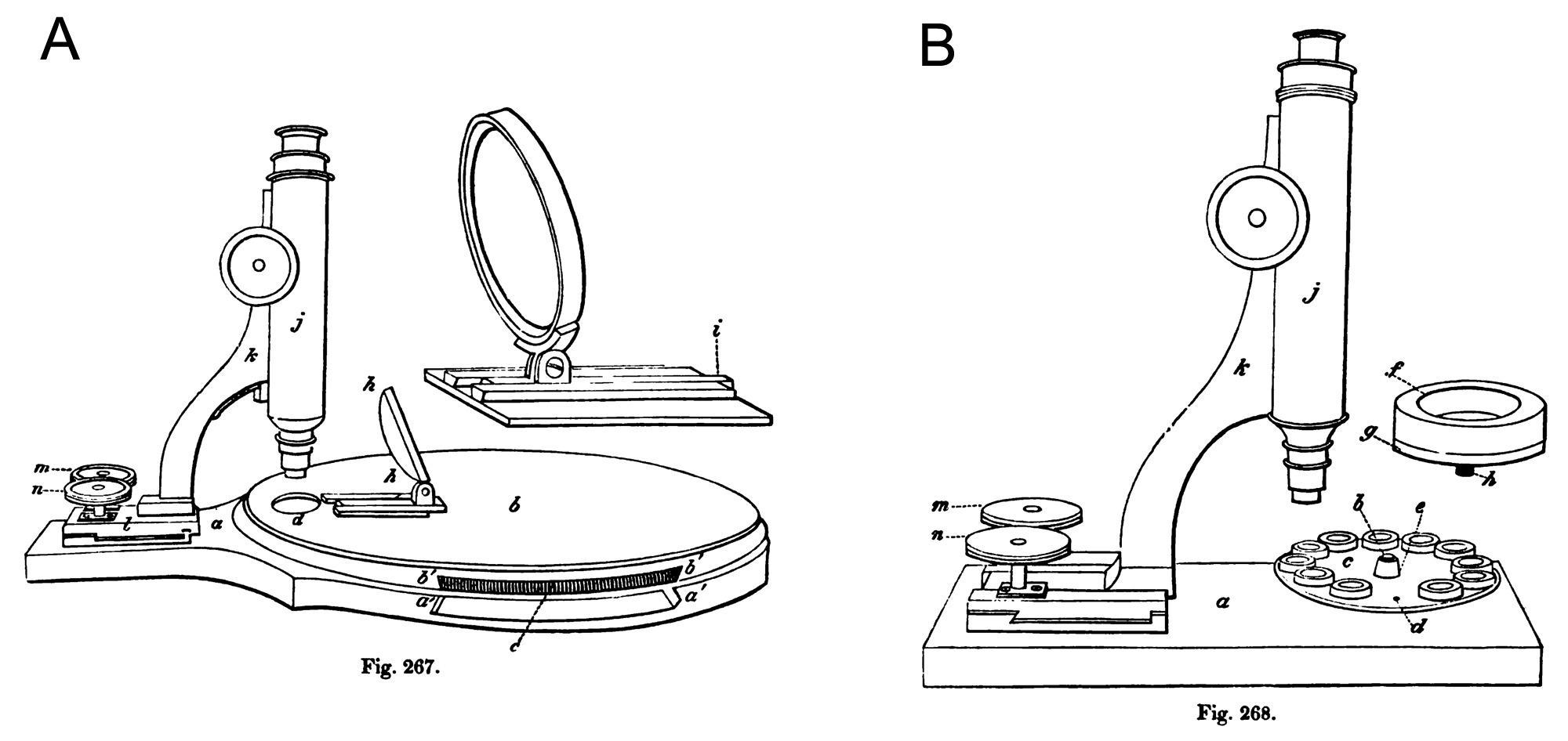
Figure 5. Drawings of “Mr.
Hett’s microscopes for injections, etc.”, adapted from J. Quekett, ‘A Practical
Treatise on the Use of the Microscope’, 1852. Microscope “A” matches
descriptions of the instrument used by Hett at the 1851 London Exposition. Both
were probably made by modifying stock Smith and Beck microscopes. Quekett
described these instruments as follows: “The first of these instruments,
represented at fig. 267 (panel A),
consists of a slab of polished mahogany, of the form shown at a, a' a', into
which, beneath b, is fixed a metal pin, on which a brass wheel revolves,
carrying forty cells with their enclosed objects; these are protected from
dust, and the action of light by the cover, b, b' b', which is fastened to the
slab, a, in such a manner as to admit of being readily removed, a portion of
the slab between a and a' is cut away, as is also part of the cover between b'
and b, so as to admit the finger, for the purpose of moving the wheel, the
milled edge of which is shown at c. A circular opening is made through the
cover at d, immediately under the object-glass, and through which the
preparations may be examined as they alternately present themselves at this
point; this opening is covered with a circular piece of thin glass, to protect
the cells beneath from dust; A A is a bull's-eye, mounted on a dove-tailed
sliding piece, i, seen on a larger scale at fig. 267, i showing the sliding
piece. The body, j, and the limb, k, are made in the usual way, the latter
being attached to a stage, l, with right angled rack-work fittings, and so
constructed that by means of the milled-head pinions, m n, the object-glass may
be made to traverse the entire space formed by the opening, d, and thus to
bring the whole of the object beneath it into view. (panel B) Fig. 268 shows another form of Mr. Hett's instrument; the
body, j, limb, h, and rack-work fittings, l, m, n, are similarly arranged to
those in the instrument previously described; but the mahogany slab, a, is of a
different form, having a brass pin fitted into its upper surface, the free end
of which is seen at b; on this pin a small brass disc, e, turns, carrying 12
objects. Through the disc, c, near to its margin and at regular distances,
twelve small holes are drilled and tapped, one of which is shown at d; a cell
having been removed for the purpose, a larger hole is drilled through the boss,
e, at its centre, through which the pin, b, passes. The glass cells employed
are made in a mould, the bottom and sides being of one piece; each cell is
mounted with marine glue on a small disc or button of brass, furnished with a
solid shank, which is tapped to form a screw, so as to admit of its being
fastened into the small hole, d, in the disc, c. One of these cells, mounted as
just described, is shown at l, the cell being represented at g, and the brass
disc or button, with its shank, at h. This is an exceedingly elegant and most
convenient method of mounting injected and other opaque objects, and will be
found very useful whenever it is required to demonstrate to classes any
particular set of organs, as cells mounted in this way can easily be removed,
thus enabling the teacher to arrange the objects on the disc in the order in
which he may wish to present them, and the rapidity with which any one of them
can be brought under the object-glass, will assist the student in ascertaining
the distinctive differences existing between them. When not in use the discs
may be kept in drawers, each drawer having a number of pins, similar to that
fixed on the slab, screwed on the bottom of it, to receive them. Mr. Hett
employs mahogany boxes, about the size of those used to hold twelve slides, but
shallower, a brass pin being fixed into the centre of the bottom of each box:
this pin is somewhat longer than that fastened to the microscope, so that it
projects beyond the boss on the disc, the projecting portion is furnished with
a thread, the disc being dropped into the box is secured by means of a nut.
These discs may be used with a microscope of the usual form, the only
additional apparatus required being a brass clip, furnished with a pin to
receive them; this clip should befitted to the stage in the same manner as an
ordinary object-plate, or the object-plate itself may have a pin attached to it
for the reception of the discs.”
Hett also displayed his injected microscopical objects at the 1855 Paris Universal Exposition and the 1862 International Exposition of London.
The 1861 census recorded Hett and his family as living at Horne House, Surrey, a farm of 133 acres. Hett’s occupation was listed as “farmer”, in addition to his medical qualifications of M.R.C.S., M.D. and L.S.A. Presumably a gentleman farmer, Hett employed four men and two boys.
Noting the modified microscopes he made (Figure 5), it is no surprise to learn that Alexander Hett was a skilled mechanic and an inventor. On January 23, 1840, a patent was issued to “Alexander Hett, of Gower-street, Bedford-square, in the parish of St. Giles in the Fields, and county of Middlesex, surgeon, for certain improvements in the arrangement and construction of fire-grates, or fireplaces, applicable to various purposes”. Thirteen year later, he was awarded another patent for “certain improved means or arrangements for the prevention of smoke and the economizing of fuel in furnaces”. During September, 1863, an patent was awarded to “Alexander Hett, of the city of London, Doctor of Medicine, and Frederick William Basset, of Camberwell, in the county of Surrey, Civil Engineer, for the invention of improvements in preserving animal substances, and animal and other substances used for food”.
Alexander Hett died at the age of 63, during early 1870. At that time, he and Lydie had lived in Lewisham, Kent. The next year’s census recorded Lydie, widow, as being a “professor of French language”.
Hett’s death escaped the notice of many people. Beale’s 1880 edition of How to Work the Microscope still recommended Hett as a supplier of slides. The 1888 Calendar of the Royal College of Surgeons of England still listed Alexander Hett as a member.
Acknowledgements
Many thanks to David Walker for generously providingslide images, and Howard Lynk and Pete Hodds for helpful comments.
Resources
Beale, Lionel S. (1865) How to Work the Microscope, third edition, Lindsay and Blakeston, Philadelphia, pages 108 and 263
Beale, Lionel S. (1880) How to Work the Microscope, fifth edition, Harrison, London, page 519
Bracegirdle, Brian (1987) A History of Microtechnique, second edition, Science Heritage Ltd., Lincolnwood, Illinois, pages 291-292 and plate 21
Bracegirdle, Brian (1998) Microscopical Mounts and Mounters, Quekett Microscopical Club, London, pages 51 and 204-205
British Farmer’s Magazine (1852) review of the Great Exposition, New series, Vol. 22, page 161
Bulletins and other State Intelligence (1852) The Great Exhibition – Prize Medals, page 625
Calendar of the Royal College of Surgeons of England (1865) Members, Taylor & Francis, London, page 162
Calendar of the Royal College of Surgeons of England (1888) Members, Taylor & Francis, London, page 56
Christening record of Alexander Hett (1807) accessed through familysearch.org
English census, birth, marriage and death records, accessed through ancestry.co.uk
The Gentleman’s Magazine (1833) Marriages, Vol. 103, page 558
Hannover, Adolphe (1853) On the Construction and Use of the Microscope, ed. by J. Goodsie, Sutherland and Knox, Edinburgh, advertisement from Hett at back of the book
Hayes, Boon (1853) Histological anatomy and microscopical manipulation, Medical Times and Gazette, Vol. 27, pages 155-158
Hett, Alexander (1830) Outrage by Messrs. Key and Cooper at Guy’s (letter), The Lancet, Vol. 1, pages 114-115
International Exhibition: Official Catalogue of the Industrial Department (1862) 2910 Hett, A., page 47
The Lancet (1828) Letter, Vol. 2, pages 29-30
The London Gazette (1863) Patent 2099, Sept. 11, page 4422
The London Journal of Arts and Sciences and Repertory of Patent Inventions (1841) Vol. 18, pages 73-76
Mechanics' Magazine and Journal of Science, Arts, and Manufactures (1853) patent 2453, Vol. 59, page 398
The Medical Times (1851) The Great Exhibition-reviews, vol. 24, page 72
Official catalogue of the Great Exhibition of the Works of Industry of All Nations (1851) 249, Hett, A., page 63
Paris Universal Exhibition: Catalogue (1855) 784 Hett, Alexander, MD, Great Britain Imperial Commission, page 34
Power, D'Arcy (1937) A urological cause célèbre, British Journal of Urology, Vol. 9, pages 330–338
Quekett, John Thomas (1848) A Practical Treatise on the Use of the Microscope: Including the Different Methods of Preparing and Examining Animal, Vegetable, and Mineral Structures, 1st Edition, H. Bailliere, London, back pages
Quekett, John Thomas (1852) A Practical Treatise on the Use of the Microscope: Including the Different Methods of Preparing and Examining Animal, Vegetable, and Mineral Structures, 2nd Edition, H. Bailliere, London, pages 492-495, and back pages
Quekett, John Thomas (1855) A Practical Treatise on the Use of the Microscope: Including the Different Methods of Preparing and Examining Animal, Vegetable, and Mineral Structures, 3rd Edition, H. Bailliere, London, back pages
Quekett, John (1850) Descriptive and Illustrated Catalogue of the Histological Series Contained in the Museum of the Royal College of Surgeons of England, Vol. 1, printed by R. and J.E. Taylor, London, pages 156-159
Rainey, George (1849) On the Structure of Sudoriparous Glands, printed by T. Pettitt, Soho, page 8
Walker, David (2011) Victorian rambles - part II, three 'injected' microscope slide preparations. Micscape, May, published on-line: http://www.microscopy-uk.org.uk/mag/artmay11/dw-victorian2.html
The Zoologist (1850) Proceedings of the Microscopical Society of London, Vol. 8, page 2836