Josef Hyrtl, 1810 - 1894
by Brian Stevenson
last updated February, 2017
While a medical student in Vienna, Josef Hyrtl learned methods for anatomical studies that involved injection and corrosion. This consists of injecting blood vessels or other open organs with colored substances that solidify, then corroding away the organic tissue with acid. What remains is a three-dimensional replicate of the vessels’ networks. Hyrtl’s skill at preparing whole organs, and his investigations of the resulting preparations, provided significant insight on the anatomy of humans and other animals. He also produced small injected and corroded specimens for microscopical investigations, which were in high demand during his lifetime (Figures 1-3 and 8). Hyrtl’s sets of microscopical preparations are very rarely encountered today.
Although Bracegirdle, in his Microscopical Mounts and Mounters, dismissed Hyrtl’s work as having “very little scientific worth”, one can argue that the three-dimensional injection-corrosion specimens of Hyrtl and others provide an understanding of anatomy that cannot be easily discerned from thin-section preparations (Figures 4, 5, and 9). For example, a contemporary of Hyrtl’s noted, “His preparations, famous for many years, demonstrate by colored material injected through some of the principal arteries the presence of the microscopic arteries and veins accompanying the lacteal vessels in the minute intestinal papillae. By the same means he demonstrated in 1874 the presence of a vascular net in the cornea of the eye, and after many ineffectual attempts he succeeded in filling the arteries and veins of an infant eight days old from the umbilical vein with coloring matter so perfectly as to reach and penetrate the minute arteries and veins of both corneae”.
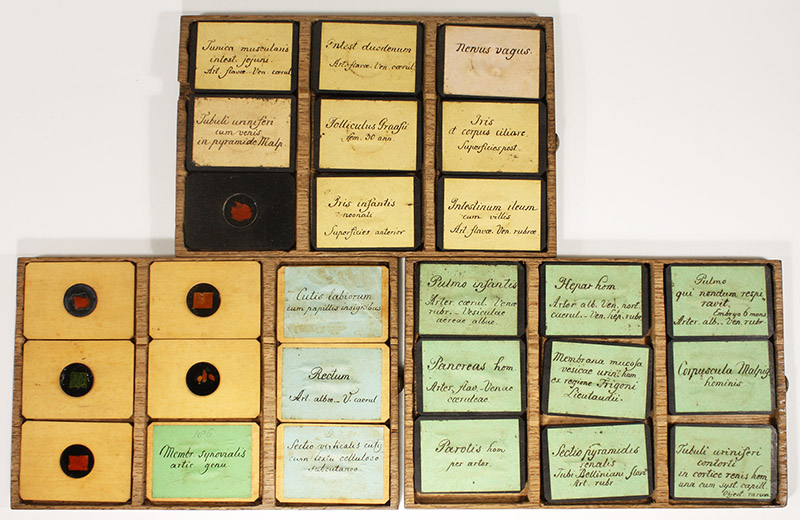
Figure 1.
A selection of Hyrtl preparations for the microscope. The injected and corroded specimens are under glass, within wooden blocks that measure 1 3/4 x 1 1/4 x 3/16 inches / 45 x 32 x 4 mm. This collection includes blocks of both dark and light hardwoods. A label covers the back of each, with the specimen description in Hyrtl’s hand.
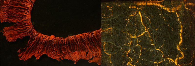
Figure 2.
3.5x magnifications of two specimens from Figure 1. Left, “Iris infantis neonati, superficies anterior”, and right, “Membrana muscularis intestini tenuis”, with arteries injected yellow and veins green.
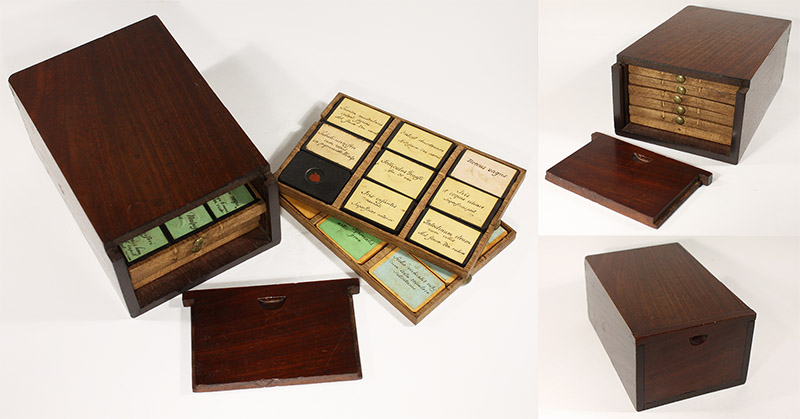
Figure 3.
Polished hardwood box, with sliding front, that holds six drawers of nine each Hyrtl microscopical preparations. Another example of a Hyrtl collection is shown below in Figure 8.
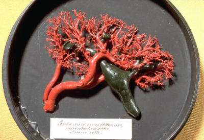
Figure 4.
Vessels of a whole organ, injected and corroded by Hyrtl. Such preparations make the three-dimensional arrangements of vessels readily understandable. Adapted for nonprofit, educational purposes from the Museum Mödling, http://www.museum-moedling.at
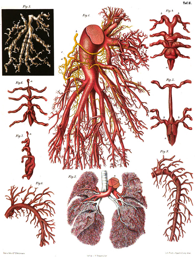
Figure 5.
Airways and blood vessels of the lungs, from Hyrtl’s 1873 “Die Corrosions-Anatomie und Ihre Ergebnisse”. The interrelationships were ascertained from micro- and macroscopic investigations of injected/corroded specimens.
Josef Hyrtl was born on December 7, 1810 in Kismarton, Hungary (now Eisenstadt, Austria). Soon afterward, he and his family moved to Vienna. There, his father played oboe in the orchestra of Nicholas II, Prince Esterházy. Young Josef became a choirboy for the palace chapel. He also received education at the government boarding school.
He began studying medicine at the University of Vienna in 1831. While a student, he became a prosector in anatomy, assisting the professor of anatomy, Joseph Berres. This job required Hyrtl to prepare specimens for lectures, etc., and Berres taught him well. After graduating in 1835, Hyrtl stayed on as prosector with Berres for another two years. A London review of Berres’ 1836 Anatomie der Mikroscopischen Gebilde des Menschlichen Korpers raved about the skills of Berres and Hyrtl in preparing specimens and using the microscope to precisely investigate anatomical structures, “Without anatomy there can be no physiology; this is now an old truth. But it is only comparatively recently that it has become generally acknowledged that there can be no rational account given of the functions of any part until the structure of its tissues is thoroughly known. Who, for example, would attempt to explain the action of the lymphatics in digestion without being familiarly acquainted with the texture of the parts, suitably injected and microscopically examined? Can the secretions of any membrane, gland, or organ, be even plausibly accounted for, without an accurate demonstration of the parts concerned? The value of microscopic investigation has long been admitted; but not universally. The sceptics have always been in the majority: they have refused to believe what they had not themselves an opportunity of seeing; and in some instances they have refused to see what, if acknowledged as real, would overturn their theories. … With the compound microscope of Plossl (an artist whom he praises in the highest terms), (Berres) was enabled to appreciate the excellence of these, but at the same time to see that they were far from being perfect. … His present criterion of the excellence of a preparation suited for microscopical purposes is this - to be able to distinguish the anastomosis of all the vessels even in the minutest twigs, and to discern their perfect detachment or separation from the surrounding organic matter. Every vessel is to be traceable continuously and without interruption through its entire course. How he and his assistant Dr. Hyrtl manage to effect these exquisite preparations is fully explained”.
In 1837, Hyrtl was appointed professor of anatomy at the University of Prague. His training with Berres, and subsequent refinement of his skills, led to Hyrtl’s classic textbook on human anatomy, Lehrbuch der Anatomie des Menschen. It was subsequently republished in over twenty editions and translated into many languages. His injection specimens continued to grow in esteem: an 1842 article stated that “those of Lieberkühn in particular … are, notwithstanding all our modern improvements, some of the finest injections in existence; they are only equaled by those of our esteemed friend, Professor Hyrtl, of Prague”.
He moved back to Vienna in 1845, to take the late Dr. Berres’ position as professor of anatomy at the University of Vienna. Another book destined to be a classic, Handbuch der Zergliederungskunst (“Handbook of Topographic Anatomy”), was published there in 1860. Many other important books and papers followed. Hyrtl’s efforts as a teacher and writer had significant impact on the importance of studying anatomy in medical school.
Hyrtl also became a tourist destination. In 1856, the author George Elliot described, “Another great pleasure we had at Vienna - next after the sight of St. Stephen's and the pictures - was a visit to Hyrtl, the anatomist, who showed us some of his wonderful preparations, showing the vascular and nervous systems in the lungs, liver, kidneys, and intestinal canal of various animals”.
Acknowledging Hyrtl’s world-wide fame, he was appointed Rector of the University of Vienna in time for their 500th anniversary, in 1865. He served for that academic year.
For many years, Hyrtl made his injected/corroded specimens available to other investigators. These sales appear to have been largely through private contacts, although he did display at expositions. He earned a Gold Medal at the 1862 London Exposition. A report of the 1867 Paris Universal Exposition stated “The anatomical preparations exhibited by Professor Hyrtl, of Vienna, are … remarkable … If the manner of preparing them is not entirely new, they are at least prepared with an astonishing skill, parts generally considered inaccessible to the scalpel being beautifully dissected. These pieces are quite worthy of the great reputation of their author. The labyrinths of the ear of different mammiferous animals exhibited by Mr. Hyrtl are some of the most curious and difficult things the anatomist can prepare”.
After the 1867 Paris Exposition, The Medical Record (USA) noted, “Hyrtl's magnificent anatomical museum has been purchased by an American College for $8,000, it having been offered for sale at the Paris Exposition. Hyrtl is the Professor of Anatomy at Vienna, and from personal observation, we are prepared to testify to its value. What college has the treasure?” (I have not located the answer to that question - does any reader know?).
The fate of another of Hyrtl’s collections is known. In 1874, the Mütter Museum of the College of Physicians of Philadelphia (USA) acquired his collection of 139 human skulls. The museum’s description, “His work was an attempt to counter the claims of phrenologists, who held that cranial features were evidence of intelligence and personality and that racial differences caused anatomical differences. Hyrtl’s aim in collecting and studying the skulls was to show that cranial anatomy varied widely in the Caucasian population of Europe. Each skull is mounted on a stand, and many skulls are inscribed with comments about the person’s age, place of origin, and cause of death” (Figure 10).
In 1873, Hyrtl published both a catalogue of his available works (Figure 7) and a book describing his methods, Die Corrosions-Anatomie und Ihre Ergebnisse. Huntington, and probably others, lamented that Hyrtl’s methods book lacked sufficient detail to replicate his works, “It is curious to note that the secrecy with which Ruysch surrounded his method of preparing corrosions seems to have been imitated to a greater or less extent by his successors. It is a matter of regret that the information which Hyrtl gives on the composition of the mass in his large work on ‘Corrosion-Anatomie’ is so unsatisfactory. Personally I look back on much time and material expended in fruitless attempts at following his instructions. In spite of the utmost care, we have found it impossible to obtain even moderately successful permanent corrosions with the mass recommended by him”.
Nonetheless, some details of Hyrtl’s methods can be ascertained. He stated that the colors for his “masses” (the waxy substances that are injected) were purchased as fine paint powders from artist supply shops. Colors were chosen for their visual impact: a visitor is quoted as declaring “mais ce ne sont pas de préparations anatomiques ce sont des bijoux” (“These are not anatomical preparations - they are jewelry”). Hyrtl followed that anecdote with the statement, “And he was right”.
In the corrosion step, Hyrtl wrote “I only use hydrochloric acid. I have never tried nitric acids, since I do not presuppose them from the very beginning, to leave certain colors, like the sulphurides and the carbonic salts, unchanged. Concentrated hydrochloric acid destroys the parenchymes so quickly that a smaller preparation can be washed out in two to three days. If we wait a few more days, very voluminous and hard organs, such as the large animal-livers, will be thoroughly corroded. Only whole parts of the body, which contain many bones, eg, heads, feet, hands, take a long time. For very small organs often already a couple of hours. Whole bodies of fish and amphibians are corroded in much more rapid time than those of warm-blooded animals”.
Hyrtl retired from the University of Vienna in 1874. His books and specimens had earned him a considerable fortune, and their continued sales allowed him a very comfortable life at his estate in Perchtoldsdorf, near Vienna.
A colleague published several anecdotes of Josef Hyrtl’s life, in 1894, providing insights on his personality:
“The famous anatomist who has just died at Vienna was always considered an eccentric man because his dress, his manners, and his mode of life differed in some respects from those of other people. It was particularly his time-worn garments that made him conspicuous, and he used in his walks the same soiled blouse which he wore while engaged in his laboratory or in gardening - his favourite hobby. Who would have thought this untidy person to be the world renowned anatomist, and a rich man? This characteristic of Hyrtl gave frequent occasion to ludicrous incidents at which he himself used to laugh most.
A few years back, when he was still in possession of his eyesight, he was accustomed to walk to Liesing, a charming Viennese suburb, where, in the beer-garden of the brewery, he refreshed himself with a glass of its well-known beverage. One afternoon he entered the garden and seated himself near a table at which a few merry Viennese burghers were engaged in diminishing the contents of a big dish of stewed fowl. These gentlemen had no idea of the identity of the newly arrived guest, and after eyeing his simple twill suit came to the conclusion that he must be an inmate of the Liesing asylum for the poor. A good portion of the dainty meal having been left uneaten, one of the guests called the waiter, saying, ‘Here, give this to that poor man; let him have a good feed for once’. which humane proposal was loudly acclaimed by his fellow revellers. The waiter, who was a new hand at the brewery, obeyed the command, placing the dish of remnants before the famous savant, who, appreciating the joke, ate a few morsels and, after expressing his thanks, left the garden. A few moments later two waiters carried in a big bowl from which the gilded heads of champagne bottles were protruding. ‘We have ordered no champagne’, cried the burghers, and their astonishment may easily be guessed on their being informed that ‘the inmate of the local asylum for the poor’ had sent them the champagne as a mark of his gratitude for the stewed fowl, and that the donor was no less a personage than Professor Hyrtl.
His sympathy with student life is well known, and many stories in this connexion are told of him. A Jewish medical student named Jerusalem was being examined, and in the hall there was waiting a small crowd of his co-religionists, when Hyrtl came out from the examination chamber. He was immediately surrounded by the crowd of young students, who asked him the fate of their comrade Jerusalem. Hyrtl, with a plaintive voice and sympathetically shaking his head, said, ‘Weep, O Israel; Jerusalem has fallen’.
Once a student was unable to reply to one single question at his examination. Hyrtl then asked him, ‘Perhaps you can tell me where you live ?’ The student named the street, of which Hyrtl professed his ignorance, and said, ‘You see how science is divided: you know nothing of anatomy and I do not know the locality where you live’.
An anecdote respecting his marriage is worth repeating. It was in the year 1866, when one day Hyrtl approached the porter of the Anatomical Institute, Andrew Swetlin by name. ‘Andrew, have you a prayer-book handy?’ asked Professor Hyrtl. Andrew fetched the book, and Hyrtl went away. An hour later he returned and handed to the porter the prayer-book. The porter, unable to suppress his curiosity, asked, ‘You do not need it any more, Sir ?’ Whereupon Hyrtl replied. ‘No, thanks; I only needed it for a moment. I have just been at my own wedding’.”
Hyrtl died in his sleep during the night of July 16-17, 1894, at his estate at Perchtoldsdorf. He had founded an orphanage in Mödling and, since he had no children, made this institution the heir to his fortune. The Museum Mödling includes an exhibit on Hyrtl.
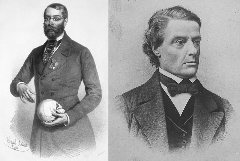
Figure 6.
Josef Hyrtl.
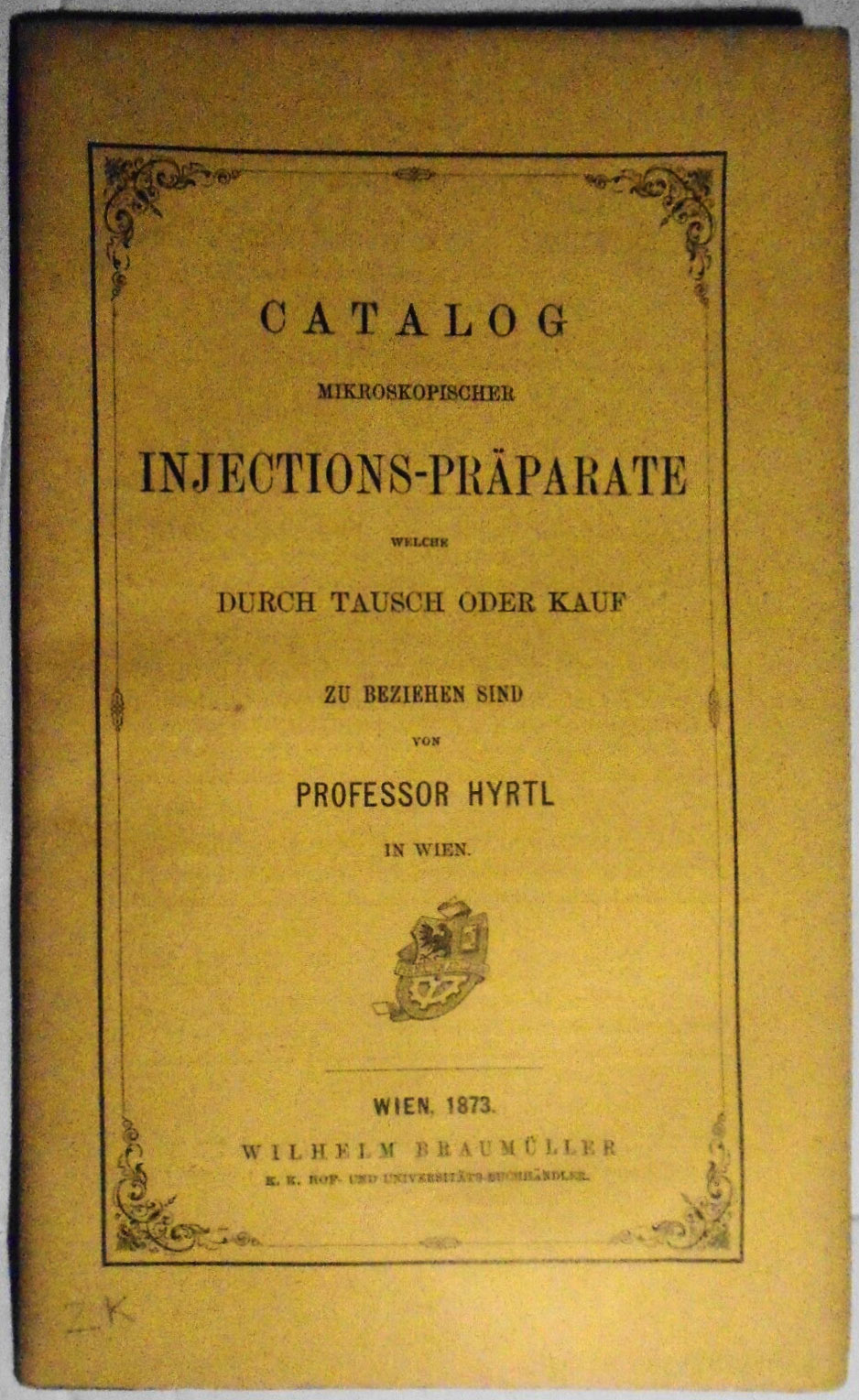
Figure 7.
Cover of Hyrtl’s 1873 catalog of injected preparations. The complete catalog can be freely downloaded for personal use from http://www.microscopy-uk.org.uk/Little-Imp/.
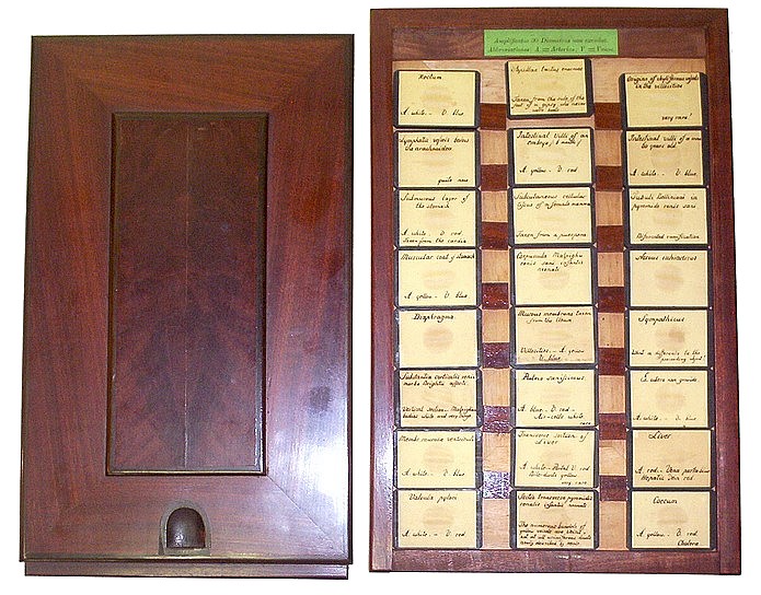
Figure 8.
Another cased set of Hyrtl injection/corrosion specimens for the microscope. Adapted with permission from http://www.antique-microscopes.com/photos/hyrtl.htm.
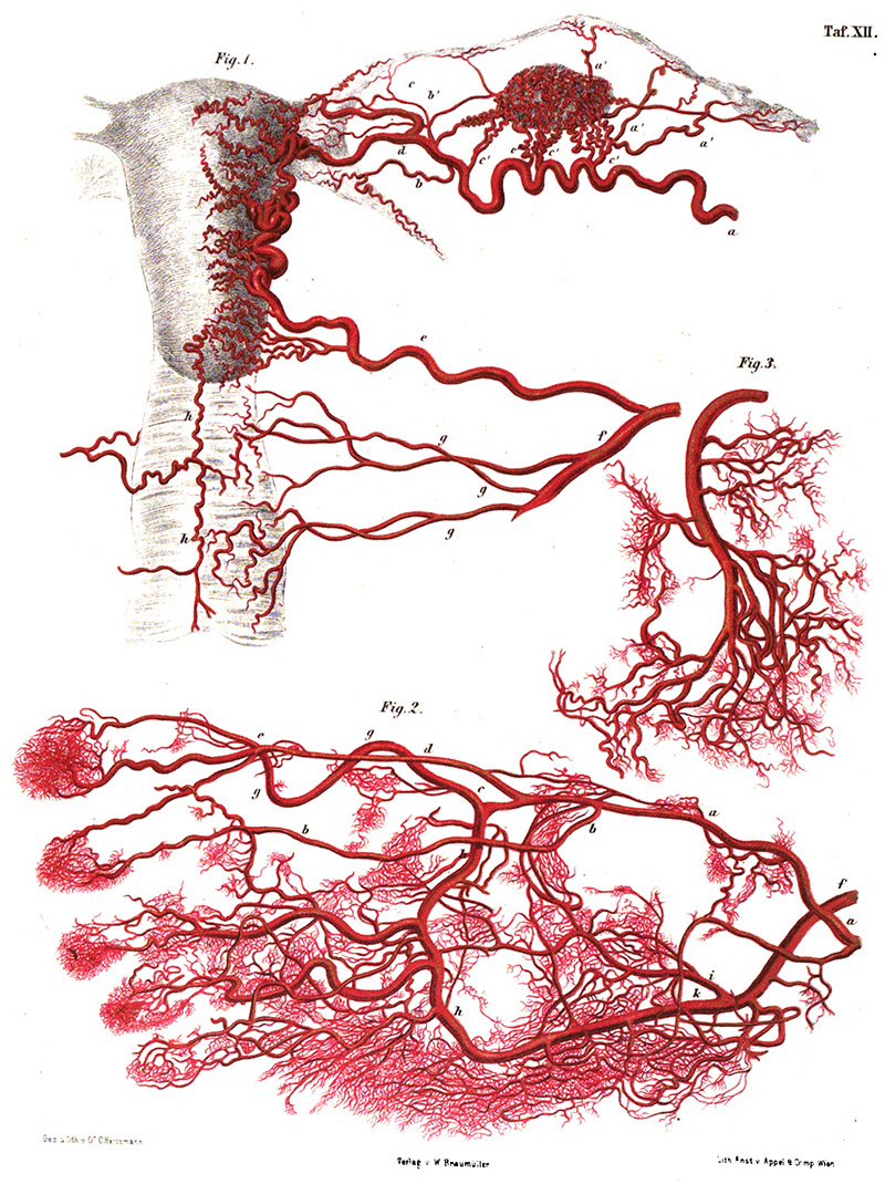
Figure 9.
Blood vessels of the human female reproductive organs, from Hyrtl’s 1873 “Die Corrosions-Anatomie und Ihre Ergebnisse”.
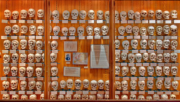
Figure 10.
Josef Hyrtl’s skull collection, on display at the Mütter Museum, College of Physicians of Philadelphia. Adapted for nonprofit, education purposes from http://muttermuseum.org/exhibitions/hyrtl-skull-collection.
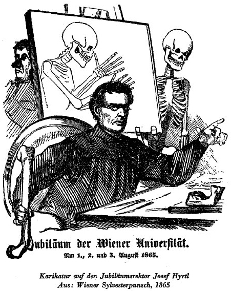
Figure 11.
Caricature of Josef Hyrtl, 1865.
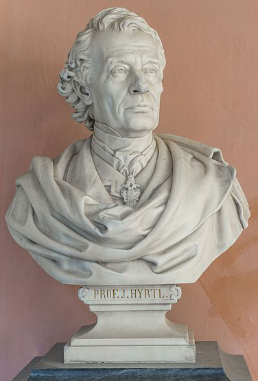
Figure 12.
Bust of Josef Hyrtl, at the University of Vienna.
Acknowledgements
Thank you to Allan Wissner for sharing images of his Hyrtl collection, and to Steve Gill for providing free access to the Little Imp Collection, including the 1873 Catalog of Josef Hyrtl, http://www.microscopy-uk.org.uk/Little-Imp/.
Resources
Anonymous (1894) Some reminiscences of the late Professor Hyrtl, The Lancet, Vol. 2, page 336
Blake, William P. (1870) Reports of the United States Commissioners to the Paris Universal Exposition, 1867, Vol. 5, Government Printing Office, Washington
Bracegirdle, Brian (1998) Microscopical Mounts and Mounters, Quekett Microscopical Club, London, pages 55-56, 204, and 222, plates 50 and 59
Eliot, George, and J.W. Cross (1884) Life of George Eliot: As Related in Her Letters and Journals, T.Y. Crowell, New York
Huntington, George S. (1897) Corrosion Anatomy, Technique, and Mass, Reprinted from Proceedings of the Association of American Anatomists, 1897
Hyrtl, Josef (1847) Handbuch der Topographischen Anatomie, J.B. Wallishausser, Vienna
Hyrtl, Josef (1873) Die Corrosions-Anatomie und Ihre Ergebnisse, W. Braumüller, Vienna
Hyrtl, Josef (1873) Catalog Mikroskopischer Injections-Präparate, W. Braumüller, Vienna
Knoeff, Rina, and Robert Zwijnenberg (2016) The Fate of Anatomical Collections, Routledge, London, page 155
The Lancet (1894) Memoriam of Joseph Hyrtl, Vol. 2, pages 169-170
London Medical Gazette (1837) Review of Anatomie der Mikroscopischen Gebilde des Menschlichen Korpers, Vol. 19, page 820
The Medical Report (1868) Hyrtl’s magnificent anatomical museum, Vol. 2, page 240
The Medico-Chirurgical Review (1842) Mr. Wilde on the School Of Ophthalmic Surgery In Vienna, Vol. 40, page 262
Museum Mödling web site (accessed February, 2017) http://www.museum-moedling.at
Mütter Museum web site (accessed February 2017) http://muttermuseum.org/exhibitions/hyrtl-skull-collection
Steudel, J. (2008) Complete Dictionary of Scientific Biography, C. Scribner’s Sons, New York
Walsh, J.J. (1910) Joseph Hyrtl, The Catholic Encyclopedia, R. Appleton Co., New York


