William Jones, 1813 - 1896
by Brian Stevenson
last updated June, 2020
William Jones was a physician and pharmacist in Newburgh, New York (located about 60 miles / 100 km north of New York City, on the Hudson River). He wrote of the importance of microscopy in medicine, especially for examining sputum, urine, and other samples. Jones practiced a branch of medicine known as “eclectic medicine”, which relied heavily on botanical remedies. Accurate identification of plant species was necessary for Jones’ medical specialty, as well as for his pharmaceutical business.
Jones also prepared microscope slides for sale or trade (Figure 1). It is likely that he sold them at his pharmacy, which was a common practice of the time. He also requested to exchange slides during the early 1880s. As with many American slide-makers, Jones’ microscope slides are not frequently encountered, probably due to lack of awareness among Americans of the value of such slides.
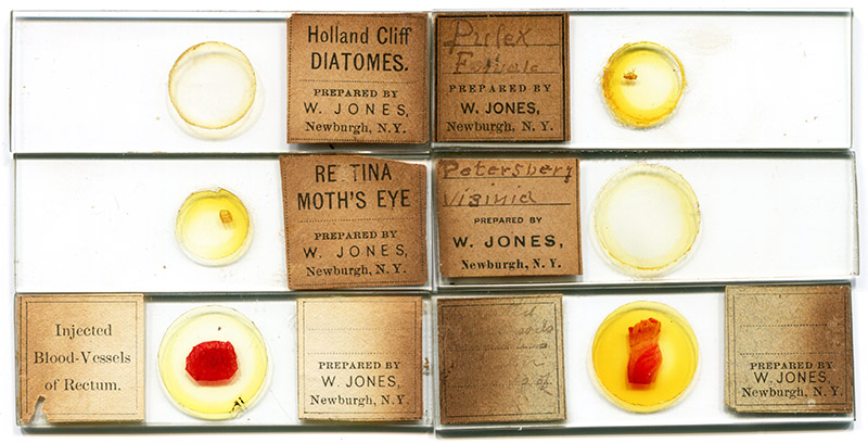
Figure 1.
Microscope slides by William Jones, probably ca. 1870s-1880s. He used several variants of printed labels (one of his printers seems to have been unfamiliar with some scientific words). Specimen descriptions were either typeset or handwritten. Jones work spread around the world, probably from exchanges with other microscopists - I acquired some of the illustrated slides from a French collection.
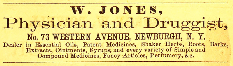
Figure 2.
An 1860 advertisement from William Jones.
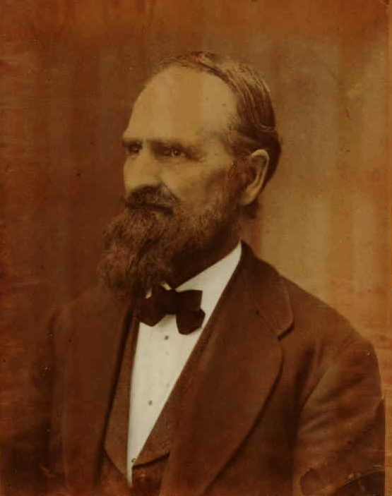
Figure 3.
An undated photograph of William Jones. Adapted for nonprofit, educational purposes from https://www.ancestry.co.uk/family-tree/person/tree/11058982/person/739176369/facts.
William Jones was born on November 29, 1813, in Vermont. His father, also named William, died the following summer, only 28 years old. Despite that setback, young William was able to receive an education and become a doctor of medicine.
Jones settled in Newburgh, New York, and married Charlotte Wheeler. They had 8 children together.
In 1879, Jones gave a speech to his medical society on the value of the microscope to medicine, beginning with, “So far as I know, microscopical observations and investigations in the eclectic school of medicine have been limited, and very little desire has been manifested by those who were familiar with the science, and the benefits to be derived from it in every-day practice, to extend the knowledge. Knowing, as I do from practical experience, the advantages that are to be obtained from a knowledge of the use of the microscope, I will endeavor to give you a brief historical sketch of the instrument, and also what I know about a microscope as applied to medicine. … So far as I have observed, the medical profession has manifested the least interest, and for that neglect I am very much surprised, for there is no profession in which the microscope is, or can be, made of such special use and benefit as in the profession of medicine. With it we can diagnose disease that it is impossible to do in any other way. And I believe the time is fast approaching when a physician will consider a microscope as necessary for his outfit as he now does a diploma. ... I am almost forced to repeat the assertion that it is to me surprising that physicians of this enlightened age are seemingly so little interested in the microscope - so indispensable in the practical part of their profession. It disciplines the mind, it instructs while it amuses, and it fills a niche in the practice of medicine that no intelligent and enlightened physician can afford to leave unfilled”. Then, several practical applications of the microscope in medicine were presented, including “I need hardly tell you, after saying what I have in favor of the microscope, that I consider it indispensable to a correct diagnosis in many cases. Take a case of trichiniasis. In this it is an impossibility to diagnose correctly without a practical knowledge of the use of the microscope. And I have great reason to believe that the disease is much more frequent than has been supposed. I think that there is scarcely a doubt that many a person has sickened and died with trichiniasis, that has been supposed to have been typhus fever, dysentery or inflammation of the bowels. The symptoms are so nearly like those of the above-named diseases, that the physician who is not familiar with the nature of trichinae, would be quite liable to mistake”.
In 1881, he wrote on Addison’s disease, “The disease to which I would call your attention is one which I do not remember having seen mentioned in any of the journals or text-books of our school. It is a remarkable and rare affection, one which some of us, perhaps, have not become familiar with, although called to treat it. It is a disease sufficiently obscure for very clever physicians to make mistakes in its diagnosis. I refer to the peculiar affection of the suprarenal capsules, called ‘Addison's Disease’, from Dr. Addison, who first described it in 1849. The discovery, like many other valuable discoveries, has been scarcely recognized until the last few years. It is worthy of note that it was through the aid of the microscope that this increased interest in diagnosis has been brought about. There is no evidence that Dr. Addison was familiar with the advantages of this instrument. But, in 1855, Dr. Wilkes was testing blood for white corpuscles, after J. Hughes Bennett's and Virchow's discovery of leukaemia, when his attention was directed to the similarity in the condition of the blood in leukaemia and Addison's disease”.
I did not find records of William Jones joining any microscopic or other scientific society. However, beginning in 1883, he provided his name and address to The Naturalists’ Directory, specifically mentioning that he was interested in collecting specimens of diatoms and histological tissue for microscopic examination. He last contacted the publishers of that annual book in 1885; the 1886 edition listed Jones with a “?”, indicating that they had not received recent communication. He was not listed in the 1888 edition.
Those listings in The Naturalists’ Directory suggest that many of Jones’ known microscope slides were produced during the early- to mid-1880s. He appears to have distributed them far, as several of the Jones slides in my collection were purchased in France.
William Jones died on June 12, 1896.
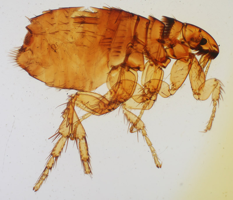
Figure 4.
Circa 1883 preparation of “Pulex, female” (flea species), by William Jones (see Figure 1).
Resources
Jones, William (1878) History of the microscope, Transactions of the Eclectic Medical Society of the State of New York, pages 220-225
Jones, William (1881) Addison’s disease, Transactions of the Eclectic Medical Society of the State of New York, pages 453-456
The Naturalists' Directory (1882) S.E. Cassino, Boston (William Jones was not listed)
The Naturalists' Directory (1883) “Jones, William, M.D., 61 Broadway, Newburgh. Diatoms, Mic., Histol. C, NY”, S.E. Cassino, Boston, page 66
The Naturalists' Directory (1886) “Jones, William, M.D., 61 Broadway, Newburgh, N.Y. Diatoms, Mic., Histol. C?”, S.E. Cassino, Boston, page 86
The Naturalists' Directory (1888) S.E. Cassino, Boston (William Jones was not listed)
Newburgh Directory (1860) Advertisement from William Jones
Newburgh Directory (1869) “Druggists … Jones, William, 73 Western Ave.”
US census and other records, accessed through ancestry.com