Charles Henry Kain, 1840 - 1909
by Brian Stevenson
Last updated October, 2025
School teacher C. Henry Kain was one of his era’s leading authorities on the diatoms of North America. In 1889, the Editor of The Microscopical Bulletin and Science News, wrote of Kain’s knowledge about diatomaceae, “it would be hard to name a more careful student of these forms, or a better authority upon them”. After his death, the United States National Museum (i.e. Smithsonian Institute) acquired “the extensive collection of diatom material of the late Professor C. Henry Kain, of Philadelphia, representing the principal fossil deposits throughout the world as well as a large number of recent gatherings made in this country and abroad”. Yet, Kain’s slides can be found outside the museum, as he gave away or traded a good number of them. His diatom preparations contain either one or a few select specimens, or consist of strews that exhibit varieties found in a particular location (Figures 1 and 2). Bracegirdle’s Microscopical Mounts and Mounters illustrates a C.H. Kain slide, an indication that some of his preparations crossed the Atlantic.
Kain was already a skillful slide-maker by 1878, when he and several colleagues founded the Microscopical Society of Camden (New Jersey, USA). An 1880 report of the meeting stated, “An eager and studious audience surrounded Prof. C. Henry Kain, at the close of business, to witness his illustration of some of the latest and most approved methods of mounting objects, the first of a series to be followed out by other members at subsequent meetings. The Prof. had his table strewn with the necessary appliances and materials, and prepared a number of beautiful slides, explaining the process the while, and answering all queries propounded as to the various steps of the process. The Society contains no more expert manipulator than Mr. Kain, as his specimens will attest, and the demand upon him for exchanges from abroad rapidly depletes his cabinet of his own work”.
Kain lived in Camden, New Jersey until 1894, when he moved across the Deleware River to Philadelphia, Pennsylvania. Those addresses help date Kain's microscope slides.
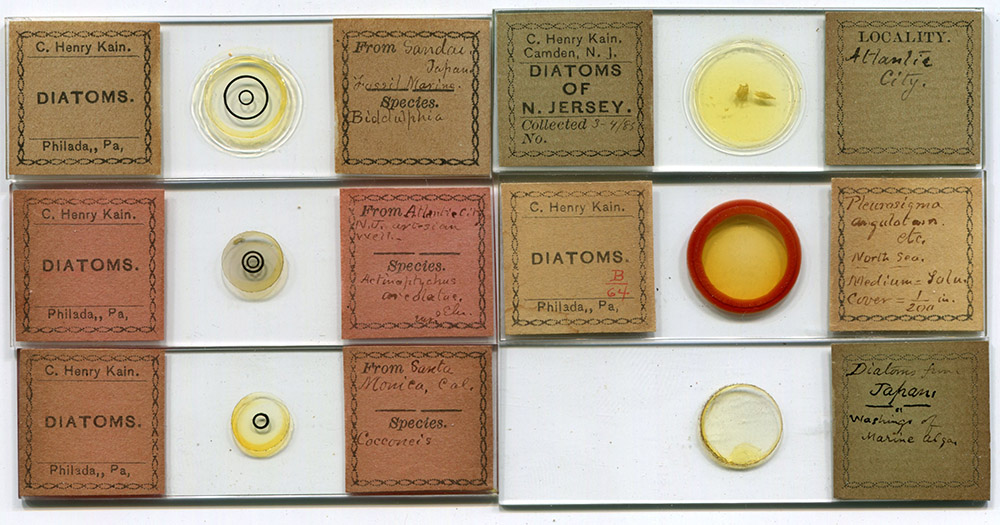
Figure 1.
Examples of diatom slides by C. Henry Kain. His address helps to date their production, with Philadelphia being after 1894 and Camden being before that date. The three slides on the left contain single diatoms of a species. Those on the right are strews: the top slide contains a pure gathering of a single species (dated 1885), while the other two slides have mixed strews. The preparations include specimens from Atlantic City, New Jersey, not far from Kain’s home, and from Sandai (Japan), Santa Monica (California, USA) and the North Sea. An illustrated essay on a 150-diatom arranged slide by Kain can be read at http://www.microscope-antiques.com/kaintypeslide.html. Images from the author's collection or adapted for nonprofit, educational purposes from internet auction sites.
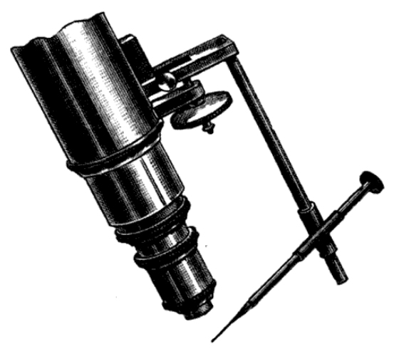
Figure 2.
Arranged diatoms, by C. Henry Kain. Images courtesy of Ian Jones.
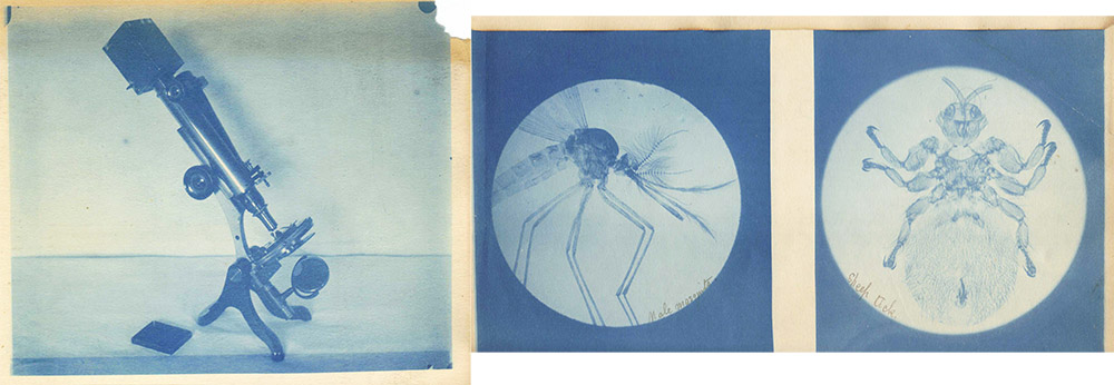
Figure 3.
“Kain’s Mechanical Finger”, as described in the “American Microscopical Journal” (1881) and the “Journal of the Royal Microscopical Society” (1882): “It consists essentially of a slotted bar, which may be firmly clamped to the upper (immovable) bar of the fine adjustment by means of a milled-headed screw. Through the end of this is fastened a round rod, at such a distance- from the objective that, when lowered, the end will not strike the stage. Over this rod slips a split tube, to which is soldered, at an angle, a smaller tube. Through the small tube passes a rod carrying a glass thread at its extremity. This rod is easily rotated by means of a milled head. The capillary glass thread is attached to the extremity by means of beeswax. … To use the finger, the point of the glass thread is first brought into the focus of the objective, or nearly so, by sliding the tube on the vertical rod, and pushing or pulling the rod carrying the thread until the desired position is attained. It is not difficult to do this, and having once been done by hand, it does not have to be repeated, as all further movements are made by the adjustments of the Microscope. Supposing now the point of the glass thread to be in focus; by means of the fine adjustment throw the focus ahead of the point, then, by means of the coarse adjustment, rack down and search for the object you wish to pick up. Having found the object desired, again bring the point of the thread into focus by means of the fine adjustment; then rack down with the coarse adjustment and pick it up. Now rack back with the coarse adjustment, remove the slip on which the material is spread, and place your prepared slip or cover upon the stage. Again, by means of the fine adjustment, throw the focus ahead of the object, rack down with the coarse adjustment, and search for the spot where you wish to deposit the object, and having found it, again focus the object, then rack down with the coarse adjustment, and when the object touches the slide and has been placed in proper position, fix it by means of a very gentle breath. I prefer this mode of fixing instead of the arrangement of tubes proposed by Professor Smith. I coat the surface of the cover or slide upon which the diatoms are to be fixed with an exceedingly thin film of gelatine, prepared thus: Dissolve 2 drachms of Cox's gelatine in 10 drachms of acetic acid by the aid of a gentle heat. When the gelatine is thoroughly dissolved, add 1 drachm of alcohol and 1 oz. of distilled water; stir well until thoroughly mixed, let stand some hours, and filter through the finest filtering paper. Keep in a glass-stoppered bottle. To coat the cover or slip I dip a small needle in the solution and wipe it once flatwise across the glass. There are many little wrinkles which the worker will acquire from time to time. One of the most important of these is the art of using the finger as a lever for moving diatoms or other objects into position when very slight movements are necessary. To do this, move the slide by hand until the point of the finger is just behind the object to be moved; then, by racking down with the coarse adjustment, the glass point pushes the object ahead of it. By a succession of pushes the object may be moved into any desired position. The coarse adjustment may be used in a similar manner for turning diatoms on edge or upside down, by pushing them against some fixed object and forcing the glass point under them. By using a point rather firmer than usual, the valves of a diatom may be separated. To do this I usually fasten the diatom on a slide which has been coated with gelatine, and when it is firmly fixed, the upper valve may be punched off without much difficulty. In studying diatoms, a mechanical finger is almost indispensable, for it may safely be said that one is not thoroughly acquainted with a diatom until he has turned it over and viewed it in all its aspects. In mounting diatoms for study it is well to mount a number of the same kind in various positions, so as to display the various spines, undulations, or other peculiarities. How often it happens, too, that in a mixed gathering of diatoms - and it is not easy to obtain pure gatherings - we find a rare frustule which we should like to preserve. By means of a mechanical finger the frustule may at once be selected and mounted. When one wishes to arrange diatoms so as to form symmetrical figures, an eye-piece micrometer will be found very useful, not only in selecting diatoms of uniform size, but also in determining their position. A circle ruled in squares and used in the same way as the eye-piece micrometer will be found still more desirable. It is a good idea to keep a number of glass points, of different degrees of fineness, ready prepared; that is, attached to little rolls of beeswax, so that if a point is unsuited for a particular work another can be substituted in a moment."
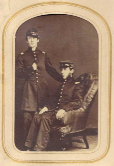
Figure 4.
Photographs by Henry Kain, of his microscope (with camera attached to one of the binocular tubes), and magnified views of a mosquito and a sheep ked (Melophagus ovinus). Adapted for nonprofit, educational purposes from https://www.ancestry.co.uk/family-tree/person/tree/66755837/person/44156395103/gallery.
The vast majority of records on C.H. Kain’s life refer to his as “C. Henry Kain”, implying that he preferred to go by the name Henry. He was born on January 4, 1840, probably in Marlton, New Jersey (not far from Camden and Philadelphia, Pennsylvania). His father, William, was a “turner”, presumably a skilled lathe-worker. Henry’s mother, Mary, died when he was only 5 years old.
Kain’s entry in The Botanists of Philadelphia states that Kain planned to “pursue classical studies with a view of entering college. This plan was frustrated by the breaking down of his health”. Instead, Kain earned a teaching degree from the University at Lewisburg (now Bucknell University). “He became principal of the North Ward Grammar School in Camden, in 1868, succeeding in that position William L. Sayre, now principal of the Central Manual Training School. When Mr. Sayre left the Stevens School in Camden, Mr. Kain took his place there. In 1874 Mr. Kain began his work in Philadelphia as Principal of the Northwest Boys' Grammar School at Fifteenth and Race Streets, and continued his work there until the close of 1886, when he accepted the position of Assistant Superintendent of Schools”.
The US Civil War broke out in April,1861. Probably deferred from major service due to his health, and also being a teacher, Kain served for a month during 1864 in Company A of the First New Jersey Militia (Figure 5). They were called up in that summer due to threats to the region from the Confederate Army. Kain served as a Lieutenant, an indication of his education and social status.
Henry married Eliza Baker on April 3, 1863. They had five children, three boys and two girls, of whom only the first two boys survived infancy.
I did not find out how, but some time during the 1860s - 1870s, Kain learned methods for preparing quality microscope slides. The Microscopical Society of Camden was formed in November, 1878, with Henry Kain as one of the original Managers. As noted above, Kain presented lessons on slide-making methods to his colleagues in January, 1880. The next month, “Professor Kain also showed a novel method of making glass cells for mounting”. At the April, 1880, Society Soiree, “The splendid cases of sides on exhibition, as the work of A.P. Brown, Prof. C.H. Kain, and H.S. Fortiner, were much admired, and showed evidence of great proficiency in the preparation and mounting of objects”.
Kain published a large number of papers on microscopical topics through the following years. Among these were “Glass cells” (1882), “Photo-micrography” (1882), a description if his “mechanical fingers (1882 - see Figure 3), an inexpensive reproduction of Adolf Schmidt's “Atlas der Diatomaceen-Kunde” (1885), "Diatoms, raising them in the laboratory” (1887), “On a fossil diatomaceous deposit from Oamaru, New Zealand” (1887), “Diatoms of Atlantic City and vicinity” (1888), “Diatomaceous deposits from the Yellowstone Geyser region” 1890), and “The Santa Monica diatomaceous deposit” (1897).
Numerous investigators used Kain’s extensive lists of diatoms identified throughout the USA, and also enlisted him to identify diatom species in their gatherings. For example, to produce the 1894 Annual Report of the State Geologist of New Jersey, Louis Woolman used lists from Kain and Charles Peticolas to identify diatomaceous material in diggings, plus, “from a preparation of diatoms from the upper portion of this bed the author made a strewn mount upon which C. Henry Kain, an authority, readily recognized the following forms, thirty-one in number. This is probably not more than one-sixth the number of species occurring in the beds”.
A hint from Kain on collecting diatoms, published in The Journal of the Royal Microscopical Society, 1889, “Mr. C.H. Kain, speaking of the bright-brown patches of diatoms frequently seen covering the surface of mud, recommends that they be collected in the following manner. Half fill a bottle with water. Touch one of the brown patches lightly with the tip of the finger, and the diatoms will adhere; then place the finger over the mouth of the bottle and shake it. The diatoms are, of course, washed off and remain. By repeating this process again and again the water finally becomes quite brown. By the time the collector reaches home the diatoms will have settled to the bottom, and the water may be poured off and the diatoms cleaned. It is worth while to examine under the collecting lens every promising patch of brown mud, for very pure gatherings of quite different species may often be collected within a few feet of each other”.
A slide of arranged diatoms by Kain, containing 150 individual specimens from the Santa Monica deposit, is illustrated and described at http://www.microscope-antiques.com/kaintypeslide.html.
Kain was also a skilled photographer, and advocated for using the camera to record microscopical images. A picture of his setup is shown in Figure 3, along with two examples of his photomicrographical work. In 1895, Kain was instrumental in the establishment of the Teachers’ Photographic Association, to aid teachers in the use of lantern projectors and other instruments.
He wrote at least two books on histories: The Military and Naval Operations on the Delaware in 1777 (1904), and The History of Old Germantown (1907).
Kain passed away in 1909, aged 69. The City History Society of Philadelphia wrote, “On June 6, 1909, C. Henry Kain, vice president, died. His death was the greatest loss the society has experienced in its whole history. He was a man of great literary and scientific attainments, a writer of note, an expert photographer and one of the best local historians in Philadelphia. He was loved and respected by all who knew him”.
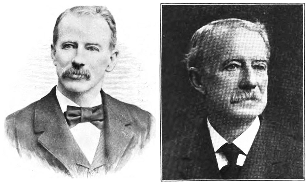
Figure 5.
Photograph of Lieutenant C. Henry Kain (standing), and colleague Edward Stratton, during their military service, 1864. Adapted for nonprofit, educational purposes from https://www.ancestry.co.uk/family-tree/person/tree/66755837/person/44156395103/gallery.

Figure 6.
C. Henry Kain, from publications of 1897 (left), and 1907 (right).
Acknowledgement
Many thanks to Ian Jones for sharing images of Kain's arranged diatom slides.
Resources
The American Journal of Microscopy and Popular Science (1880) Report of the January 22 meeting of the Microscopical Society of Camden, Vol. 5, pages 74-75
The American Journal of Microscopy and Popular Science (1880) Report of the February 19 meeting of the Microscopical Society of Camden, Vol. 5, page 100
The American Journal of Microscopy and Popular Science (1880) Report of the April 14 Soiree of the Microscopical Society of Camden, Vol. 5, pages 121-122
The American Naturalist (1879) New microscopical societies, page 201
Bracegirdle, Brian (1998) Microscopical Mounts and Mounters, Quekett Microscopical Club, London, pages 58 and 150, plate 23-G
City Directory of Camden, New Jersey (1887) "Kain C Henry, teacher, h 524 Cooper", page 343
City Directory of Philadelphia, Pennsylvania (1894) C. Henry Kain was not listed
City Directory of Philadelphia, Pennsylvania (1895) "Kain Chas H., asst supt, 713 Filbert, h 1531 Centennial av", page 964
Coville, Frederick V. (1913) Diatom collection of the United States National Museum, “the diatom collection … contains much valuable material, including the types of species accumulated by the late Professor H. L. Smith, of Geneva, New York, the specimens of all the species of the Albatross diatoms, and the extensive collection of diatom material of the late Professor C. Henry Kain, of Philadelphia, representing the principal fossil deposits throughout the world as well as a large number of recent gatherings made in this country and abroad. To the large amount of material thus brought together, there are being added the marine diatoms of the Shackleton Expedition to the South Pole, diatoms recently secured in the Panama Canal Zone by the Smithsonian Institution, and the pelagic coastal diatoms of the Atlantic seaboard now being collected under the auspices of the Cambridge Zoological Laboratory and the United States Bureau of Fisheries”, Science, Vol. 38, page 748
Custis, John T. (1897) The Public Schools of Philadelphia: Historical, Biographical, Statistical, pages 281 and 599
Harshberger, John W. (1899) The Botanists of Philadelphia and Their Work, Harshberger, Philadelphia, pages 350-352
Journal of the New York Microscopical Society (1890) Slides of diatomaceous deposits from the Yellowstone Geyser region: by C. Henry Kain, pages 63064
Journal of the Royal Microscopical Society (1882) Kain's and Sidle's mechanical fingers, Series 2, Vol. 2, pages 721-722
Journal of the Royal Microscopical Society (1882) Photo-micrography, Series 2, Vol. 2, pages 726-727
Journal of the Royal Microscopical Society (1885) Kain, C. H. - Schmidt's Atlas der Diatomaceen-Kunde, Series 2, Vol. 5, page 238
Journal of the Royal Microscopical Society (1885) Kain, C. H. - Tolu for mounting, Series 2, Vol. 5, page 570
Kain, C. Henry (1885) Photo-micrography, The American Monthly Microscopical Journal, Vol. 3, page 71
Kain, C. Henry (1882) Glass cells, The American Monthly Microscopical Journal, Vol. 3, page 101
Kain, C. Henry (1887) Notes on diatoms, Bulletin of the Torrey Botanical Club, Vol. 14, pages 25-32
Kain, C. Henry (1888) Diatoms of Atlantic City and vicinity, Bulletin of the Torrey Botanical Club, Vol. 15, pages 128-131
Kain, C. Henry, and E. A. Schultze (1889) On a fossil marine diatomaceous deposit from Atlantic City, N.J., Bulletin of the Torrey Botanical Club, Vol. 16, pages 207-210
Kain, C. Henry, and E. A. Schultze (1889) Collecting diatoms, Journal of the Royal Microscopical Society, page 137
Kain, C. Henry (1892) Letter on fixing diatoms to slides, The Microscopical Bulletin and Science News, Vol. 9, page 23
Kain, C. Henry (1904) The Military and Naval Operations on the Delaware in 1777, The City History Society of Philadelphia
Keyser, N.H., C.H. Kain, J.P. Garber, and H.F. McCann (1907) The History of Old Germantown, H.F. McCann, Philadelphia
Prowell, George R. (1886) The History of Camden County, New Jersey, L.J. Richards & Co., Philadelphia, pages 150, 339, and 501- 502
Publications of the City History Society of Philadelphia (1917) Vol. 1, page xviii
Schultze, E.A., and C.H. Kain (1897) The Santa Monica diatomaceous deposit, Journal of the New York Microscopical Society, Vol. 13, pages 77-86
Swan, Charles W. (1891) Diatoms, The American Monthly Microscopical Journal, Vol. 12, pages 53-54
US census and other records, accessed through ancestry.com
Woolman, Lewis (1892) Fossil diatoms in Philadelphia beneath the Girl’s New Normal School building, The Microscopical Bulletin and Science News, Vol. 9, pages 33-34
Woolman, Lewis (1894) Annual Report of the State Geologist for the Year 1894, pages 167-169 and 187