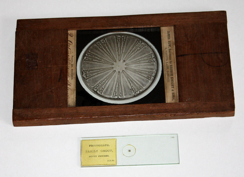
Figure 1. The two extremes of the union of microscopy and photography: a lantern slide of a photomicrograph by R.L. Maddox, alongside a microscope slide of a microphotograph by J.B. Dancer. Both date from approximately 1860.
Richard Leach Maddox, 1816 – 1902
by Brian Stevenson
last updated November, 2019
The many advances in technology during the 1800s included enhanced optics for microscopy and development of photographic processes. The marriage of those two technologies yielded results that fascinated our ancestors, and continue to amaze us today (Figure 1). Some Victorians, such as John Dancer, John Stovin, Alfred Reeves, and René Dagron, produced miniature photographs of large objects that required a microscope to see. Dagron adapted that technology to develop microfilm. Others of the time labored to develop methods to photograph tiny objects through the microscope. That technology, photomicrography, is still widely used in many branches of science. Richard Maddox played important roles in the development of photomicrography, and his works were highly regarded in his time. Maddox’s high-quality lantern slides of microscopic objects appear nowadays at auctions with moderate frequency (Figure 2).
Figure 1. The two extremes of the union of microscopy and
photography: a lantern slide of a photomicrograph by R.L. Maddox, alongside a
microscope slide of a microphotograph by J.B. Dancer. Both date from approximately 1860.
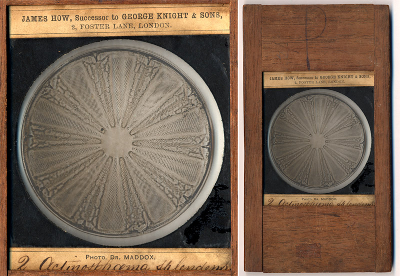
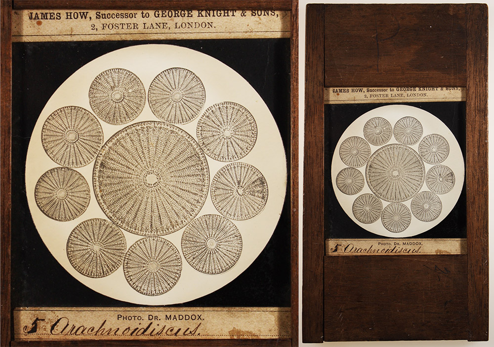
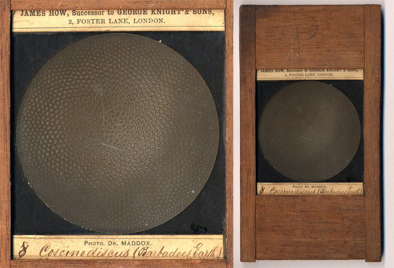
Figure 2. Three lantern slides of diatoms, produced by Richard Maddox and sold by James How. The slides measure 3 x 3 inches (75mm x 75 mm), with original mahogany wood frames that are 3 ¾ x 7 inches (95 x 175 mm). Close-ups of the photos appear to the left, and full views of the wooden-framed slides are shown to the right. Two of these subjects are known to have been exhibited in 1864 by James How to the Photographic Society of London, “Mr. How . . . exhibited . . a series of beautiful microscopic photographs taken by Dr. Maddox . . . Actinosphenia splendens . . . Coscinodiscus (Barbadoes earth)” (see below for the full quote).
Presentations of Maddox’s photomicrographs frequently elicited amazement and praise from the audience. Following are excerpts from period publications:
“Mr. How . . exhibited, on a disk of about 7 feet diameter, by means of the oxyhydrogen lantern, a series of beautiful microscopic photographs taken by Dr. Maddox, some of them with the highest-power object-glasses. These marvellous productions excited universal admiration . .The following are among those which were most admired: Vegetable. - Seed of Eccremocarpus. Diatomaceae. - Actinosphenia splendens; Aulaeodiscus formosus; Auliscus moronensis; Coscinodiscus (Barbadoes earth); Liemophora flabellata; Navicula didyma. Echinodermata. - Echinus, section of spine, tr.; Synapta digitata (spicules and plates from skin). Crustacea. - Canthocamptus furcatus; Cyclops. Insecta and Arachnida. - Parasite of Beetle Parasite of Field-Mouse; Parasite of Mole Parasite of Sparrow, female; Acarus of Fig; Itch-insect; Flea of Mole, male; Flea of Mole, female; Flea of Pigeon, male; Eye of Beetle; Foot of Fly; Gizzard of Cricket; Hygrotus elegans (Water-Beetle); Leaf-insect; Scale of Hawk Moth; Spiracle of Cockchafer; Tongue of Drone Fly; Tongue of Rhingia; Tongue of Hornet. Vertebrata. - Blood-corpuscles, Human. The Chairman said that he regarded it as a great achievement in photography to render, with such perfection as they had seen on the screen, objects so small in size that four millions of them would be required to cover one square inch. Those Members who wished to see more of them might do so at Mr. How's place, in Foster Lane. There were a hundred of them, and only twenty had been shown”. Journal of the Photographic Society of London, 1864.
“The Chairman called attention to a series of photographs of microscopic objects by Dr. Maddox, which were exhibited by Mr. How. They were, he thought, the best photographs of the kind he had ever seen. Photographs of microscopic objects had hitherto generally been failures; but these struck him as being really the finest in the world. If Dr. Maddox were in the room, it would be very interesting if he would give the Meeting some details of tbe method by which such beautiful results were obtained. If he were not present, possibly some other evening he would do so, as it could not fail to be interesting to Members to know how to produce such excellent results. Ho would recommend any Member who happened to have a lens with him to examine the prints carefully, as they would well repay such an examination”. Journal of the Photographic Society of London, 1864.
“The splendid microphotographs of Dr. Maddox, brought within the range of the general public by the ability and enterprise of Mr. James How, of Foster Lane, London, were arranged in the bird-room, and attracted a large number of discerning visitors”. Report of the 1866 Conversazione of the Liverpool Chemists’ Association, Pharmaceutical Journal, 1866.
“Dr. Maddox, who is at present in London, has favoured us with an inspection of his latest efforts in photomicrography. They are without doubt the most wonderful specimens we have seen, several of them being magnified to the great extent of three thousand diameters. The least enlarged is eight hundred and thirty-eight diameters. When we use the word enlarged we do not do so in the ordinary photographic meaning, for all these specimens are taken direct, or with one operation, and are not subsequently enlarged. They are on plates 7x5. Among the collection were several stereoscopic views of diatomaceae and similar objects belonging to the 'world of small." The great success achieved by Dr. Maddox in this direction renders it annoying to photo-microscopists of this country that a silver medal, to which he would have been entitled at the French Exhibition, was not awarded in consequence of the trivial blunder of some one who, in presenting the pictures for exhibition, had forgotten to insert the name of the artist. This informality might surely have been overlooked, seeing the jurors were well aware by whom they were done. The consequence, however, was that the silver medals for this class of work went to foreign countries”. British Journal of Photography, 1867.
“In England the name of Dr. Maddox will be well-known to every photo-micrographer, for during the past thirty years he has done more photo-micrographic work, and laboured more to bring the claims of the art before the scientific world, than any other man. As the inventor of gelatino-bromide plates, strange to say, his name is not so generally known - at least, in this country - for the great authorities on modern photography on the Continent (Dr. Eder and Dr. Vogel) have given due honour to Dr. Maddox for his invention. In England, the writer thinks, Dr. Maddox has never received sufficient recognition for an invention of such value - an invention which has revolutionised the whole science and practice of photography. The photo-micrographs of Dr. Maddox are well known: perhaps among the best are his photograph of part of the frustule of P. angulatum x 3,000, his photographs of various Coicinodisci and other diatoms. A large series of slides for the lantern was made from Dr. Maddox's negatives, and this series had a worldwide fame. Dr. Maddox still continues the practice of photomicrography, and his photographs of bacteria, illustrating papers which he has recently contributed to the Royal Microscopical Society, have been pronounced by competent authorities to be unsurpassed even by the splendid productions of Koch”. I.H. Jennings, How to Photograph Microscopic Objects, or, Lessons, in Photo-micrography for Beginners, 1885.
Being an important early photographer, Maddox’s life and works have been the subject of many publications over the years. The following biography of Richard Maddox is from his obituary that appeared in The Transactions of the American Microscopical Society:
“On Sunday, May 11, 1902, there passed away at Portswood, Southampton, England, Dr. Richard Leach Maddox, the pioneer of photomicrography, and an honorary member of our Society, and by his demise the scientific world is the poorer, losing as it does a steady hard worker and accurate observer, as well as a most genial and charming personality.
Richard Maddox was born at Bath in August, 1816. Of his early days, very few details are on record, beyond the fact that he was educated at a public school in Somersetshire. Then, having decided on entering the profession of medicine, he became a student at University College, London, in 1837. Always delicate, he had, even while a student, to suspend his work on account of the condition of his health, and in 1839 he left England for a voyage around the world. On his return in 1840 he resumed his studies, and obtained the diploma of the Royal College of Surgeons of England two years later. To this he added the license of the Society of Apothecaries in 1843. As might have been expected from a man with such a keen desire for work, and work for its own sake, we find him in 1844 pursuing his studies in Paris, which was then the centre of medical research, attending chiefly the practice of the Hotel de la Charite and the lectures of the late Dr. Donne. Dr. Maddox also devoted a very large amount of his time to microscopy, and in this connection it may be mentioned that he translated Dr. Dujardin's ‘Manual’ at about the time that Quekett's ‘Treatise on the Microscope’ appeared, but as it was impossible to arrange for the use of the beautiful plates illustrating the work, the translation was never published. In 1847 he appears to have visited Smyrna, proceeding afterwards to Constantinople, where for a time he practised his profession, and where he met Amelia, daughter of Benjamin Winn Ford, Esq., of that city, whom he married in 1849. In 1850 he returned to England, and the following year took the degree of M.D. of Aberdeen University. In 1852 he again settled in practice in Constantinople, and during the latter part of the Crimean War held the appointment of Civil Surgeon to the hospital at Scutari. His health again causing him some anxiety, Dr. Maddox came back to England, practising for a time at Islington, London, then at Ryde, Isle of Wight, and finally settling at Woolston, near Southampton, in 1859, where he remained for fourteen years. In 1874 he left Woolston to become resident physician to the late Duke of Montrose, from whom he went to Sir William Watkins-Wynn, and then to Lady Katherine Bannerman. His wife having died in 1871, Dr. Maddox married in 1875, Agnes, daughter of George Sharp, Esq., of Newport, Isle of Wight (who survives him), and the same year he again went abroad, first to Ajaccio, and afterwards to Bordighera and Cornigliano, practising his profession amongst the English residents. Returning to England finally in 1879, he lived for some years at Gunnersbury, but from 1886 onwards resided at Greenbank, Portswood, Southampton, England, living in a most retired manner, but keeping up his interest in everything relating to scientific work, and constantly writing for various journals and papers in England, France, and the United States; indeed, within a few days of his death he contributed a letter to the papers, dealing with the controversy anent the discovery of the ‘Holy Shroud’ at Turin. On the 10th of May, 1902, his old-standing complaint, aortic aneurysm, suddenly became worse, and on the following day he breathed his last at the advanced age of eighty-five years. Dr. Maddox was interred in the Southampton cemetery on May 15. A son and a daughter by his first wife, and a son by his second wife, survive him.
From this brief outline of a busy, restless life it is not easy to see where, and when, Dr. Maddox secured the necessary time and opportunity for the more strictly scientific research work which has made his name famous, and it speaks volumes for his powers of adaptability and of steady application that he was able to accomplish so much under such unfavorable circumstances. As early as 1853, he took up the study of photography, and in a contribution to ‘Photography’, February 11, 1892, he refers to this in the following words: ‘My first lens was bought about 1846, but active professional duties prevented its being used until 1852; from that date onwards, as an amateur, I have been interested in photography’. Then, too, he was undoubtedly the pioneer in the application of photography to microscopical work, just as he was one of the very first to grasp its potentialities for the reproduction of pictures of microscopical preparations. In spite of his early failures in this direction he was sanguine of ultimate success and subsequently referring to the subject he wrote: ‘Still, I felt and trusted its day would come, and be of much assistance to the busy microscopist’. His disheartening efforts in photomicrography only spurred him on to further endeavors, and there is not the least doubt that the substitution of gelatine for collodion in the preparation of photographic plates, resulting in the manufacture of dry plates, is the direct outcome of his early photomicrographic failures. The first public recognition of his work in the portrayal of microscopical objects took the form of a medal from the then ‘Photographic Society of London’ in 1853. This was followed after a long interval by a medal from the Council of the International Exhibition of Dublin (1865) for a series of his photomicrographs, published by the late James Howe. In 1865 a reproduction of some of Dr. Maddox's photographs formed the frontispiece of Lionel Beale's ‘How to work with the Microscope’ probably the first attempt in England to employ photomicrographs as book-illustrations " (see Figure 3, below)
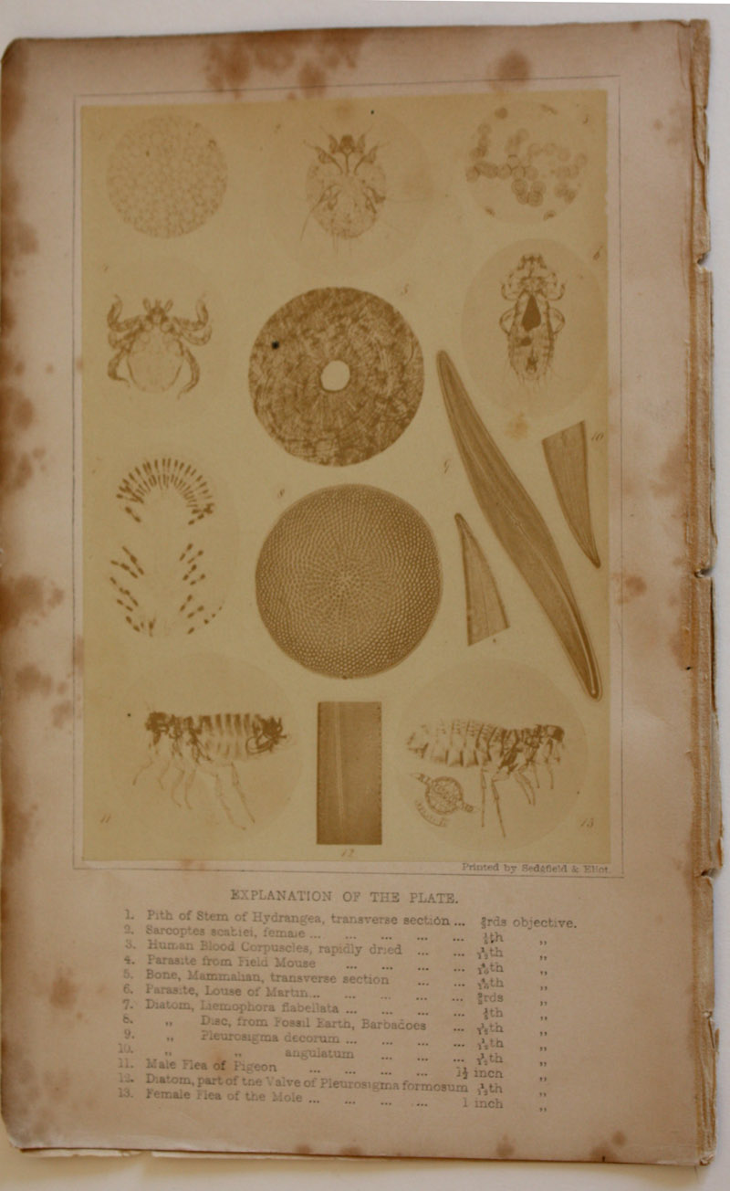
Figure 3. Frontispiece of Lionel Beale’s third edition (1865) of ‘How
to Work with the Microscope’. In the preface, Beale wrote, “For the beautiful photograph which forms the
frontispiece the author is indebted to his friend Dr. Maddox, who has also
afforded him very great assistance in writing the chapter on photography. This
is one of the most valuable chapters in the book. It contains the results of
many years' most earnest work, by one of the most successful workers in this
department of photography. The detail of some of the photographic illustrations
is so very minute, that many points cannot be seen by the unaided eye. A lens
of low magnifying power has therefore been appended to the volume, to enable
the reader to see the beautiful microscopical details which have been obtained
by this mode of illustration, in which Dr. Maddox is striving to achieve still
greater success”.
"The periodical attacks of ill-health to which he was subject, and which so frequently drove him from England in search of more genial climes, were often due to over-work; at these times, overwork in a vitiated atmosphere, charged with ether vapor from the collodion emulsions of the ‘wet’ photographic plate of that period, made its effects painfully apparent, and, combined with the desire to obtain a less cumbersome and troublesome method of securing his photograms of microscopical objects, caused Dr. Maddox to somewhat restrict the scope of his research work. The result of his experiments became apparent in 1871, when he published in the ‘British Journal of Photography‘ an account of the compounding of a practicable gelatino-bromide emulsion, and its employment as a ‘dry’ photographic plate. The Royal Microscopical Society of England immediately recognized the value of his work by electing him an honorary Fellow in 1871. Later on, he became a student of the then infant science of bacteriology (see Figure 4, below), and among other researches upon which he was subsequently engaged, was one upon the micro-organisms present in the air, in which he used a piece of apparatus of his own invention, the ‘aeroconiscope’, practically a multiple funnel set up as a vane. The wind passing through this apparatus deposited its contained organisms upon a thin coverglass prepared for its reception by being coated with a layer of gelatine; the organisms were then cultivated and the results accompanied by many careful figures, published in the current monthly Microscopical Journal. He gave up much time also to microscopical drawing, and examples of his skill may be found in the work of the late Dr. Parkes on ‘Hygiene’, and also in Dr. Nayler's ‘Skin Diseases’ (see Figure 5, below). Many of his colored drawings, however, of Diatomaceae, when subjected to the action of various reagents, and figures of the various yeasts in beer deposits, have not been published."
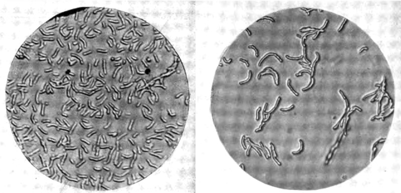
Figure 4. Two photomicrographs the
‘cholera bacillus”, Vibrio cholerae, by Richard L. Maddox. From the
Transactions of the American Microscopical Society, 1899.
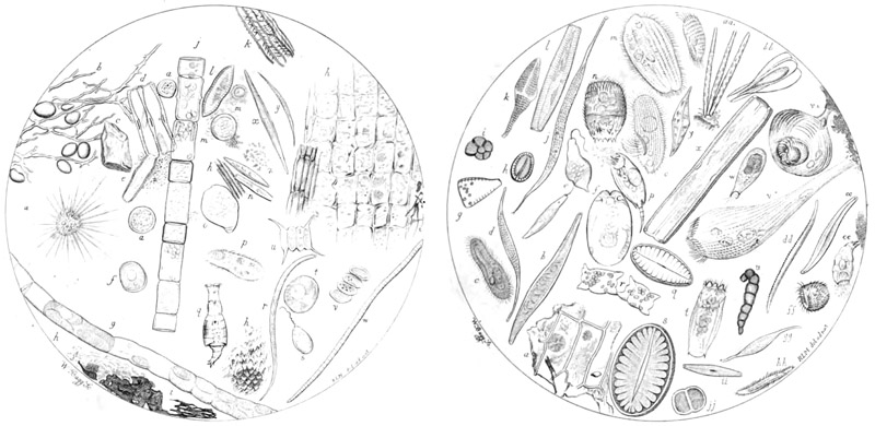
Figure 5. Two drawings of microscopic water-borne life forms, by R.L. Maddox for E.A. Parkes ‘A Manual of Practical Hygeine’, 1866.
"General public recognition of the value of Dr. Maddox's work was, as is too often the case in the world of science, delayed till late in life. In 1885 he received the gold medal of the Inventions Exhibition, at which he exhibited the earliest specimens of gelatine-bromide negatives made, in 1871, and after this many honors reached him. The Scott Legacy medal and premium from the Franklin Institute in Philadelphia, U.S.A., was awarded him in 1889, whilst in the autumn of 1891, as it was reported that he had lost heavily through a defaulting trustee, a sum of between £500 and £600 was raised for him in contributions from photographers in England, France, Germany, and America, in recognition of the value of his work. A gold medal from Antwerp, numerous diplomas, and finally the Progress Medal of the Royal Photographic Society of England (1901), were in turn conferred upon him.
Although Dr. Maddox's experiments in emulsifying silver in gelatine do not entitle him, as many erroneously claim, to the credit of having invented the gelatine dry-plate, there is not the least doubt that he pointed the way for other workers. This is not the time to go into the acrimonious discussions that have raged around this distinguished worker's name - discussions which were rendered acrimonious by the claims and counter-claims of others, for Dr. Maddox himself seems to have troubled very little about the dispute. Indeed, on his part there was throughout a conspicuous absence of assertiveness of virulence; he was one of that very high type of investigator who works for the love of his subject and for the sake of truth, without any ulterior motive, and certainly with no thought of pecuniary reward. Perhaps the most pleasant trait of his character was his readiness to help to the fullest of his capabilities everyone who sought his advice on photographic or photomicrographic work, holding as he did, that the claims of science for her advance were, ‘If freely ye have received, freely give’."
The American Amateur Photographer, in 1891, elaborated on Maddox’s financial and physical situation at that time: “Dr. R. L. Maddox. We think very few amateurs are acquainted with the early originators of the Gelatine Bromide Process, - a process so complete to-day as to make the practice of photography nearly universal, extending to every branch of industry. Dr. R.L. Maddox is acknowledged the world over as the first to suggest and practically demonstrate the use of gelatine as a vehicle for holding the sensitive salts and to discover that it aided in increasing their sensitiveness. While many who make important discoveries, retain their secret for personal profit, he freely gave the process to the world, and thereby enabled others to perfect it, until to-day it stands unrivaled, opening fields of work that were thought to be impossible. Vast industries have been built up, and successful manufacturers of dry plates have reaped liberal profits.
Twenty-one years ago Dr. Maddox was in comfortable circumstances having an excellent standing in his profession; he has now reached the age of threescore and ten and has lately been placed in an embarrassing condition, financially, in consequence of a breach of trust by a trustee, now deceased. Besides this he has been troubled with a chronic disease of a painful character, yet it has not prevented him from carrying on his investigations.
No honor is great enough to bestow upon a discoverer who acts so generously in giving his process to the world, and now that the time of necessity has arrived it seems eminently fitting that the photographic public should rise in behalf of the man, and see that his surviving days be pleasant ones. We therefore cordially agree with the rest of our contemporaries in urging upon all photographers, professional or amateur, to contribute something in aid of this discoverer. He is worthy of it; any subscriptions sent to us for the fund will be at once forwarded to the proper parties. In England one firm of dry plate manufacturers has made the first contribution of $500. Other sums of $25 and upwards have been subscribed by professionals and amateurs; a decided interest in the matter is shown. Let our American manufacturers do something handsome. They can afford it. We shall cheerfully publish a list of contributors in our columns. We understand Mr. J. Traill Taylor, of the British Journal of Photography, is taking a leading part in promoting the movement, and very appropriate it is, too, since it was in his Journal that the process was first published”.
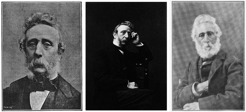
Figure 6. Photographs of Richard L. Maddox, left to right, ca. 1894, date unknown, ca. 1901.
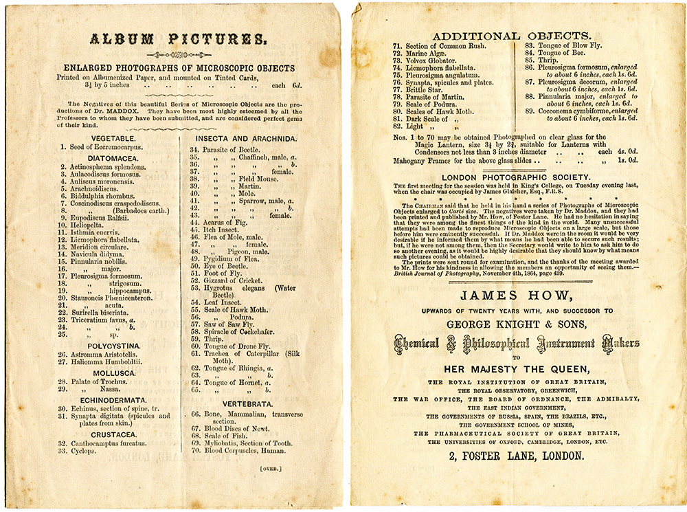
Figure 7.
A ca. 1863 list of R.L. Maddox’s photographs that were available for sale at the shop of James How.
Resources
American Amateur Photographer (1891) Aid for Dr. R.L. Maddox, Vol. 3, pages 481-482
Beale, Lionel S. (1865) How to Work With the Microscope, third edition, Harrison, London
British Journal of Photography (1867) Maddox’s Photo-micrographs, Vol. 14, pages 478
British Journal of Photography (1903) Obituary of the Year, Richard Leach Maddox, M.D., Vol. 18, pages 422-423
Hannavy, John (2008) Encyclopedia of Nineteenth-Century Photographers, Vol. 1, CRC Press, pages 884-885
Harrison, W. Jerome (1888) A History of Photography, Percy Lund & Co. London
Jennings, I.H. (1885) How to Photograph Microscopic Objects, or, Lessons in Photo-micrography for Beginners, Piper and Carter, London
Journal of the Photographic Society of London (1864) Nov. 1, 1864 Ordinary general meeting, Vol. 9, page 133
Journal of the Photographic Society of London (1864) Dec. 6, 1864 Ordinary general meeting, Vol. 9, page 155
Maddox, Richard L. (1863) On the photographic delineation of microscopic objects, Quarterly Journal of Microscopical Science, Vol. 3, new series, pages 9-12
Maddox, Richard L. (1864) On photomicrography, Journal of the Photographic Society of London, Vol. 9, pages 150-155
Maddox, Richard L. (1871) A silver salted gelatine emulsion, The British Journal of Photography
Maddox, Richard L. (1889) Hints on photomicrography, The International Annual of Anthony's Photographic Bulletin, Vol. 2, pages 156-158
Maddox, Richard L. (1898) On photographing the pollen threads of the evening primrose, Oenothera biennis, Photographic Times, Vol. 30, pages 534-536
Maddox, Richard L. (1899) Experiments in feeding some insects with cultures of comma or cholera bacilli. Transactions of the American Microscopical Society, Vol. 20, pages 75-80
Microscopical Bulletin and Science Times (1892) Proposed testimonial to Dr. R.L. Maddox, Vol. 9, page 2
Parkes, Edmund A. (1866) A Manual of Practical Hygiene, second edition, J. Churchill & Sons, London
Pharmaceutical Journal (1866), Liverpool Chemists’ Association Conversazione, Vol. 7, Second series, page 566
Photographic Times (1894) Fathers of photography, IX, Dr. R.L. Maddox, Vol. 25, page 21
Photographic Times (1901) Editorial notes (includes a photograph of R.L. Maddox), Vol. 33, page 170