Norman Nelson Mason, 1837 - 1914
by Brian Stevenson
last updated April, 2020
Norman N. Mason, of Rhode Island, USA, was a pharmacist and a professional preparer of microscope slides (Figures 1, 2, 8, and 9). In the course of his work, he developed a sectioning apparatus that came to be known as the “Providence microtome” (Figure 3). Numerous biologists of the late 1800s employed Mason to prepare and mount specimens for their investigations, especially specimens that required particular staining techniques and serial sectioning.
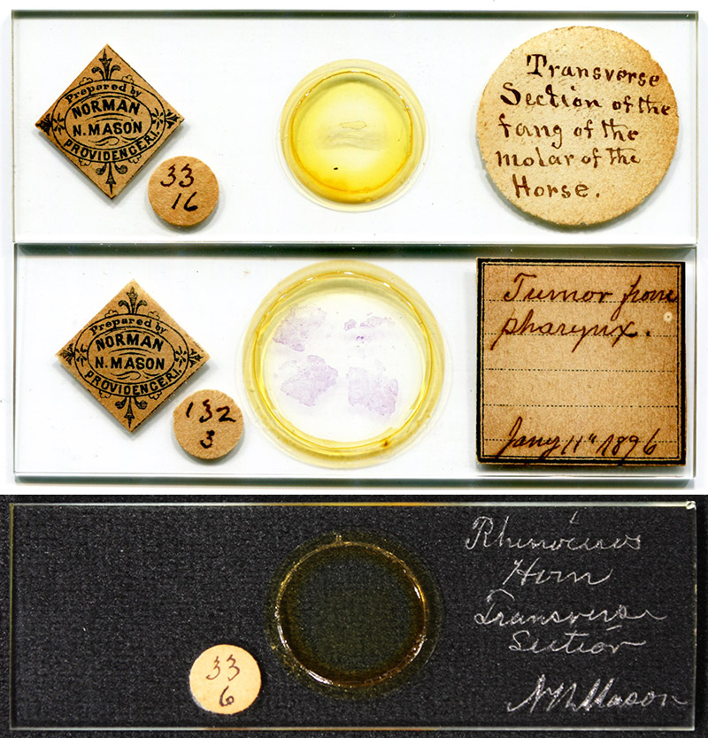
Figure 1.
Examples of microscope slides that were prepared by Norman N. Mason. The center slide is dated 1896. Mason mounted a wide variety of items, from botanical and histological sections to diatoms to thin sections of minerals. Two distinct methods would have been used in the preparation of the illustrated slides: the pharyngeal tumor and rhinoceros horn would have been cut with a microtome, whereas the horse tooth would need to be ground with abrasive.
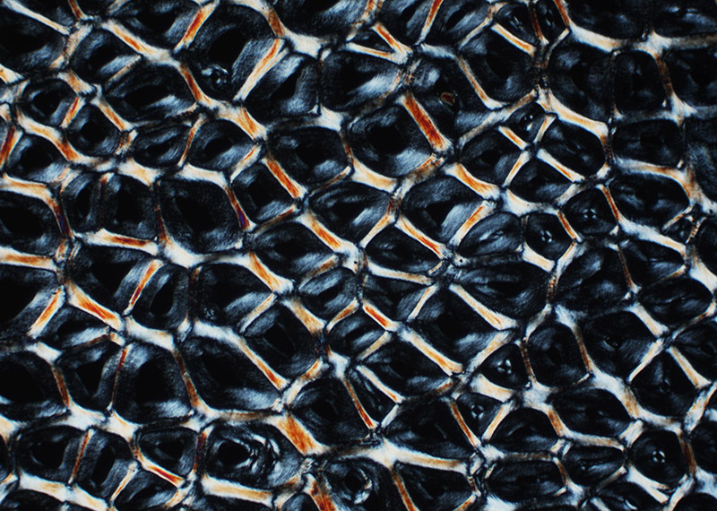
Figure 2.
Section of rhinoceros horn, prepared by Norman Mason (see Figure 1). Photographed with a 10x objective, C-mounted digital SLR camera, and crossed polarizing filters.
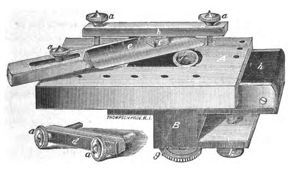
Figure 3.
The “Providence microtome”, originally designed by Norman Mason. W.J. Behrens wrote in 1885, “So called from being the joint invention of several gentlemen of Providence, R.I. The original form was first designed by Mr. N.N. Mason of that city, who has used it for several years in zoological work. The instrument was perfected by Rev. J.D. King, Microscopist of the Martha's Vineyard Summer Institute, one of the gentlemen referred to, by whom it is manufactured and sold. In its present form it is perhaps equaled by no microtome made, for extreme precision of movement and consequent accuracy of performance in cutting sections. With a good knife in good order, sections of 10µ to 25µ thick can be made without difficulty, and all alike.” .A biographical essay on King can be read elsewhere on this site.
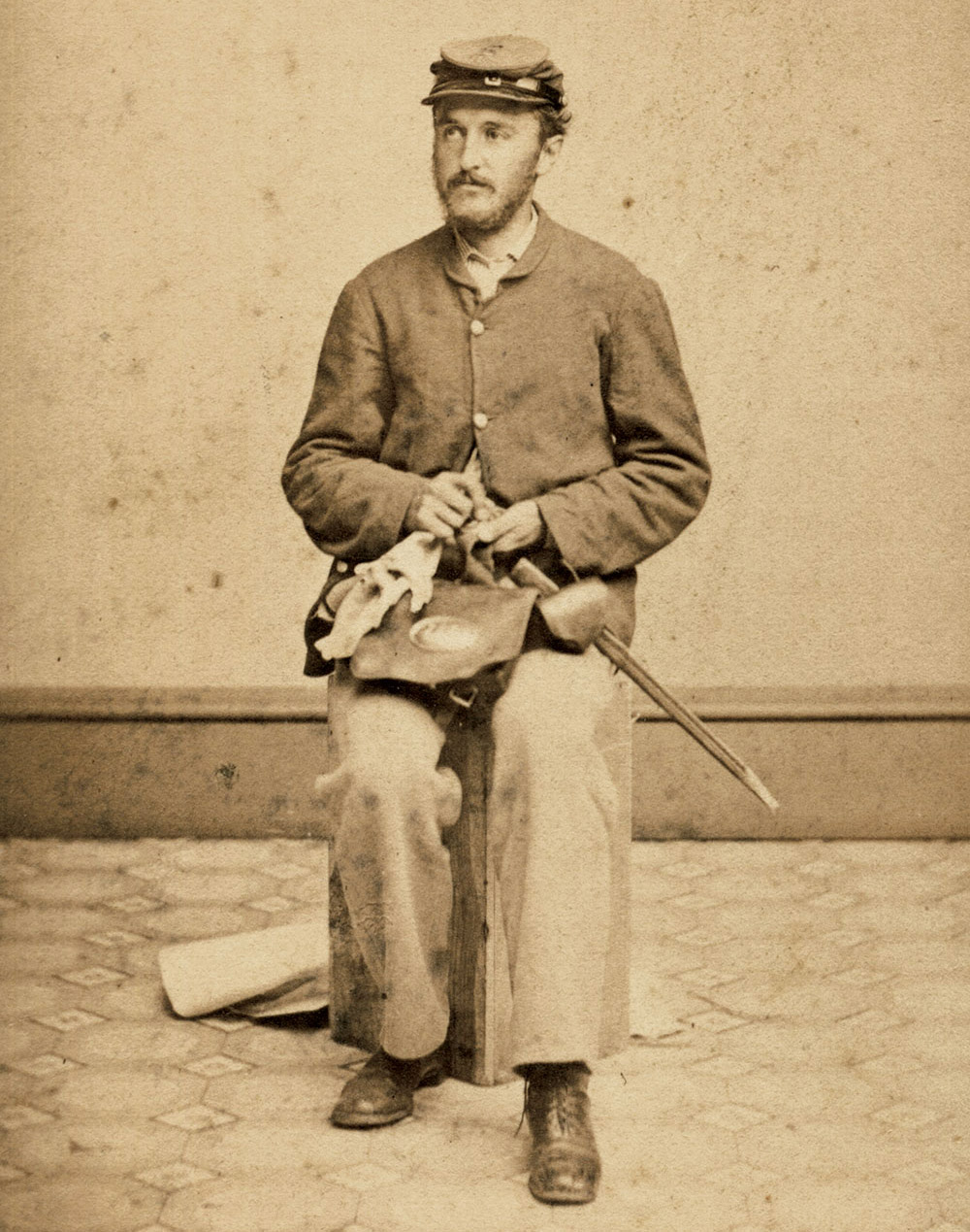
Figure 4.
Norman N. Mason in 1862, as an Army private during the US Civil War.
Norman Nelson Mason born in Lowell, Massachusetts on January 6, 1837. He was the second of two children of Norman and Caroline Weaver Mason. Caroline died in 1841, and Norman senior died in 1843; early deaths that suggest tuberculosis. After their parents’ deaths, Norman and his sister were raised by their maternal grandparents on a farm in Connecticut.
The 1860 US census shows Norman on the Connecticut farm. Around 1861 he moved to Providence, Rhode Island. The 1862 edition of that city’s directory lists him working as a “salesman”.
The U.S. Civil War spanned April, 1861 until April, 1865. Mason enlisted as a Private in the 10th Rhode Island Infantry Regiment in May, 1862. The regiment was mustered out in September, 1862, and Norman Mason returned to his sales job in Providence.
During the next few years, Mason studied pharmaceutical sciences, presumably under the tutelage of an established pharmacist. That person’s identity is not known. The 1863 city directory listed Mason as a “salesman”, in 1864 he was a “clerk” for the Providence Tool Company, in 1865 he was a “clerk”, then in 1866 he was listed as a “druggist”. His shop was then located at 116 North Main Street.
Also during that period, Norman married Susan Eliza Holmes, on June 7, 1864. They had two children, both boys. Susan died in 1873, when 31 years old.
During 1870, Mason moved his shop (or the street was renumbered), to 129 North Main Street. He moved again (or was renumbered) in 1900, to 128 North Main.
Mason played important roles in pharmaceutical education. A history of the Rhode Island College of Pharmacy states, “The first suggestion of establishing a college of pharmacy dates back to about 1870, at the time of the organization of the State Board of Pharmacy. At that time Norman N. Mason, a member of the Board of Pharmacy, in answer to a considerable demand for instruction in sciences related to pharmacy, and at the request of John Day Smith, the head master of the old Fountain street evening high school, Providence, organized a class in pharmacy. This class continued its work for one school year. Mr. Mason thus became the pioneer in the movement to establish a college of pharmacy in Rhode Island. On July 25, 1874, a number of pharmacists met in the rooms of the Franklin Society and organized the Rhode Island Pharmaceutical Association, which received its charter from the General Assembly in 1875. At a meeting of the Association held October 11, 1875, James H. Taylor of Newport suggested that a committee be appointed to examine into the expediency of establishing a school of pharmacy in Rhode Island, and James H. Taylor of Newport, Norman N. Mason of Providence and Enoch W. Vars of Niantic were appointed as members of that committee.”
Norman Mason was a member of the Microscopical Department of his local scientific society, the Providence Franklin Society. The Microscopical Department formed in 1874. Mason served as club Librarian.
It was around this time that Mason developed his microtome, which evolved into the “Providence microtome” that is illustrated in Figure 3. He would have used this to prepare the thin sections of tumor and horn shown in Figure 1, 2, and 8, as well as for the contracted preparations discussed below.
Over the years, Mason sent tips on specimen preparation and mounting to microscopy magazines. A simple clip for holding a cover slip onto a slide while fixing was published in 1876 (Figure 7).
An 1889 method for cleaning diatoms from sand was widely published, “After removal of the organic matter with acid, by the usual methods, add to the diatoms and sand in a large bottle thirty, forty or fifty times the quantity, by measure, of water, and gently shake until they are mixed. This water, with the diatoms and sand kept suspended by an occasional shake, is slowly poured in a small stream upon the upper end of a strip of clean glass, three feet long by three inches wide and securely supported. The upper end of the glass should be from one-eighth to one-quarter of an inch higher than the lower end, and the glass should be level transversely. Beneath the lower end place any convenient receiver. The water and diatoms will pass into the receiver. The sand, which will form little bars on the glass, must be removed occasionally, as it gradually creeps towards the lower end of the glass, and there would eventually pass into the receiver. The loss of diatoms will be very small. Usually one pouring is sufficient for cleaning. The sand can be re-washed if necessary, or a little clear water, run over the sand on the glass strip, will carry forward almost the last diatom ; but this will scarcely pay for the trouble. A short piece of glass will cause a failure, and too great an incline will be found almost as bad”.
In 1893, Mason published methods for “Mounting leaves and flowers entire”. This was based, in part, on techniques that he had learned from Dr. J.G. Hunt, of Philadelphia (a biographical essay on Hunt is available on this site).
Mason’s methods were often included in reports of studies that used his specimens. Some examples follow.
During 1880, Mason provided assistance to Alpheus Spring Packard’s studies of crustacean fine anatomy, including “a study of the structure of the lobster's eye from preparations of very great beauty and delicacy, kindly made for us by Norman N. Mason, Esq., of Providence, who has also made beautiful sections of the Limulus eye, after treating them in various ways”, and studies of “the brain of the horse shoe crab (Limulus polyphemus) … Mr. Mason has kindly made sections, both transverse and horizontal, stained with osmic acid; also sections of the brain of the supra-oesophageal ganglion of the lobster, stained with picrocarmine, for comparison. The following results, then, are based on over two hundred sections of the supra-oesophageal ganglion of Limulus, but more especially on one brain, which was cut by Mr. Mason into fifty-six sections, from 1/1000 to 1/500 of an inch in thickness, and another cut into over forty”.
The following three paragraphs are from 1880 and 1883 U.S. Government reports on locusts and other insect pests:
“The method of examination employed by us has been to cut the brain into a number of thin sections by means of the microtome, previously hardening the tissues in alcohol. The labor of cutting these sections has been performed by Mr. Norman N. Mason, of Providence, R.I., who has brought to his work an unusual degree of skill and care in preparing such delicate sections. In all these sections the brain was not previously removed from the head, but the entire head was cut through, having previously been hardened in absolute alcohol for twenty-four hours or more, and then soaked in gum arabic for one or two or more days. The objects were then embedded in a preparation of paraffine and sweet-oil and wax, or, in some cases, in soap and oil. After the sections were cut they were stained with picrocarmine, or partially stained with osmic acid, and then treated with picrocarmine. Finally the slices were mounted in glycerine jelly for study under the microscope”.
(Locust eggs) “were preserved in alcohol of ordinary strength, not having been hardened by any reagent, such as chromic acid, and the sections were cut by Mr. N.N. Mason, of Providence, who, after having hardened the eggs in absolute alcohol, then placed them in dissolved gum arabic, having previous to cutting stained the eggs in picrocarmine”.
“The embryo was cut into thirty-four sections, some of them passing through the limbs …, which shows … the procephalic lobes, the mouth parts and legs”.
I have not found any direct advertisements from Mason, but did locate examples of him providing slides to members of other microscopical societies. That was a common method for advertising sales, or, at least, exchanges.
From the minutes of the April 15, 1875 meeting of the Memphis (Tennessee) Microscopical Society, “The Society met at the usual hour, with a full attendance of members and visitors. Donations were received; several handsomely prepared slides of various subjects from Mr. Frank Miller, of New York, and specimens of a somewhat rare species of insect, from Mr. N.N. Mason, of Providence, Rhode Island, for all of which a vote of thanks was passed”.
From the May 17, 1889 meeting of the New York Microscopical Society, “The Corresponding Secretary presented the following communication upon his exhibit, the Crustacean Parasite, Argulus, sp., prepared and loaned for the occasion by Mr. Norman N. Mason, of Providence, R.I.”
Mason contributed slides to the “Postal Club”, a U.S. version of the U.K.’s Postal Microscopical Society. The first box of 1894 included, “by Norman N. Mason, Providence, R.I., … a nicely ground section of graphic granite showing to eye the appearance of Persian cuneiform characters, written by the quartz in the feldspar”.
Assistance was provided to Providence’s Brown University. The 1896 Report of the President of that university included, “Mr. Norman N. Mason has frequently donated expensive chemicals and microscopical reagents”. In 1903, he was acknowledged for service on visiting committees that examined on botany and on zoology/geology.
Around 1906, Mason moved to Plattsburgh, New York, to live with his son, Robert, and family. According to the 1910 census, the 73 year-old Norman Mason was still working on his own account as a “chemist”.
Norman N. Mason passed away on July 20, 1914, from a cerebral hemorrhage. He was buried with his wife, Susan, in Providence.
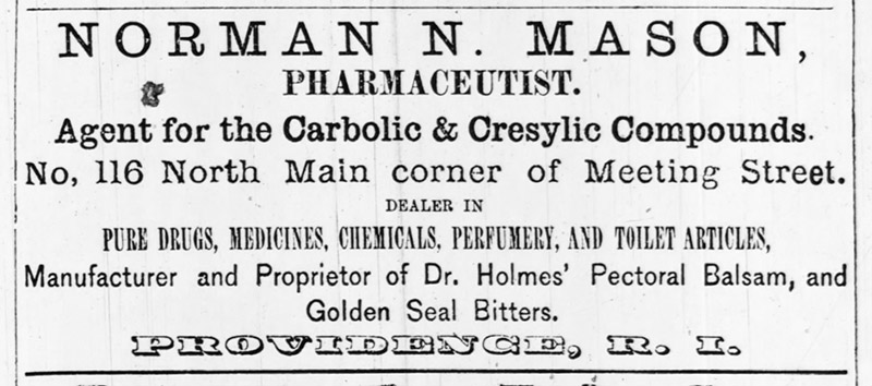
Figure 5.
A medicine bottle label, with Mason’s 1866-1870 business address. Adapted for nonprofit, educational purposes from http://tmacsribottleinfo.blogspot.com/2014/10 .
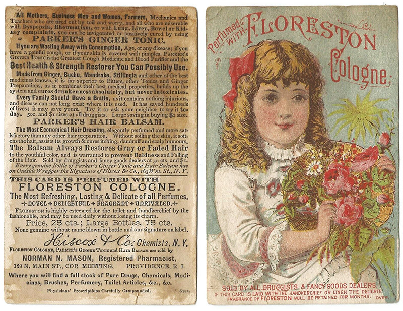
Figure 6.
An advertising card, with Mason’s 1870-1900 address of 129 North Main Street, Providence.
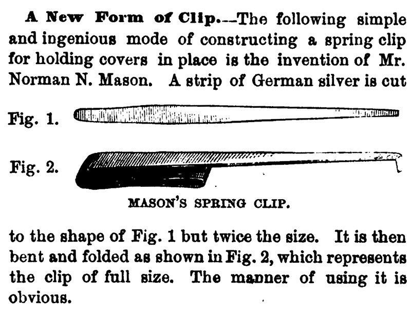
Figure 7.
A simple device that was developed by Norman Mason in 1876, to hold a cover slip against a slide while the mountant sets. Microscopists could easily make such devices at home, from scrap material.
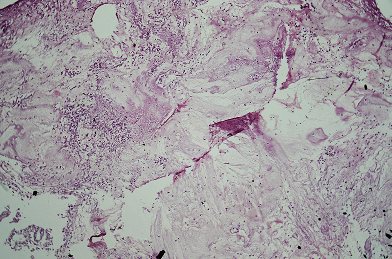
Figure 8.
Section of a “tumor from pharynx”, stained, sectioned, and mounted by Norman Mason on January 11, 1896 (see Figure 1). Photographed with a 10x objective and C-mounted digital SLR camera, digitally processed to enhance contrast.
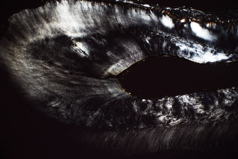
Figure 9.
“Transverse section of the fang of the molar of the horse”, by Norman Mason (see Figure 1). Photographed with a 3.5x objective, C-mounted digital SLR camera, and crossed polarizing filters.
Resources
American Druggist and Pharmaceutical Record (1914) Deaths, Vol. 62, page 63
The American Journal of Microscopy and Popular Science (1876) A new form of clip, Vol. 1, page 50
The American Journal of Microscopy and Popular Science (1876) Providence Franklin Society, Vol. 1, page 73
Annual Report of the President to the Corporation of Brown University (1896) pages 66-67
Annual Report of the President to the Corporation of Brown University (1903) page 53
Annual Report of the State Board of Education (1921) Rhode Island College of Pharmacy and Allied Sciences, page 106
Behrens, Wilhelm Julius (1885) The Microscope in Botany, translated and edited by A.B. Hervey, S.E. Cassino & Co., Boston, pages 188-189
Clark-Mason Family Papers, 1775-1981, Vermont Historical Society
The Defier and other Union Archetypes by a Master Lensman (accessed April, 2020) https://militaryimages.atavist.com/the-defier-autumn-2018
The Druggists' Circular (1903) Rhode Island Association, Vol. 51, page 131
Journal of the New York Microscopical Society (1889) meting of May 17th, Vol. 5, pages 114-115
Mason, Norman N. (1893) Mounting leaves and flowers entire, The Microscope, Vol. 12, pages 28-29
Military record of Norman N. Mason (accessed April, 2020) ancestry.com
The Monthly Microscopical Journal (1875) Memphis Microscopical Society, page 104
Packard, Alpheus Spring Jr. (1880) On the internal structure of the brain of Limulus polyphemus, Annals and Magazine of Natural History, Vol. 5, pages 29-30
Packard, Alpheus Spring Jr. (1880) Structure of the eye of Limulus, American Naturalist, Vol. 15, pages 212-213
The Pharmaceutical Era (1914) Norman N. Mason, page 425
Pharmaceutical Record (1891) Rhode Island Board of Pharmacy, Vol. 12, pages 227-228
Providence, Rhode Island City Directory (1862) “Mason Norman N. salesman, 35 N. Main, boards 14 Wheaton”, page 98
Providence, Rhode Island City Directory (1863) “Mason Norman N. salesman, 35 N. Main, boards 14 Wheaton”, page 96
Providence, Rhode Island City Directory (1864) “Mason Norman N. clerk, Prov. Tool Co. armory, bds 65 Congdon”, page 103
Providence, Rhode Island City Directory (1865) “Mason Norman N. clerk, house 27 Governor”, page 105
Providence, Rhode Island City Directory (1866) “Mason Norman N. druggist, 116 N. Main, h 186 Benefit”, page 115
Providence, Rhode Island City Directory (1868) “Mason Norman N. apothecary, 116 N. Main, h, 34 Meeting”, page 133
Providence, Rhode Island City Directory (1870) “Mason Norman N. apothecary, 116 N. Main, h, 34 Meeting”, page 158
Providence, Rhode Island City Directory (1871) “Mason Norman N. apothecary, 129 N. Main, house 127 do”, page 165
Providence, Rhode Island City Directory (1882) “Mason Norman N. registered pharmacist, 129 North Main, house 70 Congdon”, page 261
Providence, Rhode Island City Directory (1900) “Mason Norman N. 129 North Main”, page 245
Providence, Rhode Island City Directory (1901) “Mason Norman N. 128 North Main”, page 1086
Providence, Rhode Island City Directory (1904) “Mason Norman N. chemist 128 N Main rm 11 bds 49 Waterman”, page 493
Report of the United States Entomological Commission for the Years 1878 and 1879, Relating to the Rocky Mountain Locust and the Western Cricket (1880) page 226
Report of the United States Entomological Commission, Relating to the Rocky Mountain Locust, the Western Cricket, the Army Worm, Canker Worms, and the Hessian Fly (1883) page 274
Sylvester, W.H. (1894) Notes on the Postal Club, The Observer Magazine, Vol. 5, page 177
The Tenth Regiment Rhode Island Volunteers (accessed April, 2020) ancestry.com
U.S. census and other records, accessed through ancestry.com