Johann Diedrich Möller, 1844-1907
by Brian Stevenson
last updated July, 2025
J.D. Möller initially planned to be a professional artist. During his apprenticeship in Hamburg, Germany, he became curious about microscopes and sought assistance from the top optical worker in that city, Hugo Schröder. Möller was given access to Schröder’s workshop and learned how to grind lenses. At the termination of his art training, Möller worked for Schröder, decided that he preferred microscope-making to painting, and moved back to his family’s home in Wedel to establish a microscopy business. Initially, he manufactured lenses and other optical supplies for Schröder and other retailers, then branched into producing microscope slides of a wide range of subjects. Diatoms became a fascination, and it is for his diatom slides that he is best known to microscopy enthusiasts. Möller’s diatom mounts can be grouped into three classes: arrangements of diatom frustules into geometric designs; rectilinear arrangements that illustrate species from a geographic site or are used as test objects; and strews of mixed diatoms that are either rich in a particular species or specimens collected from a specific location.
Johann’s son Hugo continued the business after his father’s death. The business continued to produce microscope slides well into the twentieth century. They also manufactured many other optical products, and the current iteration, Möller-Wedel Gmbh, produces microsurgical equipment.
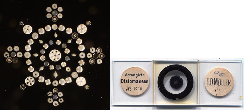
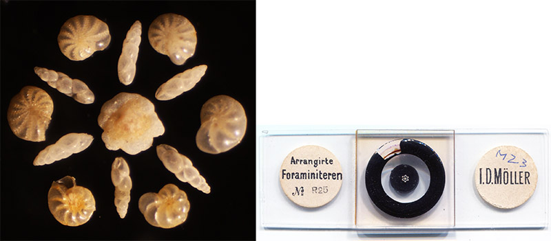
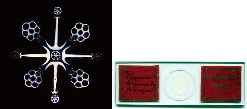
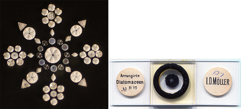
Figure 1.
Arranged diatoms (top two slides), foraminifera (third slide), and sponge spicules (bottom slide), by Johann D. Möller, ca. 1870s. His training as an artist led him to see the aesthetic possibilities of arranging these tiny objects into works of art.
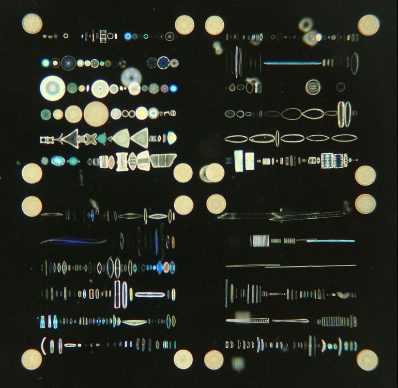
Figure 2A.
Magnified details of a circa 1868 400-diatom Möller “typenplate” (species type slide). An 1868 description of such a slide noted that “specimens of Eupodiscus argus are placed as marks to indicate the commencement of lines”. Adapted for nonprofit, educational purposes from an internet auction site.
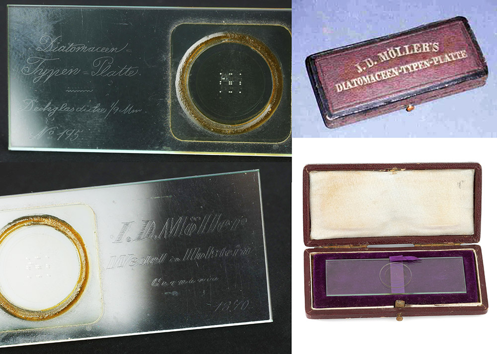
Figure 2B.
Möller's earliest 400-diatom Typenplatten have descriptions handwritten in the glass with a diamond, and include the production number and date of creation. The example on the left is number 195, which was made in 1870. Möller made 597 of these 400-diatom slides. The large, round Eupodiscus argus guide diatoms are easily seen in the upper left image. Adapted for nonprofit, educational purposes from internet auction sites.
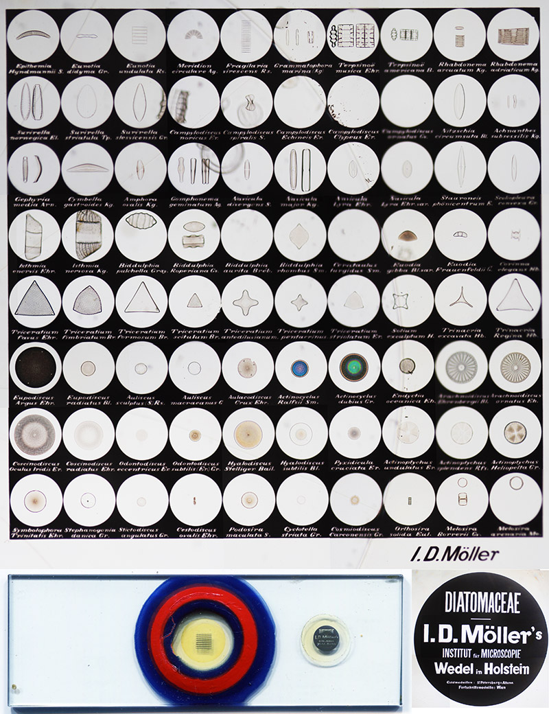
Figure 3.
An 80-diatom typenplate, circa 1874. The diatoms are mounted above a microphotograph that states their genus and species. Some of the specimens have drifted over the years. A microphotograph of Möller’s address, etc. is affixed to the slide, under a glass coverslip (a magnified image is shown beside the slide). In 1874, ‘The Quarterly Journal of Science’ reported, “Herr Möller has introduced a very ingenious modification of his celebrated Diatomacean typenplatte … The new arrangement consists of a photograph about 4 millimetres square, of eighty circles, ten in a longitudinal and eight in a vertical direction; beneath each circle is the name of the object and its author, and in the centre of each of these circles is a diatom, and in many cases two are mounted in order to show front and side views. The whole collection independently of its great value to the student of Diatomaceae is a marvel of manipulative skill”.
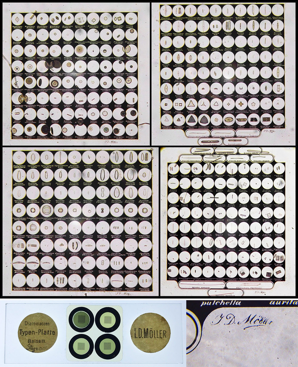
Figure 4A.
A Möller typenplatte with 325 different species of diatoms. Adapted for nonprofit, educational purposes from an internet auction site.
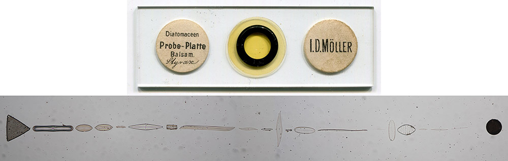
Figure 4B.
A Möller probe-platte with 21 different species of diatoms, for use as test objects.
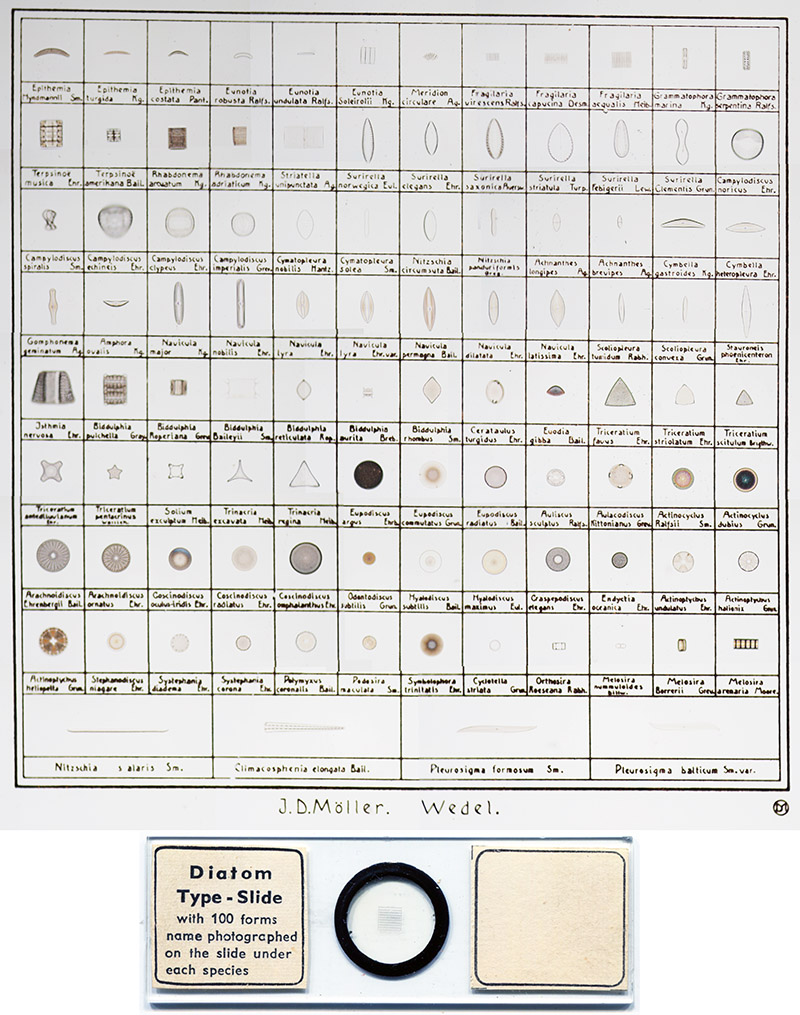
Figure 5.
A later, 100-diatom typenplatte.
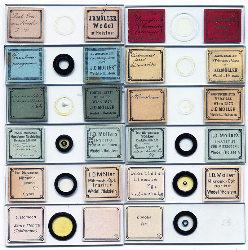
Figure 6.
Microscope slides of diatoms by J.D. Möller and his descendant companies. Notices of prize medals on some can help date their production. The slides on the lower row, identified with a “JDM” monogram, date from the twentieth century. Most such Möller diatom preparations are strews of mixed species, although some are of selected specimens (e.g. the three with dark circles on the coverslips, which indicate the locations of the diatoms). Möller described his business as “Institut für Mikroscopie” from the early 1870s until after the turn of the twentieth century, so that phrase on a label does not do much to identify production date.
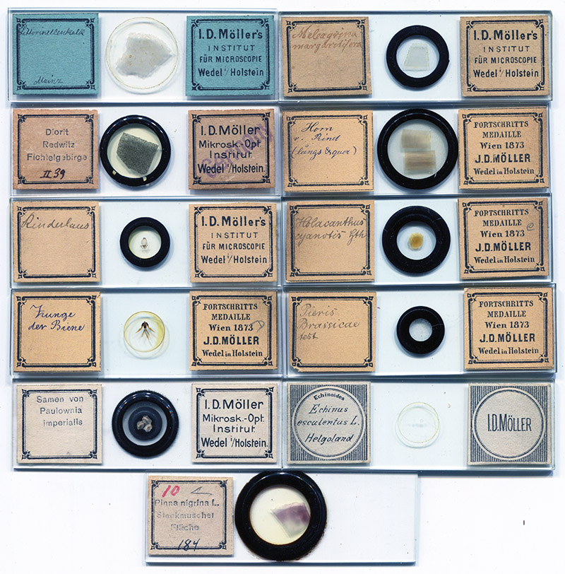
Figure 7.
Möller mounted a wide variety of specimen types, not just diatoms. Shown are some examples of minerals, insect, plant, and other specimens. Möller’s 1868 catalogue, and the 1951 catalogue of J.D. Möller Gmbh, listed several pages of non-diatomaceous preparations.
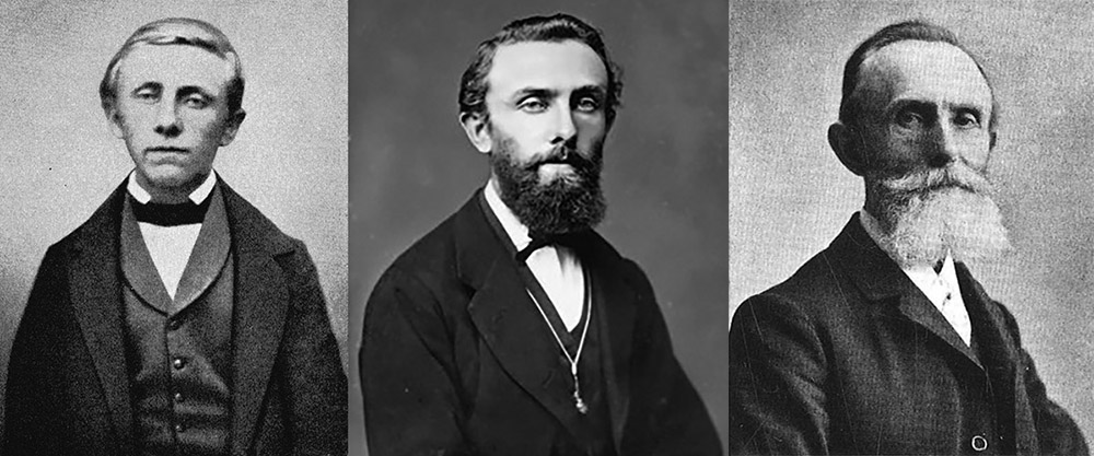
Figure 8.
Johann Diedrich Möller, at age 20 (left) and at unknown older ages (center and right). Adapted for nonprofit, educational purposes from https://www.wedel.de/wirtschaft-branchen/branchenbuch/alle-branchen/unternehmensportraits/moeller-wedel/das-jd-moeller-wasserwerk.html, https://www.wedel.de/wirtschaft-branchen/branchenbuch/alle-branchen/unternehmensportraits/moeller-wedel/jd-moeller-historische-serie-teil-1.html, and Burba (2007).
Johann Möller was born on March 16, 1844 in Wedel, Holstein (Germany), a city in which he would spend the majority of his life. He was the second son in a family of four children. His father was a linen weaver, who also painted as a side-business.
When he was fifteen, Johann moved to Hamburg to study painting. His master there sent Möller to the drawing school of the Patriotischen Gesellschaft (Patriotic Society). In the Society’s library, he came across a book on optical instruments by Johann Prechtl (1778-1854). Möller used information in that book to build a grinding machine, then polished some lenses, and made himself a microscope.
Möller was not satisfied with those home-made lenses, which used ordinary glass. Hoping to do better, he sought out Hugo Schröder, who was the most renowned maker of optical instruments in northern Germany. Schröder was reported to be “greatly astonished at the painter's wish … but was impressed … to an extent that he not only showed Möller his excellent microscopes, but also allowed him unrestricted access to his workshop”. Soon afterwards, Möller built himself a satisfactory microscope.
After Möller’s art apprenticeship ended in the spring of 1864, he worked part-time as a lens-maker for Schröder. After a few months, Möller decide to give up painting and go into business as a maker of microscopical and optical equipment. He established his workshop in his hometown of Wedel in 1864. Initially, he made lenses, calcites for polarizing apparatus, and pictures for the magic lantern. He also prepared microscope slides, which were well-received, including “preparations of insects, snails, sea urchins, etc.”.
At an early point, unable to determine a diatom’s species, Möller sent one of his type slides to Rabenhorst for help. Unexpectedly, Rabenhorst promptly ordered a similar slide, and made his collection of diatomaceous material available to Möller. Other diatomists soon learned about Möller’s work. Albert Grunow (1826-1914) of Vienna ordered a slide with 400 diatom species, and supplied his own material for its construction.
Minutes of an 1868 meeting of the Quekett Microscopical Club (London) reported exhibition of “a very remarkable collection of diatoms … from J. D. Möller, of Wedel, in Holstein; they were in number about 400, and were mounted on one slide, called by the preparer a “Typen-platte,” and were accompanied by a key or catalogue … the impression produced is that our German fellow microscopists are very far advanced in manipulation. The slide is a most exquisite specimen of microscopic skill, and better still, it forms a most useful typical collection of diatomaceae … The slide we have examined contains 403 specimens and 387 distinct species; these are mounted in balsam in a circular glass cell, covered by a disc, the first measuring .010, and the last .006 of an English inch in thickness. It would appear from the uniformity of focus that the diatoms are placed on the inner side of the covering glass. A 1/12th objective of large angular aperture can easily be used upon them. Fine specimens of Eupodiscus Argus are placed as marks to indicate the commencement of lines. The slide is accompanied by a written catalogue, neatly bound, in which all the specimens are entered, and numbered in their proper order … By slowly passing the slide across the field each specimen is brought into view in succession, and can thus be named from the written catalogue. M. Möller also prepares a test slide, containing twenty examples of diatoms, arranged in a straight line, and similarly mounted to the one described. The idea of type slides is evidently a very good and useful one. It would be far more convenient if typical specimens could thus be mounted for reference, instead of being compelled to keep a separate slide for each”.
Möller’s son, Hugo, reported that J.D. Möller sold 597 copies of the 400-diatom Grunow arrangement, 1009 copies of a 100-diatom slide, and 2162 copies of a 20-diatom preparation. Beginning in 1874, Möller produced slides in which select diatoms were arranged over photographs that stated the genus and species names (Figures 2,3, and 4) (his earlier preparations required a printed booklet to identify each diatom). Special orders led to production of even larger arrangements, such as the 720-diatom slide for the U.S. Army Medical Museum, an 860-diatom slide for Columbia College, New York, a 1715-diatom slide for the Emperor of Brazil in 1880, and, presumably, the 3215-diatom slide shown in Figure 4. A masterwork contains 4026 specimens, and took Möller five years to produce.
Möller produced an important series of diatom slides between 1877 and 1882, in collaboration with Swedish diatomist Per Teodor Cleve (1840-1905). A total of 324 preparations were produced, released in six parts, along with detailed annotations by Grunow. Slides from the Cleve and Möller sets are seen occasionally, although they may not be immediately recognized as Möller’s work, being labeled “Cl. a. M.” (Figure 9).
Möller mounted a variety of specimens at international exhibitions, and won numerous prizes. Three of these are listed on some Möller slide labels, and can help identify probable dates of production (Figures 6 and 7). Gold medals were received at the 1869 exhibitions in Saint Petersburg, Russia, and Altona, Germany. A display of 800 botanical, zoological, and geological preparations earned him a Medal of Progress at the 1873 Vienna World’s Fair.
In 1875, Möller announced his intentions to publish a book that would describe his methods of cleaning, separating, and mounting diatoms (Figure 11). Publication was to be contingent on a sufficient number of pre-sales. The goal was not met, and Möller’s procedures remained secret. Henri Ferdinand van Heurck (1838-1909) later wrote, “Mr. Moller employs a specially constructed microscope to make his type slides, and he adopts his own particular methods, which he has hitherto disclosed to but three or four persons, of whom I am fortunately one. I have now known it for a long time but am under an obligation not to reveal anything in connection with the subject”.
In addition to distributing slides through his own business, Möller’s productions were sold by other retailers, both with and without Möller’s labels (Figures 10 and 13-14)
Other optical products remained major parts of the Möller business. During the 1890s, Möller developed a new process for silver-plating. This produced durable mirrors that reflected 96% of light (Figure 12). Möller also produced glasses with fine divisions, for use as micrometers, crosshairs, etc (Figure 15).
Möller also built water and beer businesses. He put in an asparagus farm adjacent to the optical works, and developed improved varieties. He received a prize at the Hamburg horticulture exhibition of 1897. He also grew berry bushes, and made commercial quantities of berry wine and brandy.
Johann Möller died from pneumonia on October 29, 1907.
His second-eldest son, Hugo Möller (1880-1959), operated the business after Johann’s death. The company continued to make microscope slides well into the twentieth century; they, and binoculars, were a major source of revenue during the post-World War I depression, and the 1951 catalogue of slides was published as a microphotograph slide (Figure 16).
Zeiss purchased 51% of J.D. Möller in 1925, but Hugo bought them out in 1931. Following Hugo’s death, his sons Alfred (1910-1995) and Hans (1912-1982) took control of the business, which continued to grow in the post-war boom. After Hans’ death, the Möller-Wedel business was acquired by Haag-Streit, and became Möller-Wedel Optical GmbH in 1999.
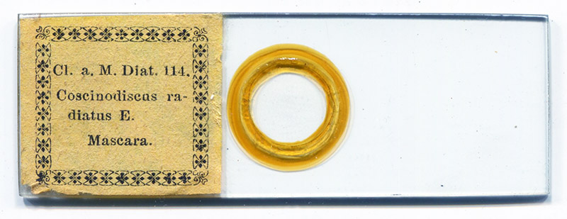
Figure 9A.
Specimen 114 from Cleve and Möller’s 1877-1882 diatom series. It is a strew of mixed diatom species, which contains examples of the identified diatom, Coscinodiscus radiatus.
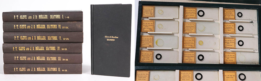
Figure 9B.
Volumes 1-6 of Cleve and Möller’s diatom series, with accompanied book. Each volume contains 4-5 dozen microscope slides. Adapted for nonprofit, educational purposes from an internet auction site.
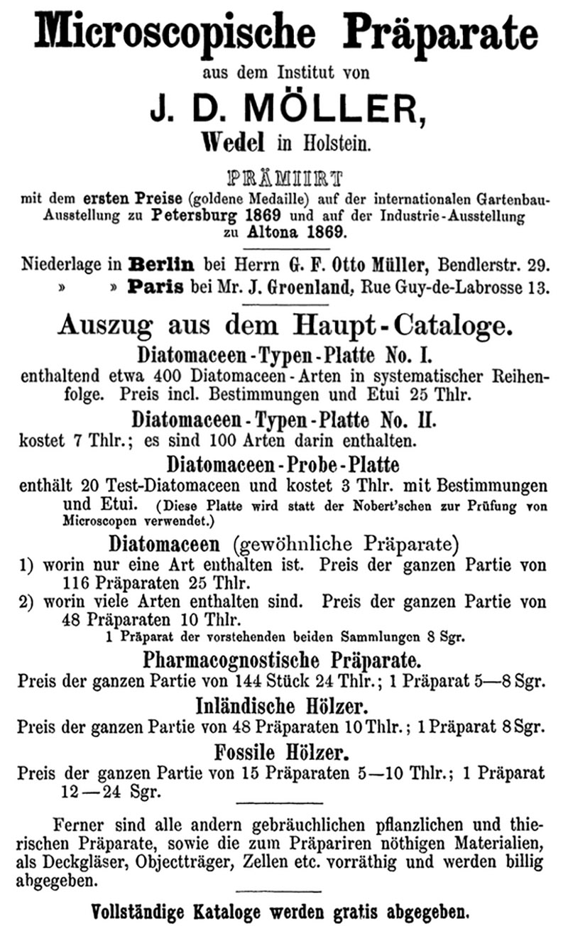
Figure 10.
An 1870 advertisement from J.D. Möller. His products were available through publisher Georg F Otto Müller in Berlin and slide-maker Johannes Groenland in Paris.
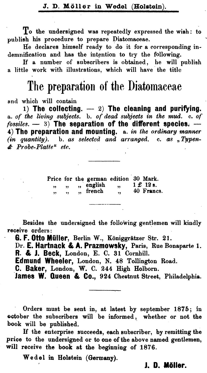
Figure 11.
An 1875 advertisement from Möller, to drum up interest for his proposed book on preparing diatoms. He did not receive enough subscribers to make the project financially worthwhile, and the book was never published.
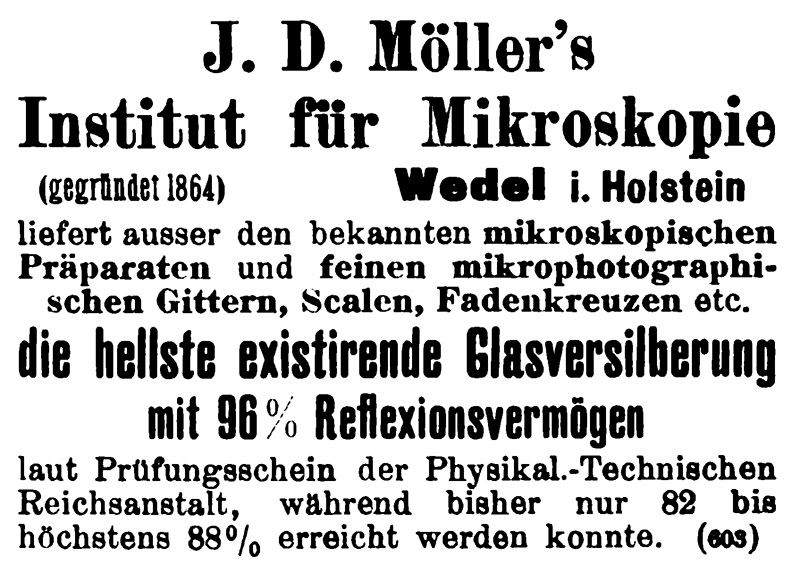
Figure 12.
A 1902 advertisement, focused on Möller’s microscopical preparations, photomicrography equipment, and highly-reflective mirrors.
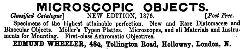
Figure 13.
An 1876 advertisement from London microscope retailer Edmund Wheeler, announcing that he was selling Möller’s typenplatten.
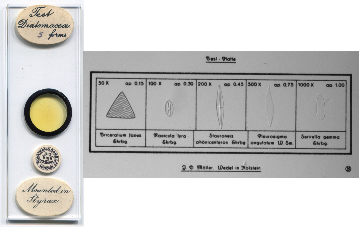
Figure 14.
A five-diatom test slide (probe platte) by Möller, with labels from London retailer Watson and Sons.
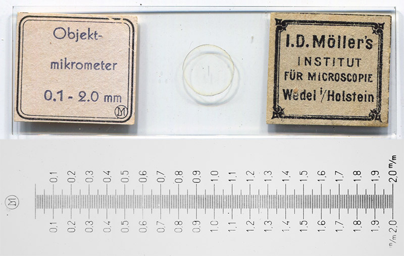
Figure 15.
A Möller stage micrometer. It is a precisely-made microphotograph. The combination of old- and new-style labels suggest that it was made during the early twentieth century.
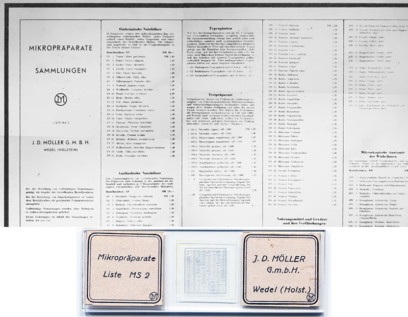
Figure 16.
An excerpt from the 1951 J.D. Möller Gmbh microscope slide catalogue microphotograph.
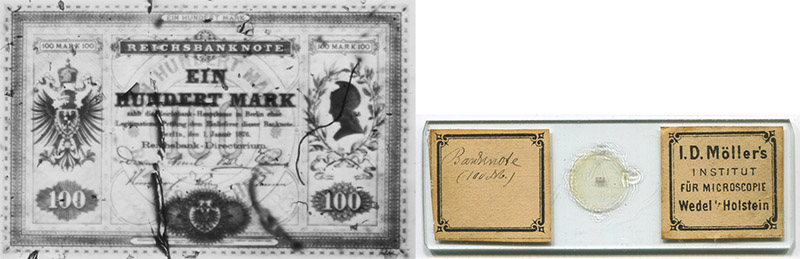
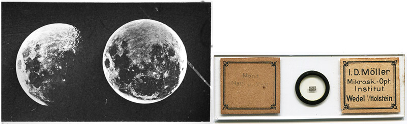
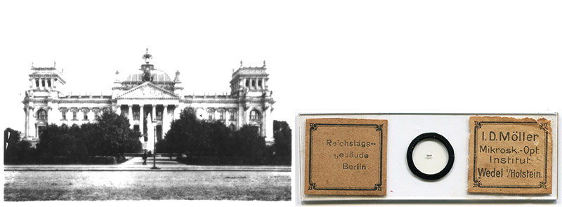
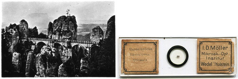
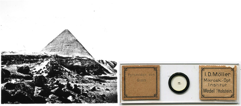
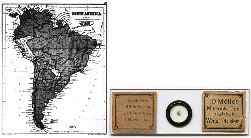
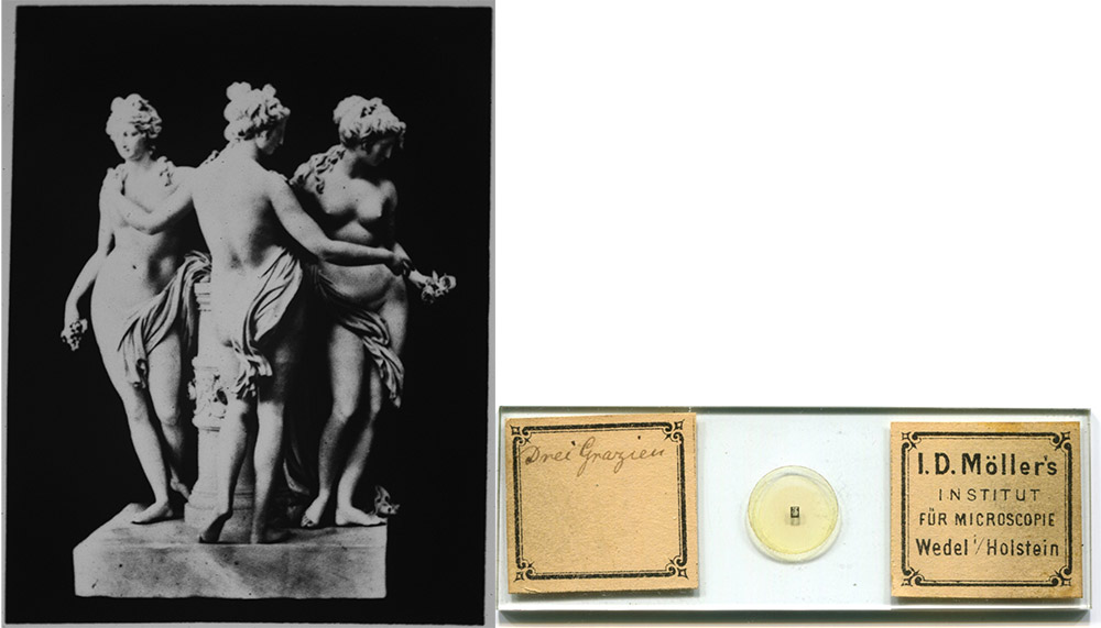
Figure 17.
Some microphotograph slides by J.D. Möller. He was a skilled photographer, and may well have produced the negatives himself. Many are of German subjects, or with German language in the image. The top slide is a 100 Mark banknote that bears the date 1876.
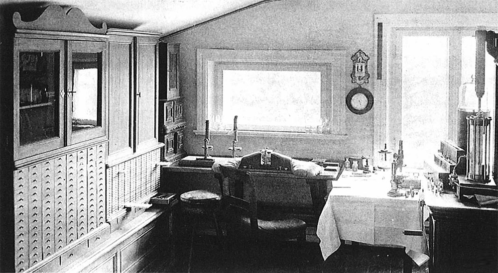
Figure 18.
J.D. Möller’s laboratory, 1890. Adapted for nonprofit, educational purposes from Burba (2007).

Figure 19.
A 1949 advertisement from "Mikrokosmos", which reads "There is nothing you can do! When Möller sends him a new shipment of micro-preparations, he forgets dinner, his mistress, and everything around. Those who collect will understand."
Acknowedgement
Thank you to Ulrike Hausl-Hofstätter for calling attention to Matthias Burba’s article on Möller.
Resources
Bracegirdle, Brian (1998) Microscopical Mounts and Mounters, Quekett Microscopical Club, London, pages 68, 156, 184, 194, and 216, and plates 26-P, 26-Q, 40-M, 45-A through 45-S, and 56-C
The British and Foreign Medico-Chirurgical Review (1876) Advertisement from Edmund Wheeler, Vol. 57, January advertiser section
Burba, Matthias (2007) Johann Diedrich Möller (1844-1907) - Über die Kunst, Diatomeen zu Iegen, Mikrokosmos, Vol. 96, pages 7-17
de Wall, Rudolf (accessed August, 2017) Johann Diedrich Möller - der Begründer des Industriezeitalters in Wedel, https://www.wedel.de/wirtschaft-branchen/branchenbuch/alle-branchen/unternehmensportraits/moeller-wedel/jd-moeller-historische-serie-teil-1.html and other, linked pages
Deutsche Mechaniker-Zeitung (1902) Advertisement from J.D. Möller, April issue advertiser
Diebes, E. (1908) Memorial of J.D. Möller, Zeitschrift für Angewandte Mikroskopie, Vol. 13, pages 289-292
Hardwicke's Science-Gossip (1875) The preparation of diatomaceae, Vol. 11, page 184
Hager, Hermann (1870) Das Mikroskop und Seine Anwendung, Springer-Verlag, Berlin, advertisement from Möller at the rear of the book
van Heurck, Henri F. (1896) A Treatise on the Diatomaceae, translated by W.E. Baxter, W. Wesley & Son, London, pages 78 and 110
The Journal of Botany, British and Foreign (1875) Advertisement from J.D. Möller, Vol. 13, back pages
Journal of the Quekett Microscopical Club (1866) Type slides, Vol. 1, page 141
Journal of the Quekett Microscopical Club (1875) “The Secretary announced that Herr Möller's proposed work on the preparation and mounting of diatoms, to which attention had been called upon a former occasion, would not be published, in consequence of the number of subscribers for it being deemed insufficient”, Vol. 4, page 132
Mikrokosmos (2001) Historische Anzeige, Vol. 90, page 196
Möller, Johann D. (1868) Preiz-Verzeichniss Mikroskopischer Präparate, Utensilen und Materialien zur Anfertigung Derselben, G.F. Otto Müller, Berlin
Möller, Otto (1907) Memorial of J.D. Möller, Berichte der Deutschen Botanischen Gesellschaft, Vol. 25, pages 57-60
Quarterly Journal of Science (1874) “Herr Möller has introduced a very ingenious modification of his celebrated Diatomaceen typenplatte ; the old form had about four hundred species neatly arranged in the space of about a quarter of an inch square, each slide being accompanied by a manuscript catalogue. The new arrangement consists of a photograph about 4 millimetres square, of eighty circles, ten in a longitudinal and eight in a vertical direction; beneath each circle is the name of the object and its author, and in the centre of each of these circles is a diatom, and in many cases two are mounted in order to show front and side views. The whole collection independently of its great value to the student of Diatomaceae is a marvel of manipulative skill”, New series, Vol. 4, page 547
Welt-Ausstellung 1873 in Wien Officieller General-Catalog (1873) “Möller J. D., Wedel, Holstein. Ein Glaskasten, enthaltend circa 800 mikroskopische Präparate der Botanik, Zoologie und Geologie für wissenschaftlichen Gebrauch”, page 434