"Mura"
by Brian Stevenson
last updated February, 2021
Microscope slides are occasionally seen that have brightly-painted decorations around the cover slip, and which generally have labels with handwritten descriptions and a typeset "Mura - England" (Figure 1). Many bear dates, ranging from the late 1930s to the early 1940s. Other slides have "Mura" labels but are not decorated (Figure 2).
Brian Bracegirdle, in Microscopical Mounts and Mounters suggested that "Mura" were "professional mounters of histology". Extensive searching failed to identify a business or a person named Mura who was involved with microscope slides, histology, or pathology. It was also puzzling to think that a professional business would hand-paint microscope slides in the 1940s.
The answer to "Mura's" identity turned out to be simple, yet unexpected. "Mura" was a line of labels and other stationery supplies that was produced by Cooper, Dennison, and Walkden, Ltd. The "Mura" brand was trademarked by them in 1924. The makers closed their doors in 1983. Figures 3 and 4 show "Mura" labels in other contexts, as a box of unused labels and for a handwritten price tag on the box of an antique tea set.
Thus, the answer is that an unknown slide-maker used "Mura"-brand labels.
All of this creates a new question: who was that person? He/she had excellent sectioning and staining technique, and the majority of these slides are of animal histology.
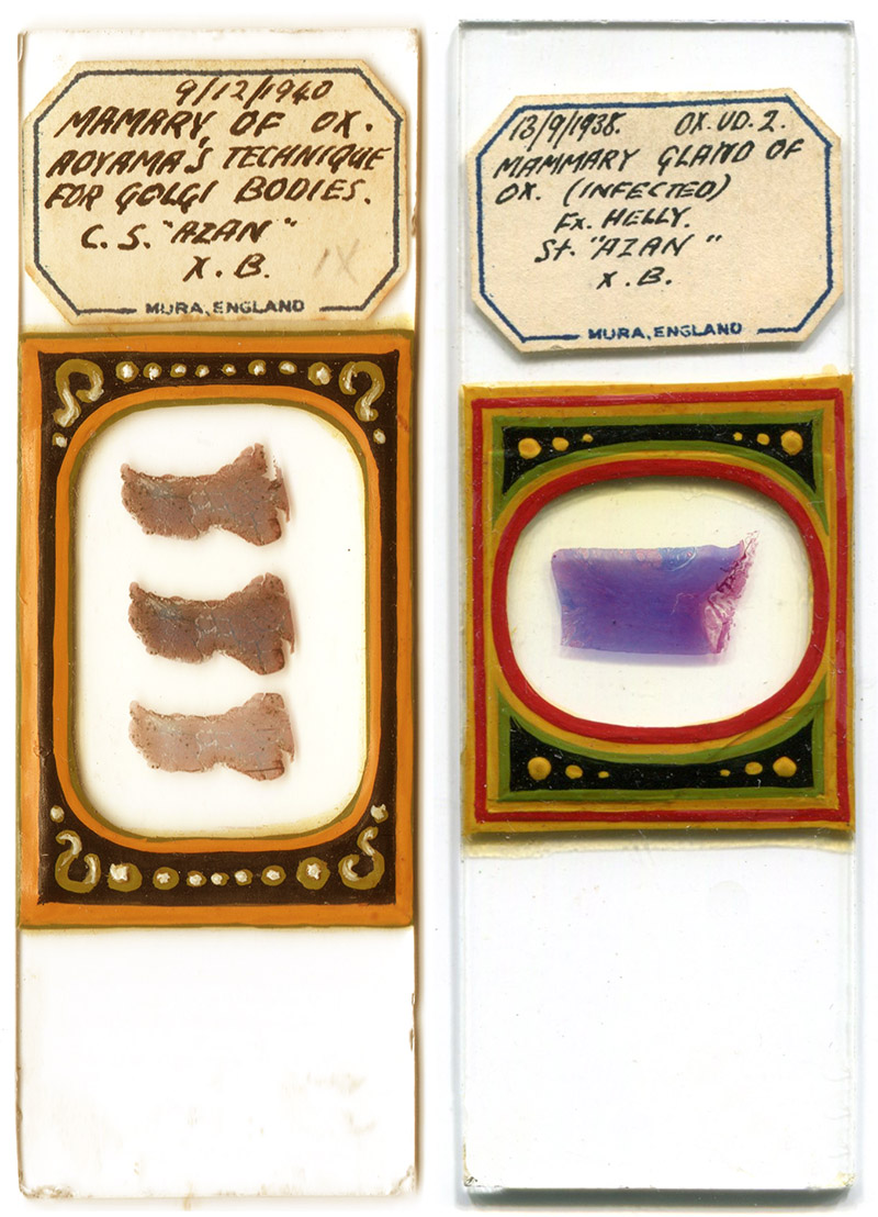
Figure 1.
Two microscope slides with hand-painted decorations and "Mura" labels. The left slide is dated December 9, 1940, and the right one is dated September 13, 1938. The slide on the left is also illustrated in Brian Bracegirdle's "Microscopical Mounts and Mounters" on Plate 27-R.
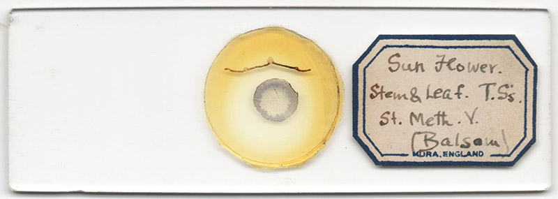
Figure 2.
A very different type of microscope slide with a "Mura" label. It is not decorated, and the specimens are sections of sunflower.
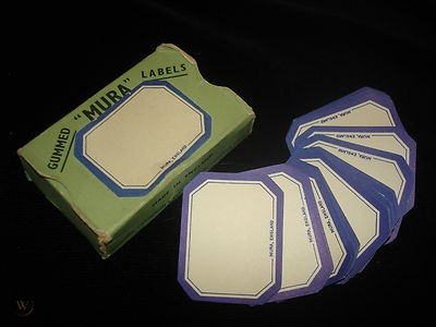
Figure 3.
A box of unused "Mura" labels. Adapted for nonprofit, educational purposes from an internet auction site.
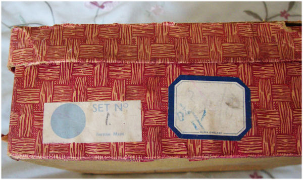
Figure 4.
A "Mura" label that was used as a price tag on the box of an antique tea set. Adapted for nonprofit, educational purposes from https://www.collectorsweekly.com/stories/87654-childs-vintage-4-pc-tea-set-in-the-shape.
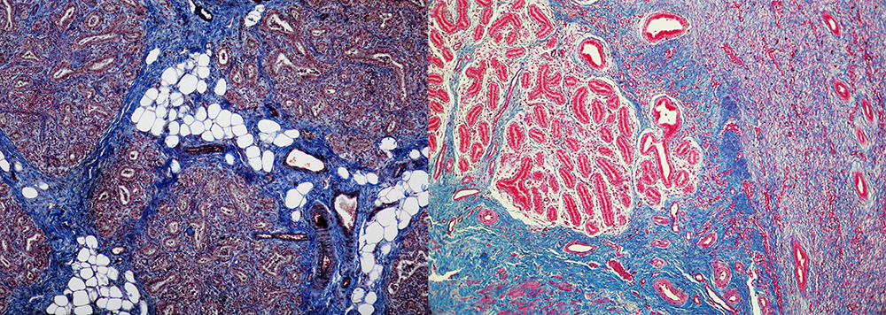
Figure 5.
Photomicrographs of the bovine mammary gland specimens in the slides shown in Figure 1.
Acknowledgement
Thank you to Sandie Pearce for generously sharing images and for helpful discussions.
Resources
Bracegirdle, Brian (1998) Microscopical Mounts and Mounters, Quekett Microscopical Club, London, pages 69 and 158, and Plate 27-R
Progress is Fine, But It's Gone on for Too Long (accessed February, 2021) Vanished makers: Cooper, Dennison & Walkden Ltd., London, England, http://progress-is-fine.blogspot.com/2015/02/vanished-makers-cooper-dennison-walkden.html