James White Neville, 1840 - 1900
by Brian Stevenson and Howard Lynk
last updated April, 2010
James
Neville’s microscope slides are distinctively styled, and easily recognized by
the ornate patterns he applied to ringing cement. Neville was a professional japanner (lacquer painter), and
his artistic abilities obviously extended to many aspects of his life. Figure 1 illustrates examples of
Neville’s handiwork, ranging from simple to ornate. It is rare to find two slides on which he used the identical
pattern and color combination.
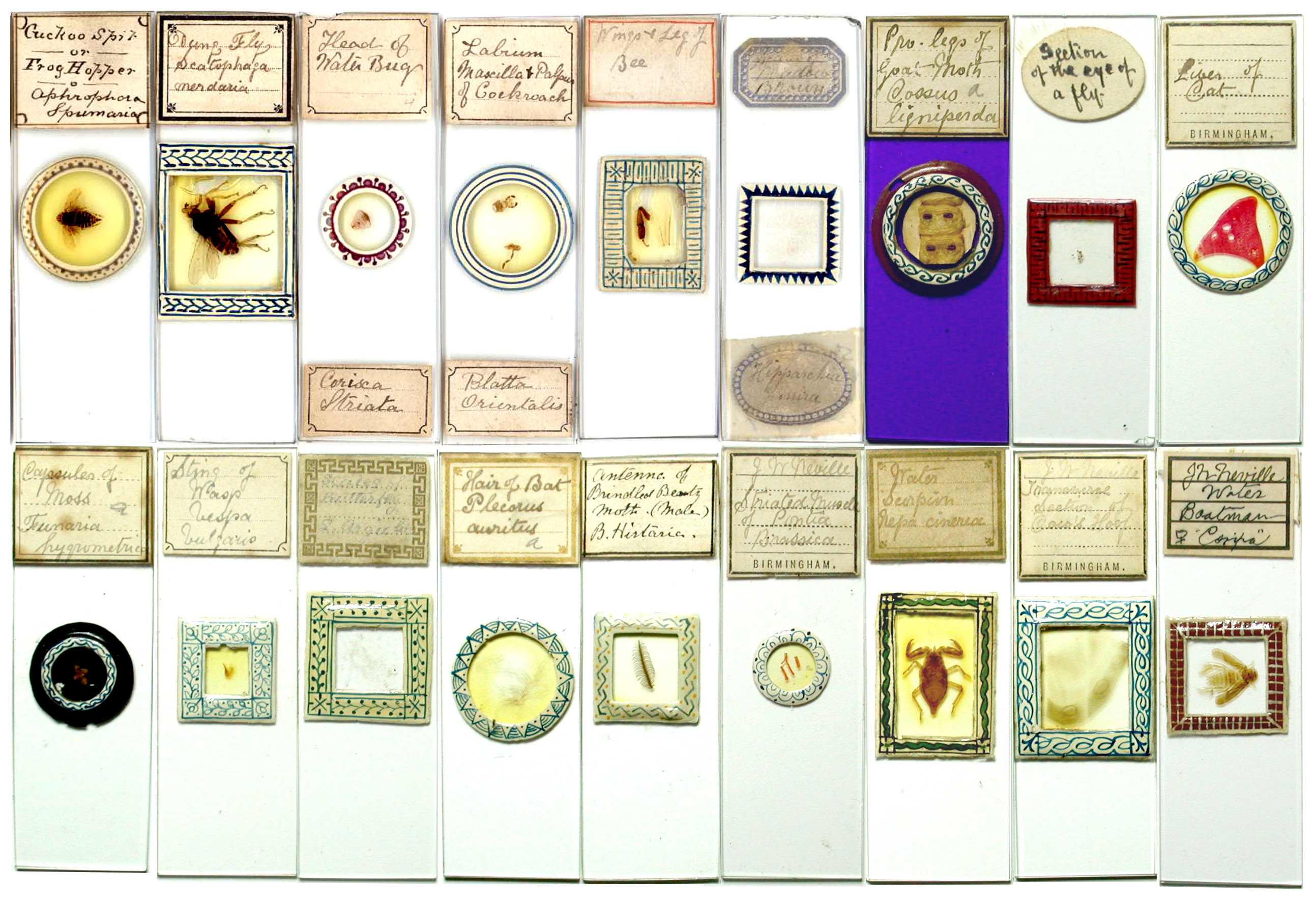
Figure 1. Examples of microscope slides
produced by James Neville. The
fourth slide from the left, top row, is ringed in a simple pattern, but can be
identified as being by Neville from his handwriting on the label. The seventh slide from the left, top
James White
Neville was born in Birmingham, England, during mid-summer, 1840, the third child
of Charles and Judith Neville. Father Charles was recorded on the 1841 census as being an artist,
although James’ marriage record described Charles as having been an optician. Between 1841 and 1849, the family moved
to Wolverhampton, Staffordshire. Charles died in 1849, leaving Judith to care for James, two
brothers and two sisters. The 1851 census records Judith as working as a
confectioner. The family’s economic situation at that time must have been reasonably
decent, as they employed a live-in general servant.
By the time of
the 1861 census, James was working as a japanner. He and most of his family
lived together in Aston, Warwickshire. Mother Judith did not report being
employed. Younger brother William worked as an optician. A third sister appeared
on this census, Helen, aged 5.
James married
Margaret Neill in 1866, at the Parish Church of Handsworth, Staffordshire. The
couple then moved in with Margaret’s mother, Hannah, in Birmingham. Hannah was
a pawnbroker, an occupation also attributed to Margaret on the 1851 census.
Hannah was recorded as being married, but her husband does not appear with the
family on any census. Margaret was born in Dublin, Ireland, so it is possible
that her father was still in that country, or travelling. James and Margaret
had seven children over the next decade.
Neville
mentioned in an 1883 article that he had made slides using a particular method
seven years ago, indicating that he was seriously involved with microscopy by
1876. The earliest detailed record we found of James Neville’s interest in
microscopy were two 1878 exchange advertisements in Hardwicke’s Science-Gossip (Figure 2). He did not advertise in
earlier issues of this magazine, suggesting that had only recently achieved the
competence to produce high quality slides and the confidence to exchange them
with strangers. The descriptions he used for his slides, “well-mounted” and “neatly-finished”,
may indicate that he was already painting his slides with ornate ringing. As a
caveat, though, these were common phrases used by other slide makers to
describe their work, and may simply mean that he produced good-quality slides.

Figure 2.
Exchange offers from James Neville appearing during 1878 in “Hardwicke’s Science-Gossip”.
The following
year brought requests to exchange for slides of a dissected water beetle and a
whole-mounted spider. By 1880, Neville had joined the Birmingham Microscopists’
and Naturalists’ Union. For the next 11 years, The Midland Naturalist reported proceedings of the club’s meetings,
including descriptions of specimens exhibited under their microscopes. James
Neville was frequently noted as having exhibited microscopic, and macroscopic,
objects to his club. A list of microscopic specimens reported to be shown by
Neville at club functions may be found as Table 1, at the end of this essay.
This is an abbreviated description of his output, as many were listed as large
groups. For example, at the 1880
Annual Conversazione, Neville exhibited “150
slides of insects of the district”, and showed “100 slides of insects, mounted whole, and in dissected parts, also
tongue of spider” at the 1882 Conversazione. Moreover, Neville continued to
request slide exchanges for many years, so many of his works would have entered
the collections of other microscopists throughout the country.
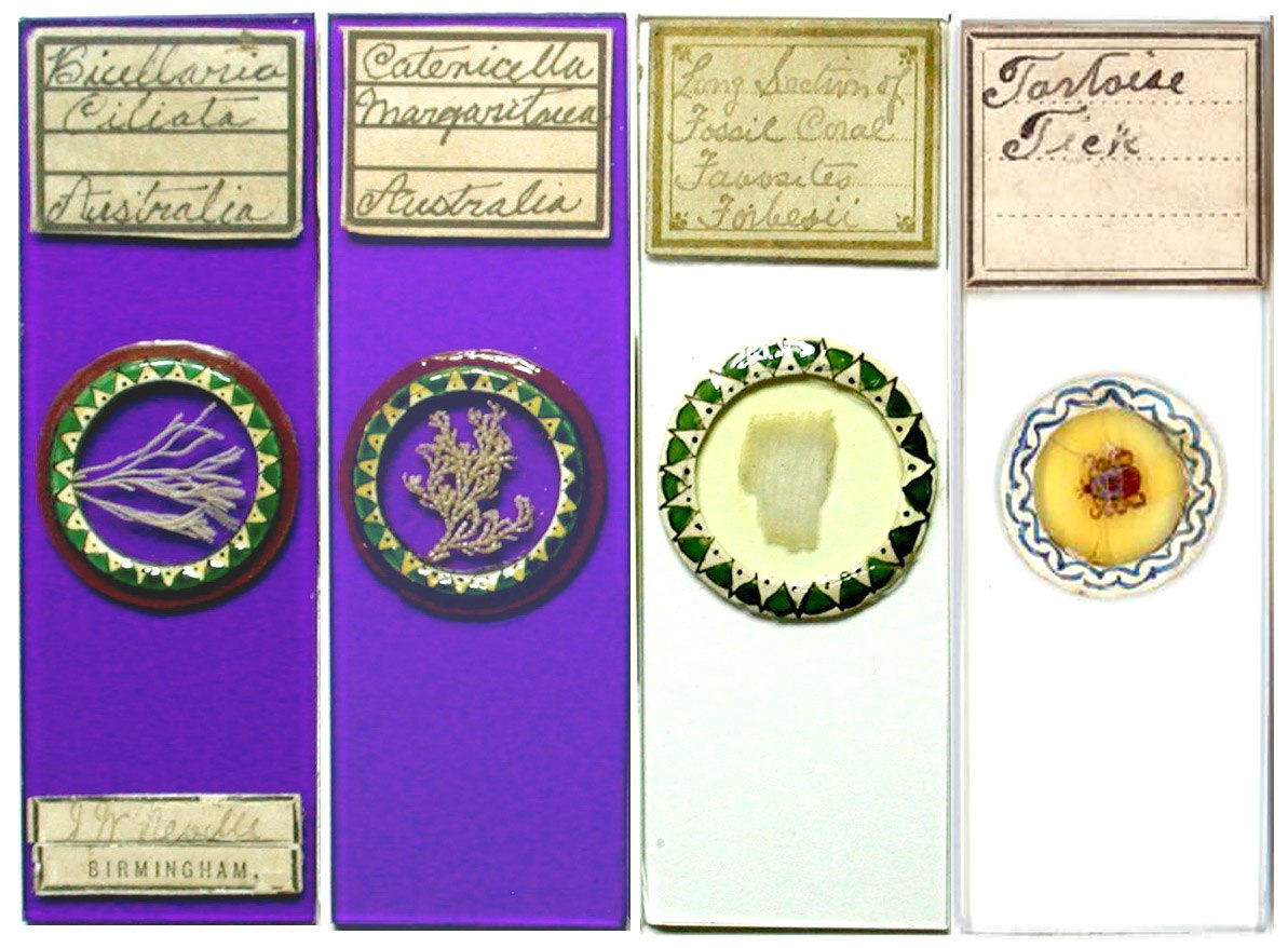
Figure 3. Four microscope slides whose label descriptions correspond
with those known to have been exhibited by James Neville to the Birmingham
Microscopists’ and Naturalists’ Union. Left to right (descriptions are verbatim
from the “The Midland Naturalist”):
Bicillaria ciliata and Catenicella margaritacea, Australian polyzoa (both shown
Jan. 19, 1885); Section of Favosites forbesii, a fossil coral (Sept. 19, 1887); Parasite of tortoise (Oct. 26, 1891).
Neville served
as President of the Birmingham Microscopists’ and Naturalists’ Union in
1884. The following year, he
stepped down to become a Vice President. He also acted as the club’s Secretary
for many years. Neville’s’ presidential retirement address, "The Offensive and Defensive Weapons of Insects," was summarized in The Midland Naturalist,
and gives a good idea of James’ depth of knowledge: "remarking that as the subject was a wide
one, he should only take that part of it that referred directly to ourselves.
It might appear necessary to some to apologise for introducing into respectable
company some of the insects to which he should have occasion to refer. But
these would not be naturalists, for naturalists studied living objects as they
were, whether they pleased the sentiments or no. The offensive weapons of the
following insects were then referred to: Pediculus capitis, Nepa cinerea,
Notonecta glauca, Cimex lectularius, and Pulex irritans. The peculiarities of
their mouth organs were described and compared with a typical insect's mouth,
that of the ground beetle, Carabus, when the remarkable departure from a
probable original type was made apparent. In the latter part of the subject the
sting of the wasp was described with its complicated mechanism of poison bag,
duct with chitinous rings, pistons for ejecting poison, lancets, etc. The
address concluded by regretting that the labours of microscopists were often of
a desultory character, and pointing out the advantages of more special
pursuits. The use of the various forms and ornamentation of pollen grains was
suggested as good ground to be worked by microscopic botanists. The
Entomostraca, Diatomaceae, and Desmidiaceae of the district were mentioned as
fields of labour where good and useful work was required, and local catalogues
much needed. The address was illustrated by diagrams.”
Although he
was an amateur microscopist, James Neville developed considerable skill as a
slide mounter. He frequently
lectured to his club and wrote articles on mounting techniques. On October 11, 1885, “Mr. J.W. Neville demonstrated
the simplicity of preparing whole insects for investigation by describing and showing the processes they pass through.
Several objects were prepared and mounted, and afterwards exhibited to the meeting”. On September 22, 1890, "Mr. J.W.
Neville gave a short address on plant crystals and how to prepare them for the
microscope. The speaker said there was no royal road to mounting, and it was
impossible to mount a leaf to show its whole structure satisfactorily. In
mounting crystals we generally lost other cell contents, and in trying to
preserve the protoplasm of the cell we lost the crystals. The process
recommended was to bleach the leaves in chlorinated soda, and when quite
transparent mount in balsam through carbolic acid, and afterwards view with
polarised light. A collection of slides mounted in this manner was shown under
the microscope”. On July 13, 1891 he “gave
a demonstration of the use of carbolic acid in microscopic mounting. This is a
medium through which objects can be passed directly from water into Canada
balsam. Its use was particularly advantageous for objects that shrunk and
cracked in drying, and also for those that were difficult to rid of air, and it
was perhaps the most simple of the ‘wet methods’. A number of objects were then
mounted to show the process, a minute or two sufficing to transfer them from
water into balsam”.
He
wrote to The Midland Naturalist in 1883: “Many of the readers of the ‘Midland
Naturalist’ will learn with pleasure that there is such a ready way of mounting
vegetable preparations as that mentioned by Mr. Bagnall in the July number, as
the invention of Professor Hillhouse of the Mason College. The process is very
simple, and the medium excellent; but from practical experience I would suggest
the sealing of the cover-glass with pale copal varnish, instead of dilute
balsam; it can be obtained of as light a colour, is much tougher, and not
likely to get so brittle as that medium. As regards the newness of the invention,
I can only say I have preparations by me that have been put up in this way for
seven years or more, and several of my friends have used it as long a time,
preferring it to glycerine jelly, as it does not show such a disposition to
leak. Practical microscopists will, however, be glad to learn that after this
space of time the objects show no signs of deterioration, but rather wear an
improved appearance.”
During 1886,
Neville published in Hardwicke’s
Science-Gossip on “Wilks' Cell”. This invention, by George Wilks of
Manchester, was a grooved lead ring to be used as a spacer between a slide and
coverslip, for mounting large objects. The soft lead could be compressed to
reduce the depth, if necessary. “It is
somewhat difficult to give R.S.P. the information he seeks on the manner of
using the Wilks' cell, without first knowing how advanced he may be in the
preparation of objects without pressure. It requires a considerable amount of
care to get the objects ready for the cell, but when this is done the mounting
of them is the easiest part. If R.S.P. refers only to this latter part, he will
find no difficulty if he proceeds as follows. Take a cell and see that it is no
deeper than the object requires. If it is too deep it can easily be flattened
between two pieces of glass by pressure, until the exact depth is gained. All
that remains then to be done is to place it on a clean slip, fill it with thick
balsam, and, immersing the prepared object, put on the cover glass. In using
the thick balsam, numerous air bubbles will most likely appear in the cell.
This is a small matter, for if kept in a warm place they will gradually work to
the edge and disappear. If R.S.P. wishes for anything like full information on
the preparation of objects for the cell, I fear it cannot be given without
devoting at least one article to the subject. The Wilks' cell is a very useful
introduction, and deserves to be largely used. Prior to its invention I have
frequently extemporised one from a vulcanite cell, that sufficiently answered
the purpose, although at the cost of time and trouble. Still I think the cell
is open to improvement, and would suggest that it had at least twice the number
of elevations, or, perhaps better still, was corrugated, as in its present form
the cover glass is likely to get broken unless well banked up with edging
colour, as there is so large a space between the supports.” Neville was
also familiar with Wilks’ microscope slides, and on February 23, 1885,
displayed “twelve botanical sections,
double stained, prepared by Mr. G. Wilkes (sic)” to his microscopy club.
In addition to
microscopical preparations, James Neville exhibited a wide variety of natural
objects at meetings of the Birmingham Naturalists’ and Microscopists’
Union. On December 17, 1888, he showed
the skin of a large Australian snake. This is probably the same specimen he
advertised for exchange the following year “Offered,
skin of snake, ten feet long. Wanted, exotic insects or offers”. He is also
recorded as having exhibited tropical land shells, fossils in limestone,
ammonites, “leaves of Spircea Ulmaria
infested with brand”, and “a pansy
with six petals, an abnormal growth that has been continued on all off-sets for
several years”. Neville read several papers to the club, including “Peeps into Ponds” on Sept. 30, 1895, and “Protective Devices of Larvae” on June 26, 1899. Two other well-known microscope slide-makers, Herbert Darlaston and Richard Hancock, were also members of the club during those years.
On November 17, 1890, at the Union’s Annual Public Exhibition,
Neville exhibited “a frame of lantern
slides, illustrating the ‘Wonders of a Pond’”. These may have been his own
productions. In 1895, he published a small book, The Photographic Colourist: “a
manual for the use of amateurs: giving every particular required for painting
lantern slides and other transparencies, colouring diagrams, preparing lantern
slides without the aid of photography, blacking out backgrounds, scratching in
details, tinting paper prints, colouring paper prints to imitate oil paintings,
crystoleum painting etc. etc.”
James
Neville’s artistry extended well beyond decorating microscope and lantern
slides. One of his descendants, Faith Blancquiere, provided us with pictures of
a painting and two decorated silk cushions made by Neville (Figure 4). Undoubtedly,
many examples of his decorations of furniture, etc. remain, although it is very
likely that they were unsigned and remain anonymous.
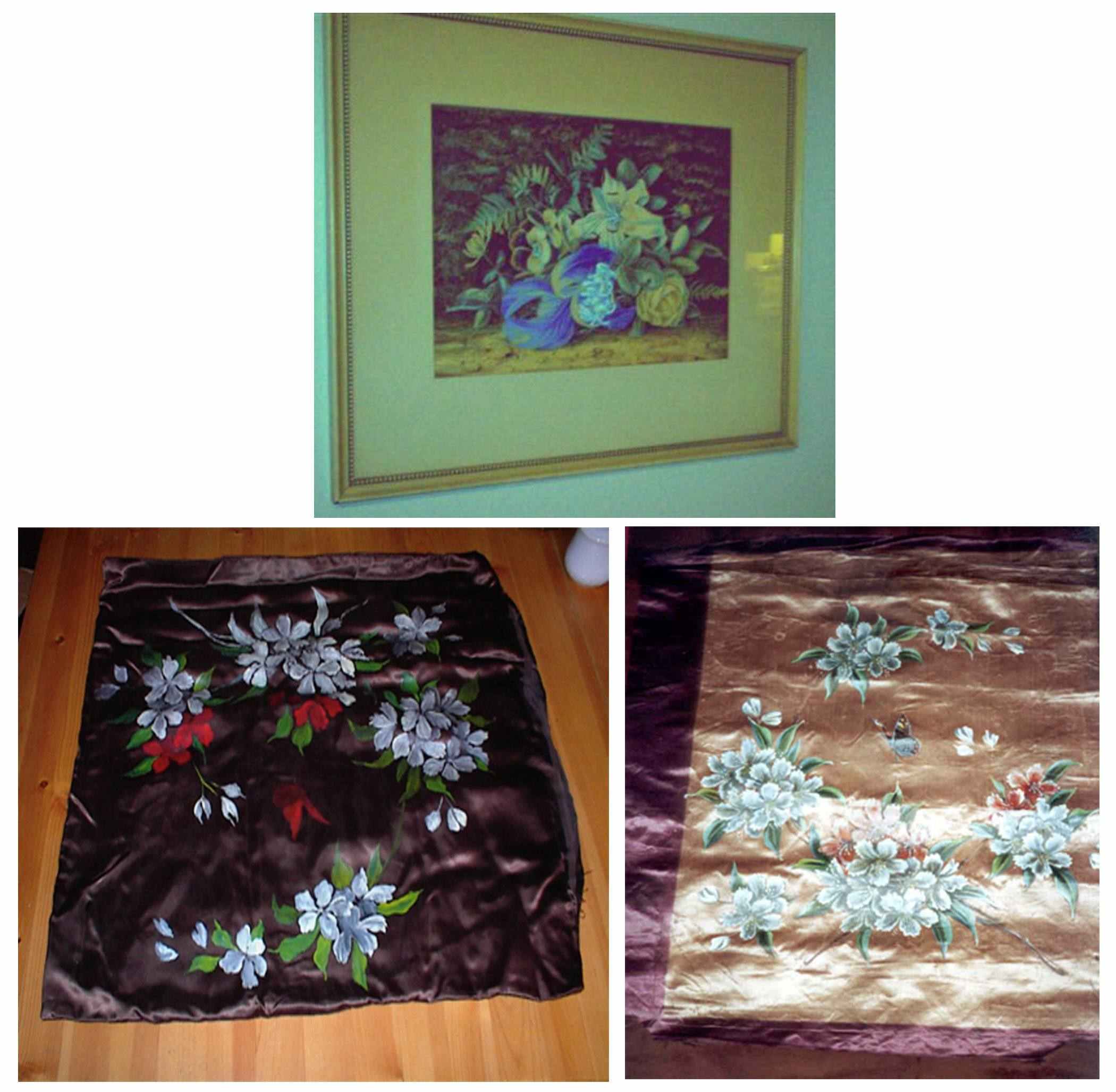
Figure 4. Artworks by James Neville.
Top, framed painting. Bottom, two painted silk cushion covers. Photographs
provided by Faith Blancquiere.
James White
Neville died at the age of 59 on June 14, 1900, at his home on Wellington Road,
Handsworth. The cause of death was
recorded as “Malignant Sarcoma, Pulmonary
Haemorrhage, Syncope”. There is a strong possibility that his cancer was
brought on by a lifetime of inhaling fumes from the chemicals he used as a
japanner. It is ironic to think that each of his elaborately-decorated
microscope slides may have contributed to their maker’s early death.
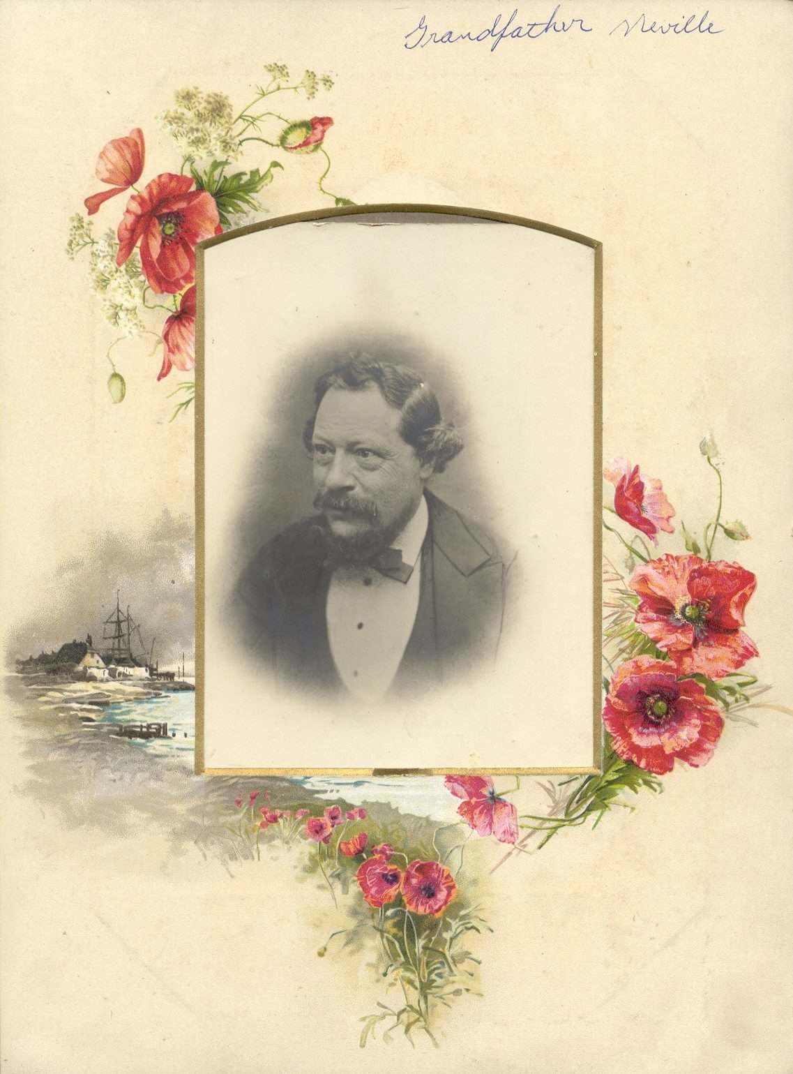
Figure 5. Photograph of James White Neville, provided
by Faith Blancquiere.
Table 1. Microscopical preparations
exhibited by James W. Neville at meetings of the Birmingham Microscopists’ and
Naturalists’ Union, between 1881 and 1891. Neville was a sub-editor for The Midland Naturalist during
that time period, and regularly reported items displayed at meetings of his
society. This list describes only
some of Neville’s slides shown at club functions, as many were exhibited in
large groups that were not individually identified. He also traded extensively with other naturalists, and so
undoubtedly made many more slides than were exhibited at the club. Nonetheless, this list shows the
breadth to Neville’s interests in natural history, with specimens of all sorts
of animals, plants and minerals being represented. Descriptions in the table
are paraphrased from the published descriptions.
|
Antennum of butterflies and moths
|
March 16, 1891
|
|
Aphids
|
November 9, 1891
|
|
Aregma bulborum
|
August 31, 1885
|
|
Aregma bulbosa
|
May 26, 1884
|
|
Aregma obtusatum
|
June 23, 1884
|
|
Arran pitchstone, section
|
February 20, 1888
|
|
Astromyelon and particular woody tissue, from coal
measures
|
June 12, 1882
|
|
Australian sundew, leaf with captive insects
|
June 18, 1886
|
|
Australian tettigoinia larvae, found on the eucalyptus
tree
|
June 21, 1886
|
|
Batachospermum,
from Keeper’s Pool
|
March 18, 1881
|
|
Bicellaria
ciliata, Australian polyzoa
|
January 19, 1885
|
|
Bladderwort, Utricularia vulgaris
|
July 14, 1884
|
|
Bombyx mori
larva, mounted whole, showing tracheal system complete
|
September 5, 1881
|
|
Bombyx mori
larva, showing two rows of hooklets in each leg
|
October 2, 1881
|
|
Bombyx pernyi,
antenna
|
November 30, 1885
|
|
Brittle starfish, Ophiocoma neglecta
|
August 17, 1885
|
|
Butterfly wing, Morpho
cypris
|
June 15, 1885
|
|
Butterfly wing, Orthoptera
rhadamanthus
|
March 10, 1884
|
|
Calamite stem
|
April 25, 1881
|
|
Calamites, fossil, transverse section
|
December 15, 1884
|
|
Carchesium
polypinum
|
March 23, 1885
|
|
Catenicella
auritia, an Australian zoophyte
|
October 26, 1885
|
|
Catenicella
margaritacea, Australian polyzoa
|
January 19, 1885
|
|
Cement-stein from Isle of Fur, Denmark, showing Trinaeria excavata and other diatoms
in situ
|
February 15, 1886
|
|
Chaleis minuta,
from Turkey
|
November 29, 1886
|
|
“Cherry-gall” flies, Cynips quercus folii, male and female
|
August 18, 1884
|
|
Chiton cinercus,
palate
|
September 15, 1884
|
|
Cluster Cup and Coleosporum
on leaves of coltsfoot
|
September 19, 1881
|
|
Coal-ball section, showing a sporangium with spores in
situ
|
December 20, 1886
|
|
Coal ball section, with transverse section of Rachiopteris oldhamium
|
November 16, 1885
|
|
Coal from Hamstead, fibrous, showing dotted vessels
|
December 9, 1889
|
|
Coal section showing excreta of insects deposited in
the tissues of plants which they had eaten, probably while in the larval
stage
|
December 12, 1881
|
|
Coal sections, showing fern sporangia with spores in
situ
|
November 21, 1881
|
|
Coltsfoot leaf infested with micro-fungus (Coleosporum)
|
August 22, 1881
|
|
Comb-footed ichneumon fly, Ophion luteum
|
October 6, 1884
|
|
Cricket, chirping file and drum
|
October 3, 1887
|
|
Cuttle fish bone, section
|
November 23, 1885
|
|
Deutzia corymbosa,
leaf
|
June 25, 1888
|
|
Deutzia scabra,
leaf
|
June 25, 1888
|
|
Diatomaceae, from deposit in Black Root pool, Sutton
Park
|
November 3, 1884
|
|
Dog rose, section through a prickle
|
June 30, 1884
|
|
Doris flammea,
palate
|
September 22, 1884
|
|
Dragon-fly (Agrion),
jaws
|
July 10, 1882
|
|
Dredgings from “Challenger” Expedition
|
January 8, 1883
|
|
Dredgings from Indian Ocean
|
January 28, 1884
|
|
Drone Fly proboscis
|
September 25, 1882
|
|
Drosera
rotundiflora leaf, with captured insects
|
May 19, 1884
|
|
Dytiscus
marginalis, alimentary canal
|
July 28, 1884
|
|
Dytiscus
marginalis, elytra
|
April 20, 1891
|
|
Emperor Moth larva skin, which had been pierced by
Ichneumon Fly
|
June 26, 1882
|
|
Euplectella
aspergillum, spicules
|
October 18, 1886
|
|
Favosites
forbesii, a fossil coral, section
|
September 19, 1887
|
|
Ferns, fossil, from Albion
|
October 20, 1884
|
|
Fern fructification, fossil in a section of coal-ball
material
|
September 10, 1888
|
|
Fern, Rachiopteris
cylindrica, stem, transverse section
|
November 21, 1881
|
|
Flea, common, gizzard
|
August 25, 1883
|
|
Flea, common, lancet
|
August 25, 1883
|
|
Flea, common, stomach
|
March 27, 1882
|
|
Flint, section showing Xanthidia
|
August 21, 1882
|
|
Floral organs, sections, showing all the parts in situ
|
January 5, 1891
|
|
Flustra
episcopalis, from New Zealand
|
January 26, 1885
|
|
Foraminifera, dredged off the coast of Galway
|
January 5, 1885
|
|
Foraminifera from Borth
|
July 6, 1891
|
|
Foraminifera from Jersey
|
March 5, 1888
|
|
Foraminifera etc., from chalk washings
|
November 9, 1885
|
|
Foraminifera from sponge sand, selected
|
May 2, 1887
|
|
Funaria
hygrometrica, capsules
|
June 16, 1890
|
|
Fungus, paper mildew, Myxotrichum chartarum
|
March 31, 1884
|
|
Fusus islandicus,
palate
|
November 17, 1884
|
|
Fusus islandicus,
palate
|
October 24, 1887
|
|
Gamasus
coleoptratorum from humble bee
|
June 7, 1886
|
|
Gorgonia from Australia, spicules
|
April 4, 1887
|
|
Grasshopper, auditory organs
|
September 5, 1887
|
|
Gyrinus natator,
double eyes
|
October 24, 1881
|
|
Haliotis
tuberculata, lingual ribbon
|
December 4, 1881
|
|
Hedge Maple, transverse section
|
September 18, 1882
|
|
Helices, jaws, series on a single slide mounted for
comparison
|
February 21, 1887
|
|
Heliolites
interstinetus, section
|
May 18, 1885
|
|
Honey bee, antenna comb
|
April 12, 1886
|
|
Horned aphis, Cerataphis
lataniae
|
February 25, 1884
|
|
House fly, Musca
domestica, teeth
|
August 11, 1884
|
|
House spider dissections, showing falces, tongue,
&c.
|
February 6, 1882
|
|
Human colon, transverse section
|
April 24, 1882
|
|
Human lung
|
April 24, 1882
|
|
Hydrodictyon
utriculatum, water net
|
August 15, 1887
|
|
Hydrodictyon
utriculatum, in four stages of growth
|
March 17, 1890
|
|
Ilispa atra,
a spiny beetle from Turkey
|
August 23, 1886
|
|
Leaf crystals, treated with chlorinated soda, mounted
in balsam/carbolic acid, various
|
September 22, 1890
|
|
Lecythea on
leaves of barren strawberry
|
August 25, 1883
|
|
Lecythea and Aregma on leaf of barren strawberry
|
September 19, 1881
|
|
Lecythea or
rust on leaf of rose
|
September 19, 1881
|
|
Lepidodendron
bark
|
April 25, 1881
|
|
Lepidopterous larvae, 12 slides
|
September 2, 1889
|
|
Maize, annular vessels
|
June 8, 1891
|
|
Marchantia,
spores and elaters
|
July 18, 1881
|
|
Megalicthys,
head plate, from Lancashire coal beds
|
June 6, 1884
|
|
Melicerta ringens
|
June 15, 1891
|
|
Membranipora
membranacea, a polyzoan from New Zealand
|
January 12, 1885
|
|
Mica, showing dendritic crystals of manganese
|
April 4, 1881
|
|
Mole cricket gizzard
|
March 1, 1886
|
|
Mosquito, mouth organs
|
May 4, 1885
|
|
Moss fruits
|
January 4, 1886
|
|
Nassa reticulata,
palate
|
April 27, 1885
|
|
Nitella
translucens
|
May 23, 1881
|
|
Oak Apple fly, Cynips
terminalis
|
February 9, 1885
|
|
Oak Spangle fly, Cynips
longipennis
|
May 5, 1884
|
|
Octopus palate
|
June 29, 1885
|
|
Ocypus olenus
beetle, mouth organs
|
September 2, 1889
|
|
Oidium moniliodes,
on leaves of grass
|
August 25, 1883
|
|
Ophiocoma
neglecta
|
May 12, 1884
|
|
Orgyia pudibunda,
larva, mounted whole, popularly known as the Hop Dog
|
February 2, 1885
|
|
Oysters, young shells
|
March 7, 1881
|
|
Parasite of Limax
flavus
|
July 30, 1888
|
|
Parasite
of tortoise
|
October
26, 1891
|
|
Pediculus capitis,
tracheal system
|
November 26, 1888
|
|
Pelopaeus
fistularis, a mason-wasp from Constantinople, mouth organs
|
January 17, 1887
|
|
Permian marl, with fern impressions
|
December 22, 1884
|
|
Phlota plumosa
|
June 26, 1882
|
|
Phragmidium
obtusum on leaves of barren strawberry
|
August 25, 1883
|
|
Pinguicula
vulgaris leaf, with insects
|
September 7, 1885
|
|
Piniperda
larva
|
August 11, 1883
|
|
Plant bug, Tingis,
from Turkey
|
September 20, 1886
|
|
Plant bug, Tingis,
from Turkey
|
October 15, 1888
|
|
Plocamium
coccineum in fruit
|
April 3, 1882
|
|
Polyxenes lagurus
|
April 19, 1886
|
|
Puccinia on
leaf of violet
|
September 19, 1881
|
|
Pulex irritans,
striated muscles
|
February 7, 1887
|
|
Ranunculus repens
leaf, showing aecidium in situ
|
June 18, 1883
|
|
Ruby sand with fluid cavities, from New Zealand
|
July 27, 1885
|
|
Sea mouse, hair
|
May 1, 1882
|
|
Shells from the Gulf of Aden
|
April 13, 1891
|
|
Sirex gigas,
ovipositor
|
July 27, 1891
|
|
Sheep tick, Ixodes
reduvius
|
December 7, 1885
|
|
Snake’s head coralline (Anguinaria spathulata)
|
February 4, 1884
|
|
Sugar-cane, transverse section
|
November 28, 1881
|
|
Synapta adherens,
skin, showing anchors and plates in situ
|
February 20, 1882
|
|
Testacella
haliotidea, palate
|
April 18, 1887
|
|
Trachea, from larva of drinker moth
|
July 21, 1884
|
|
Trochus,
palate
|
September 4, 1882
|
|
Trochus,
shell
|
September 4, 1882
|
|
Volvox globator
|
April 20, 1885
|
|
Wasp, mouth organs, mounted without pressure
|
March 9, 1885
|
|
Wasp queen, mouth organs
|
March 23, 1891
|
|
Water spider, tongue
|
October 17, 1881
|
|
Worm-eaten glass, from an old Warwickshire church
|
July 2, 1883
|
|
Yoredale limestone, section, with goniatites in situ
|
September 29, 1884
|
|
Zonites
draparnaldi, palate
|
January 3, 1887
|
Acknowledgements
We thank Faith
Blancquiere for her generosity in providing information on James Neville and
pictures of him and his work. It was a pleasure to assist her with new
information on her ancestors. We also thank Maureen Carter for providing
information on Neville and his associates at the Birmingham Naturalists’ and
Microscopists’ Union.
Resources
Bracegirdle,
Brian (1998) Microscopical Mounts and
Mounters, Quekett Microscopical Club, London.
Death record of James White Neville.
English
census, birth, marriage, and death records, accessed through www.ancestry.co.uk
Hardwicke’s Science-Gossip, vol. 14
(1878), Exchanges column, pages 168 and 216.
Hardwicke’s Science-Gossip, vol. 15
(1879), Exchanges column, pages 168 and 264.
Hardwicke’s Science-Gossip, vol. 19
(1883), Exchanges column, page 264.
Hardwicke’s Science-Gossip, vol. 24
(1888), Exchanges column, page 120.
Hardwicke’s Science-Gossip, vol. 25
(1889), Exchanges column, page 168.
Hardwicke’s Science-Gossip, vol. 27
(1891), Exchanges column, page 192.
Kelly, E.R. (1879) Post
Office Directory for Birmingham, with it Suburbs, page 245.
The Midland Naturalist,
vols. 3-14 (1880-1891) Birmingham Naturalists’ and Microscopists’ Union
proceedings, numerous pages. Details of specimens exhibited by Neville and
other club members.
The Midland Naturalist,
vol. 7 (1884) Birmingham Naturalists’ and Microscopists’ Union, page 852.
The Midland Naturalist,
vol. 8 (1885) Our sub-editors, page 28.
Neville, James W. (1883) New methods of mounting for the
microscope, The Midland Naturalist,
vol. 6, page 190.
Neville, James W. (1885) Crystals for the polariscope, Hardwicke’s Science-Gossip, vol. 21,
page 115.
Neville, James W. (1885) Abegma bulbosum, The Midland Naturalist, vol. 8, page 297.
Neville, James W. (1886) Wilks’ cell, Hardwicke’s Science-Gossip, vol. 22, page 233.
Neville, James W. (1895) The
Photographic Colourist, Iliffe and Son, London.
Yearbook of the
Scientific and Learned Societies of Great Britain and Ireland (1884) The
Birmingham Naturalists’ and Microscopists’ Union, page 71.
Yearbook of the
Scientific and Learned Societies of Great Britain and Ireland (1895) The
Birmingham Naturalists’ and Microscopists’ Union, page 105.
Yearbook of the
Scientific and Learned Societies of Great Britain and Ireland (1900) The
Birmingham Naturalists’ and Microscopists’ Union, pages 116-117.