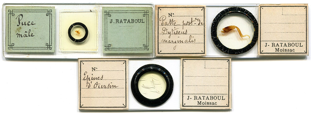
Figure 1. ca. 1880-1900 microscope slides by Joseph Rataboul.
Joseph Rataboul, 1858 - 1905
by Brian Stevenson
last updated July, 2020
Joseph Rataboul was a lawyer and microscope enthusiast who lived in Moissac, Tarn et Garonne, France. In 1881, he joined the Belgian Society of Microscopy (Société Belge de Microscopie), which was a premier French-speaking microscopical society. He became a member of the Natural History Society of Toulouse (Societe D'Histoire Naturelle de Toulouse) in 1882. Rataboul had diverse interests in the subjects he mounted and studied, although he was best known for his research on diatoms. Jules Pelletan and Julien Deby provided details of Rataboul’s method of sorting diatoms in their 1888 Les Diatomées (described in detail below). At the time of his death, Rataboul was working with noted diatomist Jacques Brun (1826-1908) on a catalogue of the diatom species found in Moissac and Geneva.
Figure 1.
ca. 1880-1900 microscope slides by Joseph Rataboul.
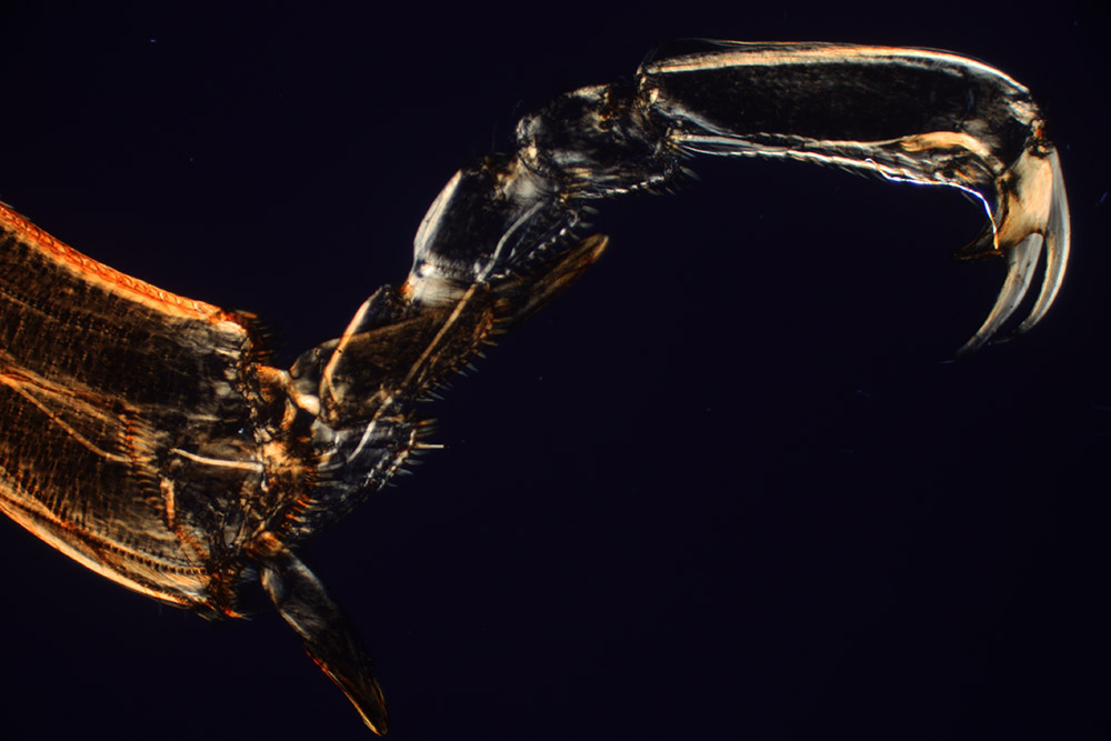
Figure 2.
“Patte post du Dytiscus marginalis” (hind leg of a Dytiscus marginalis, the “great diving beetle”) by Joseph Rataboul (see Figure 1). Photographed with transmitted light and crossed polarizing filters, 3.5x objective lens, and a C-mounted digital SLR camera.

Figure 3.
“Espines de Oursin” (spines of sea urchin) by Joseph Rataboul (see Figure 1). Photographed with transmitted light, 3.5x objective lens, and a C-mounted digital SLR camera.
Joseph Rataboul was born in February, 1858, in Moissac, to Jean Guillaume Ernest and Eilize Catherine Belbézé Rataboul. An 1862 trade directory listed a Monsieur Rataboul as a “Notaire” (notary) in Moissac, presumably Joseph’s father.
At the February 26, 1881 meeting of the Société Belge de Microscopie, “Le Conseil propose l’admission, comme membres effectifs, de MM Guior, a Dijon, et Ratabould, avocat à Moissac (Tarn-et-Garonne), presentes par MM Cornet et Coomans” (“The Counsel proposed the admission, as effective members, of Messers. Guior, of Dijon, and Rataboul, lawyer of Moissac (Tarn-et-Garonne), presented by Messers. Cornet and Coomans”).
The following year’s Naturalists’ Directory carried this entry, “Rataboul, Joseph, Prof. d'Histoire Naturelle, Moissac (Tarn-et-Garonne). Vegetable Anatomy, Microscopy, Entomology. C. Ex. Microscopic and entomological specimens, especially Lepidoptera” (note: “C. Ex.” indicated that Rataboul was interested in collecting and exchanging those specimens). It is possible that Rataboul worked as a science teacher as well as being a lawyer, but it is equally possible that intended to show a serious interest in natural history.
In 1883, Rataboul presented on “Les diatomés, récolte et préparation” (“Diatoms, harvest and preparation”), to the Societe d'Histoire Naturelle de Toulouse. His presentation was published in the society’s Bulletin, then reprinted in Journal de Micrographie. Parts of his preparation techniques were included in Pelletan and Deby’s 1888 Les Diatomées (see below).
A paper sent to the Société Belge de Microscopie on mounting anthers of flowers (“Sur la préparation des anthères”), was published in the Society’s 1883 Annales.
Diatomist Hippolyte Peragallo (1851-1921) acknowledged Rataboul for contributing specimens to Peragallo’s 1884 Diatomées du midi de la France.
Joseph married Céline Boscus on February 7, 1888. A daughter, Ernestine, was born later that year.
The 1888 Naturalists’ Directory indicated that Rataboul’s attention was then focused specifically on diatoms (an asterisk notation with his entry indicated that he had recently corresponded with the publisher): “Rataboul, Joseph, Moissac (Tarn-et-Garonne). Diatoms. C. Ex. *”. His interest broadened somewhat with is 1890 entry, “Rataboul, Joseph, Moissac (Tarn-et-Garonne). Diatoms., Geol., Min. C. Ex. Correspondence solicited.*”
The 1902 Botaniker Adressbuch listed, “Rataboul, Joseph, Micrographe, - Moissac, Tarn-et-Garonne. (Cryptogames. Diatomées. Echanges.)”.
Joseph Ratabould died when only 47 years old, in 1905. We learn this indirectly from two sources:
The Conservatoire Botanique de Geneve (Switzerland) holds a manuscript file, “Diatomees. Especes et varietes nouvelles, par J. Rataboul et J. Brun. Geneve-Moissac. 1904. Projet de publication, arrete par le mort du Mr Rataboul. J. Brun. 1905” (Diatoms. New species and varieties, by J. Rataboul and J. Brun. Geneva-Moissac. 1904. Draft publication, stopped by the death of Mr Rataboul”).
The August, 1905 issue of l’Apiculteur informs both of Rataboul’s death and that he raised honey bees, “A vendre, pour cause de décès, 40 ruches Voirnot bien peuplées, autant de ruches vides, gaufrier neuf, cire gaufrée, outillage complet, ouvrages d'apiculture, etc., dans de bonnes conditions. S'adresser à Madame Ve Rataboul, à Moissac (Tarn-et-Garonne)” (“For sale, due to death, 40 well-stocked Voirnot hives, as many empty hives, new gaufrier, embossed wax, complete tools, beekeeping works, etc., in good conditions. Contact Madame Widow Rataboul, in Moissac (Tarn-et-Garonne”).
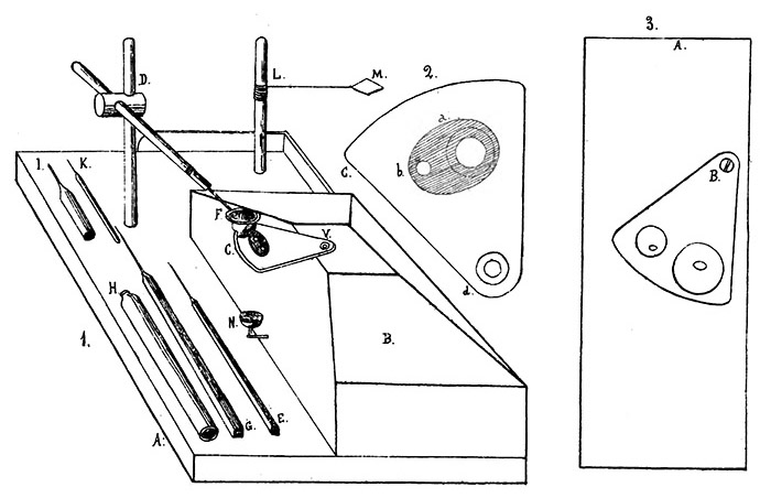
Figure 4.
Illustrations of Joseph Rataboul’s diatom sorting equipment, from “Journal de Micrographie”, 1883. It was also reproduced in Jules Pelletan and Julien Deby’s 1888 “Les Diatomées”.
(1) A, tablette avec bloc, B ; C, platine; D, support de la loupe F; L, support de la plaque M pour le chauffage des préparations; N, miroir; E, G, H, I, K, accessoires divers. (A, tablet with block, B; C, plate; D, magnifier support F; L, support for plate M for heating preparations; N, mirror; E, G, H, I, K, various accessories).
(2) Platine tournante (revolving plate).
(3) Autre système pour le triage des petites espèces (other system for sorting small species).
Extracted from Les Diatomées, 1888, by Jules Pelletan and Julian Deby:
(all diagrams are shown in Figure 4, above)
“Appareils et procédés de M. Rataboul
Mr. Rataboul has invented various small instruments that greatly facilitate these delicate manipulations and allow diatoms to be stored as regularly as with Zentmayer's mechanical finger.
The first of these instruments, which is used for sorting large species, is composed of a thick board A used to give the device the desired stability. A block of wood B, with cut sides, is attached to it and is used to support the hands, like the inclined plane supports of current simple microscopes. In a corner of the lath is a vertical brass rod D, carrying a doubly-articulated horizontal arm, at the end of which is mounted a doublet or a Coddington magnifying glass F, which can thus rise or lower and take all the directions you want.
The revolving plate is fixed onto the flat top of the wooden block. This is piece of 3 mm thick copper, blackened on its upper face. It is fixed to the block by a large flat head screw V, but loose enough that it can rotate around the screw. The revolving plate is pierced with two round holes (Figure 4-2, b and c), whose centers are located at the same distance from the center of the screw V, so that the radii V-b and V-c are equal. It is best that this revolving plate be large enough so that if, in the transport of diatoms, a frustule is dropped, it will fall onto the plate and not be lost.
Use of this small device is easy to appreciate. Diatoms to be sorted are placed on a glass cover slip that is fixed over the largest hole c of the plate, and the glass slip on which we want to place the chosen frustules is glued to the smallest hole b. This small circle makes it easy to find the center, if it is desired, for example, to mount a single Diatom. The covers are fixed onto the revolving plate with a little paraffin, which had been melted by use of a small heated metal rod. We then have the magnifying glass above the cover c. With a hair mounted on a handle, we choose a frustule; then, with the left hand, a slight rotational movement is made on the turntable until the cover b comes to be placed under the magnifying glass. We then place the frustule in the desired position - and we start again.
Ordinarily, a mirror is not used. Diatoms are seen more clearly on a black background than on a lit background. For this reason, it is advantageous to stick a little black paper under the openings b, c, of the plate. If, however, it is necessary to light from below, a small mirror can be arranged at N.
This device is very convenient whenever you have to sort large species, and that the magnifying glass, which does not reverse the images, gives sufficient magnification. But for very small species, when it takes a magnification of 100 to 150 diameters, we are forced to replace the magnifying glass by a compound microscope, always seeking to obtain above all the magnification by the eyepiece to have more space under the lens. The device must then be modified. The turntable is mounted in the same way on a copper plate A (Figure 4-3) pierced with a hole in its center. This plate is fixed firmly with screws on the stage of the microscope so that its central hole, on which the openings b, c will be brought successively, coincides with the hole in the stage of the microscope. Blocks can be attached to each side of the instrument to serve as hand rests, and we operate under the compound microscope as we did under the doublet, but the images and movements are reversed.
With the first device or with the second, sorting the diatoms freehand or with a mechanical finger, Mr. Rataboul operates as follows: The diatoms to be sorted are deposited with a pipette onto a glass coverslip that is placed on the small platinum blade M (Figure 4-1) and burned according to the ordinary process. Then, the cooled cover is fixed with a little paraffin on the hole c of the rotating sector. Another very clean cover is then attached to hole b. We then place in the center of that cover slip, with a brush, a small drop of a very pure gum solution. This solution is made with a large drop of thick gum arabic in 15 cubic cm distilled water. The gum is allowed to dry, and, if necessary, the desiccation can be hastened by bringing a red-hot metal rod near to the cover. The sorting is then done with the hair, bringing the cover c under the magnifying glass or the objective, removing a frustule and depositing it on the cover b, brought in turn into the field by a small movement of the revolving plate. We place the frustule where we want, on the dry gummy coating. To fix it we take a glass tube H (Figure 4-1), which is wide at one end and narrow at the other, and, the smallest opening facing the diatoms, we gently breath onto the cover, softening the gum enough for the frustule to stick to it.
We repeat the operation as many times as we want, and when the diatoms are stored and fixed, it only remains to mount the preparation, either with balsam or dry.
To mount it in Canada balsam, Mr. Rataboul detaches the cover and places it for an hour, the loaded side down, in a watch glass containing turpentine or lavender essence. Then, he deposits a drop of balsam on a slide. He heats this on an alcohol lamp, and when streaks appear in the balsam, it leaves to cool. If there are bubbles, he bursts them or removes them with a cold needle. The cover is then removed from the essence, drained and placed, the diatoms facing down, on the drop of balsam; using controlled heat, adhesion occurs, excess essence evaporates. It is compressed slightly and allowed to cool. It only remains to clean the preparation when it is perfectly dry, as we have indicated.
Mr. Rataboul recommends copal varnish, as easier to handle, but much longer to dry, because it is easier to avoid air bubbles. The varnish should be evaporated to a syrupy consistency, about a third of its volume, and it is enough to place a drop on the object holder and place over the cover soaked in turpentine. The preparation is then left for 2 or 3 hours on a slightly heated cast iron plate (at around 70 °). And we will not have to fear any bubbles. It is advisable to wait some time before cleaning the preparation.
For dry preparations, Mr. Rataboul does not use gum to coat the cover, but instead uses a droplet of balsam dissolved in chloroform, or copal varnish. He operates as above but, instead of using the humidity of the breath to soften the coating and make it adhesive, he uses a heated metal rod that is brought near to the cover. When the diatoms have been arranged, the cover is placed on the platinum blade and heated to red. The balsam darkens, then disappears entirely, but the frustules remain in place. The cooled cover is then gently applied, the diatoms below, on a microscope slide where one or two concentric cells have been traced with bitumen. The cells must be completely dry and prepared at least three weeks in advance. The preparation is heated very slightly to obtain adhesion which is then helped by pressing on the cover. Finally, the preparation is closed by a circle of bitumen on the edges of the cover.”
Resources
Annales de la Société Belge de Microscopie (1881) Vol. 7, pages 101 and 175
Annuaire-Almanach du Commerce, de l'Industrie, de la Magistrature et de l'Administration (1862) “Moissac .. Notaires .. Rataboul”, page 2447
L'Apiculteur (1905) Insertions diverses, Vol. 49, August issue
Botaniker Adressbuch (1902) Dörfler, Vienna, page 87
Bulletin of The Natural History Museum, Botany Series (1995) Vol. 25, page 12
Bulletin Societe D'Histoire Naturelle de Toulouse (1882) page 426
The Naturalists’ Directory (1882) Cassino, Boston, page 201
The Naturalists’ Directory (1885) Cassino, Boston, page 70
The Naturalists’ Directory (1888) Cassino, Boston, page 68
The Naturalists’ Directory (1890) Cassino, Boston, page 39
Pelletan, Jules, and Julien Deby (1888) Les Diatomées, pages 131-135
Peragallo, Hippolyte (1884) Diatomèes du midi de la France, Bulletin Societe D'Histoire Naturelle de Toulouse
Petignot, Maalan (accessed July, 2020) https://gw.geneanet.org/jtaurel?n=rataboul&oc=1&p=joseph
Petignot, Maalan (accessed July, 2020) https://gw.geneanet.org/jtaurel?lang=en&pz=geraud&nz=petignot&p=celine&n=boscus
Rataboul, Joseph (1883) Les diatomées, récolte et préparation, Bulletin Societe D'Histoire Naturelle de Toulouse, Vol. 17, pages 41-75
Rataboul, Joseph (1883) Les diatomées, récolte et préparation, Journal de Micrographie, Vol. 7, pages 644-646
Rataboul, Joseph (1883) Sur la préparation des anthères, Annales de la Société Belge de Microscopie, Vol. 7, pages 115-116