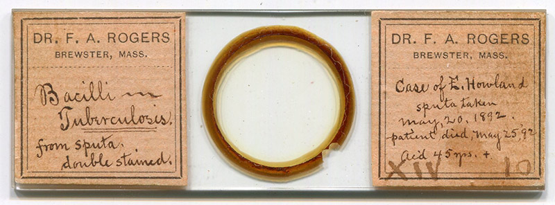
Figure 1. An 1892 microscope slide by Frank A. Rogers. Described as “bacilli in tuberculosis, from sputa, double stained”. The staining has faded over the years, although the bacteria can still be viewed with darkfield microscopy (Figure 2).
Frank Alvin Rogers, 1855 - 1940
by Brian Stevenson
last updated July, 2026
Dr. F.A. Rogers was a US physician, practicing in Maine, Georgia, Nebraska, and, from 1882 onward, in Massachusetts. He was an active member of the Massachusetts Medical Society and the American Microscopical Society. Rogers prepared professional-quality microscope slides, bearing custom-printed labels (Figure 1).
Figure 1.
An 1892 microscope slide by Frank A. Rogers. Described as “bacilli in tuberculosis, from sputa, double stained”. The staining has faded over the years, although the bacteria can still be viewed with darkfield microscopy (Figure 2).
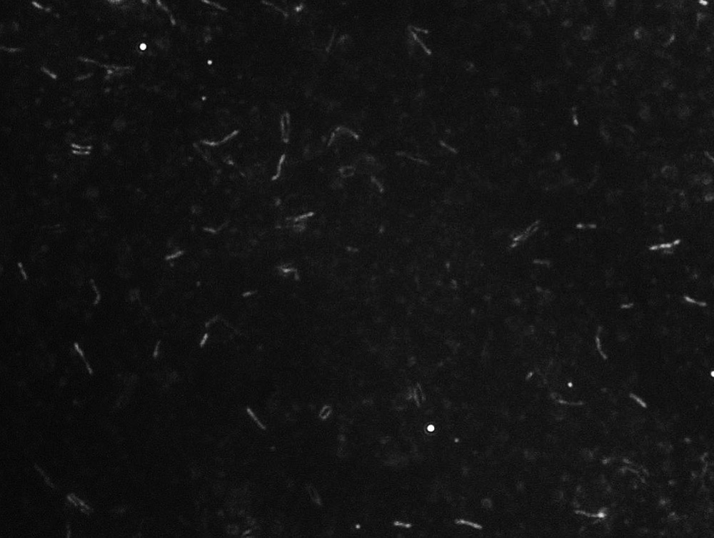
Figure 2.
Photomicrograph of Mycobacterium tuberculosis bacteria in the sputum sample from Elisha Howland, collected May 20, 1892, and mounted by Frank A. Rogers (see Figure 1). Courtesy of Nerina Jusufovic, imaged with a darkfield illumination, a 40x objective lens, and a C-mounted digital camera on an Olympus BX51 microscope.

Figure 3.
Death record of Elisha Howland, who died of “pthisis pulmonatis” (pulmonary tuberculosis) on May 24, 1892. Dr. F.A. Rogers collected a sputum sample from Howland on May 20 (Figures 1-2). The death was evidently reported to Rogers on the following day, such that he labeled the slide with a death date of May 25.
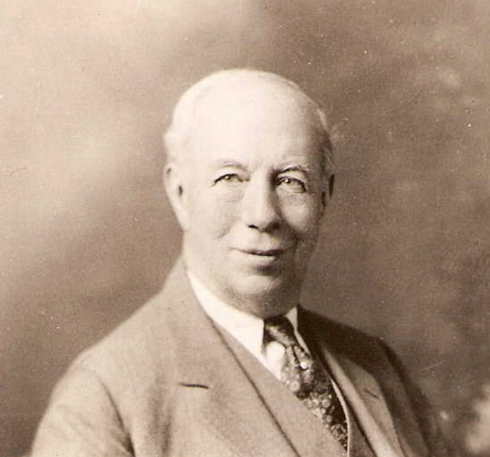
Figure 4.
Undated photograph of Frank A. Rogers.
Frank Alvin Rogers was born on October 8, 1855, in Newfield, Maine. He was the first of two childen of John A. and Julia A. (nee Nealey) Rogers. Father John was a Baptist minister. He died in 1866, and Frank and his sister were raised by the neighboring Durmells family.
Nonetheless, Frank received an excellent education, attending Limerick Academy (Limerick, Maine), and Kent's Hill Seminary (Maine Wesleyan Seminary, Readfield, Maine). He intended to become a minister, but changed his focus to medicine after entering Bowdoin College. He graduated with his M.D. in 1876, then began a practice in Bethel, Maine. Later that year, he married Lottie A. Bowker, with whom he had three children.
In 1877, Rogers sold his practice, and took on the position of Principal at Litchfield Academy, in Litchfield Corners, Maine. He moved to Atlanta, Georgia in 1879, to serve as Professor of Natural Science at Clark University. In 1880, Rogers opened a medical practice in Tekamah, Nebraska.
They returned east in 1882, setting up a medical practice and pharmacy in Brewster, Massachusetts. Rogers became a well-respected physician in the area, with an excellent reputation as a surgeon.
Rogers likely became familiar with microscopes during his medical training and practice. Microscopy was also an important skill for pharmacists, as one needed to precisely identify raw materials for use in drugs. Rogers joined the American Microscopical Society in 1890.
A local farmer named Elisha Howland developed active pulmonary tuberculosis in 1892, and Frank Rogers was evidently his physician. There was not much that could be done to treat tuberculosis at that time. Rogers collected sputum samples from Howland, hoping to observe a reduction in numbers of bacteria. The slide by Rogers that is shown above in Figure 1 notes that the specimen was “double stained”, presumably an acid-fast method such as the recently developed method of Franz Ziehl (1859-1926) and Friedrich Neelsen (1854-1898).
Rogers presented a pair of slides to the editors of The American Monthly Microscopical Journal in 1892, who then wrote: “Acknowledgment to Dr. F.A. Rogers, Brewster, Mass., for a remarkable slide, it being nothing less than a longitudinal section of a human foetus of eight weeks. The specimen was stained in bulk by borax-carmine, embedded in paraffine, sectioned on a Bausch & Lomb Student microtome to 1-2000 inch, fixed on the slide by an original and special method, stained as a contrast with sulphindigotate of soda, and mounted in balsam, using King's cement for ringing. To cut so large an object demands considerable care and skill, and the entire preparation is laborious and time consuming, but the result is worthy of attentive study. Dr. Rogers has also kindly sent a section of the hand of the same foetus, showing the developing muscle and bone. These preparations are the finest of the kind that we have ever seen.”
Makers of microscope slides often gave samples of their productions to editors of popular scientific magazines, in hopes of the positive attention mentioned above. Professional slide-makers would expect to increase sales, and amateurs could anticipate opportunities to exchange slides with other microscopists who lived great distances away. There are no indications that Rogers sold his mounts, so he was presumably looking for exchanges. That was probably what led to the slide shown in Figure 1 becoming part of a collection of works by other mounters.
He was also an excellent photographer. The Microscopical Bulletin and Science News acknowledged a donation from Rogers in 1890, “Dr. F.A. Rogers, Brewster, Mass., sends us a photograph (bromide print) which does much credit to the skill of one who only claims to be a beginner. The object, magnified about 1000, is a vertical section through the apex of the molar of a young mouse. The lens used was our one-fiteenth oil immersion; the section carmine-stained; and the plate a Carbutt special”. Rogers presumably also prepared the specimen, which would have included carefully grinding both sides of the tooth to make a thin, flat surface, and requires more than average competence.
Rogers published a two-part paper on “Photomicrography” in The Microscope in 1893.
The USA developed a club similar to the UK’s Postal Microscopical Society, in which boxes of a dozen or so slides were shipped along a circuit of numerous microscopists, so that members could examine a variety of specimens and mounting methods. Members were expected to contribute slides from time-to-time. In 1894, a group by Frank Rogers was described thusly: “The labels on a box of slides contributed to the Postal club by Dr. F.A. Rogers are beautifully etched, even to a neat border. This is the way Dr. Rogers says the work is done. Coating a clean slide with wax, heated so that it is evenly distributed, he covers the other side of the slide with vaseline after having traced the design with a needle in the wax. The slide is then placed wax side down over a vessel containing powdered fluorspar, one teaspoonful to half an ounce of strong sulphuric acid, and allowed to remain over night. In the morning remove the wax with turpentine or chloroform and the etching is done. Dr. Rogers uses a shallow lead dish, say one half an inch deep by four inches long and nearly three wide. This holds four slides at once.”
During the mid-1890s, the Rogers family moved across Cape Cod to Chatham, with Frank pursuing medical practice in that town.
Rogers had a diversity of intererests. In 1898, he contributed a paper on “The eye of Pecten irradians or the scallop” to The American Monthly Microscopical Journal. It was accompanied by drawing by Rogers, produced with a microscope and camera lucida (Figure 5).
He spoke to the Massachusetts Optical Society in 1910: “F.A. Rogers, M.D., professor of pathology and bacteriology, spoke on diseases of the conjunctiva and iris and how recognized. He took up the various diseases and gave a very interesting talk on the way to recognize them. At the conclusion of his speech, which occupied a trifle over an hour, he was given a rising vote of thanks. It might be said in passing that this lecture was one of the strongest that has ever been delivered before the Massachusetts Optical Society. It was delivered by a medical doctor who understood what he was talking about, and the members were enthusiastic over the way the lecture was given. There was talk of having him come back again to lecture on other conditions of the eye. It may be possible that Doctor Rogers will be secured again to appear before the society at an early date.”
Frank Rogers died on November 15, 1940, in Boston.
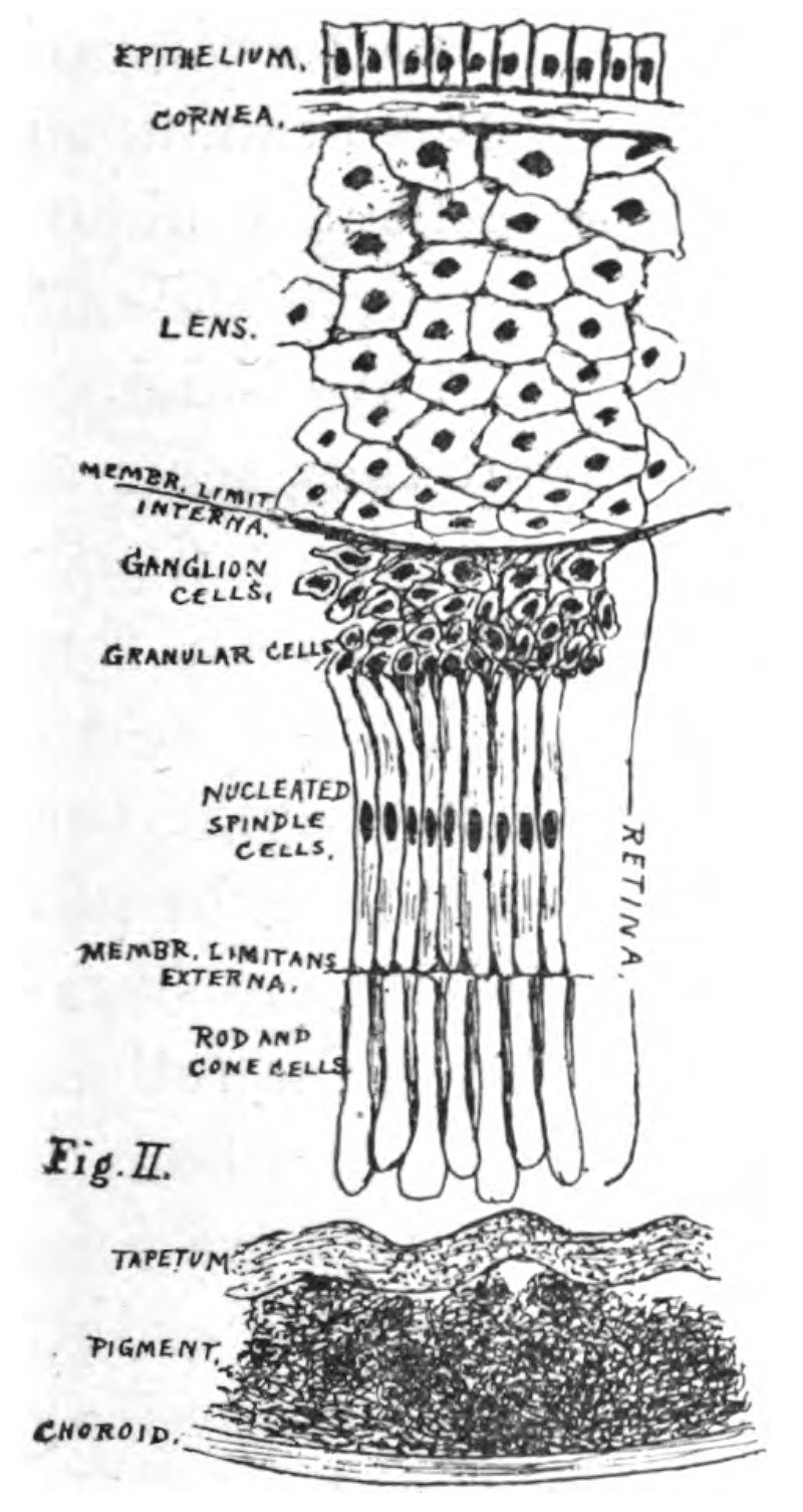
Figure 5.
“Diagramatic sketch of the arrangement of the internal parts of the eye (of a scallop), from one pole to the other”. Drawn by Frank Rogers with microscope and camera lucida, to accompany his 1898 paper on “The eye of Pecten irradians or the scallop”. From “The American Monthly Microscopical Journal”.
Acknowledgement
Many thanks to Nerina Jusufovic for her enthusiasm and patience in finding and photographing Mycobacteria tuberculosis bacteria in the Rogers-Howland microscope slide.
Resources
The American Monthly Microscopical Journal (1890) Election of new members, Vol. 11, pages 197-198
The American Monthly Microscopical Journal (1892) Acknowledgement of gift of slides from F.A. Rogers, Vol. 13, page 86
Death record of Elisha F. Howland (1892) accessed through ancestry.com
Deyo, Simeon L. (1890) History of Barnstable County, Massachusetts, H.W. Blake, New York, pages 221-222 and 241-242
The Microscopical Bulletin and Science News (1890) Acknowledgement of photograph by F.A. Rogers, page 44
The Observer (1894) Notes on F.A. Rogers’ slides for the American Postal Microscopical Club, Vol. 5, page 27
Optical Age (1910) Report of the November 15 meeting of the Massachusetts Optical Society, page 1179
Reed, Wallace Putnam (1889) History of Atlanta, Georgia: With Illustrations and Biographical Sketches of Some of Its Prominent Men and Pioneers, D. Mason & Company, Syracuse, New York, page 372
Rogers, Frank A. (1893) Photomicrography, The Microscope, pages 5-8 and 21-25
Rogers, Frank A. (1895) The histogenesis of the plasmodium malariae, The Boston Medical and Surgical Journal, pages 125-130
Rogers, Frank A. (1898) The eye of Pecten irradians or the scallop, The American Monthly Microscopical Journal, Vol. 19, pages 49-60
Stevenson, Brian (2026) A farmer, a physician, a fatal case of tuberculosis, and a historical microscope slide, Quekett Bulletin, issue 90, April, pages 10-12
US census and other records, accessed through ancestry.com