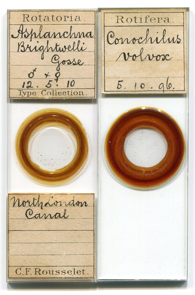
Figure 1. Microscope slides that were made by Charles Rousselet, dated 1910 and 1896.
Charles Frederic Rousselet, 1854 - 1921
by Brian Stevenson
last updated April, 2023
Charles Rousselet was an early expert in Rotifers, and developed a number of apparatus to aid with examination of those and other microscopical objects. Among these were a compressor and a live-box, both of which were commercially produced well into the twentieth century. He also developed at least two unique types of microscopes. Rousselet was an active member and officer of the Royal Microscopical Society and Quekett Microscopical Club for many years.
Figure 1.
Microscope slides that were made by Charles Rousselet, dated 1910 and 1896.
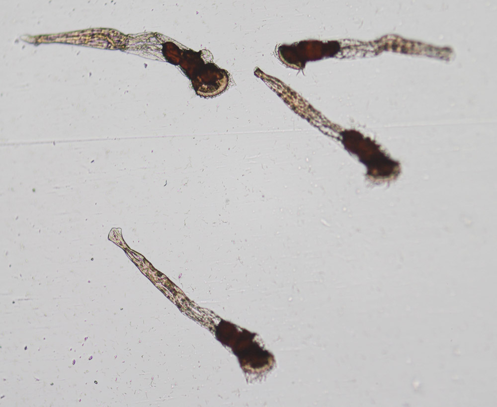
Figure 2.
Specimens of Conochilus volvox, prepared by C.F. Rousselet on October 5, 1896 (see Figure 1). Photographed with a 10x objective lens and C-mounted digital SLR camera on a Leitz Ortholux II microscope.
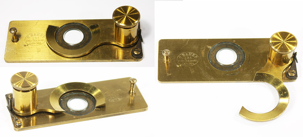
Figure 3A.
“Rousselet’s Compressorium”, original 1893 model. It was produced and sold by Charles Baker. As described in “The Journal of the Royal Microscopical Society” (1893), “The glass tablet being fixed flush with the lower part of the brass slide, which is slightly countersunk, allows high-angled condensers to be used to the very edge of the tablet for the illumination of the objects. The arm carrying the cover-glass is raised and lowered by screw adjustment, and is held in position by a spring catch, but can easily be turned aside. The arm moves parallel to the glass tablet, so that very small free-swimming animals can readily be caught and held fast between the two glasses. The thin cover-glass is cut off at the top, thus allowing reagents to be added to the drop of water while the animals are under examination; it is much larger than the glass tablet and therefore allows the highest powers to be used all over the field and to the very edges of the glass tablet, which is of great importance in practical work”. Although the glass coverslip is attached by glue, Rousselet noted that, “The removal of a broken cemented cover is … readily effected by slightly heating it over a lamp”.
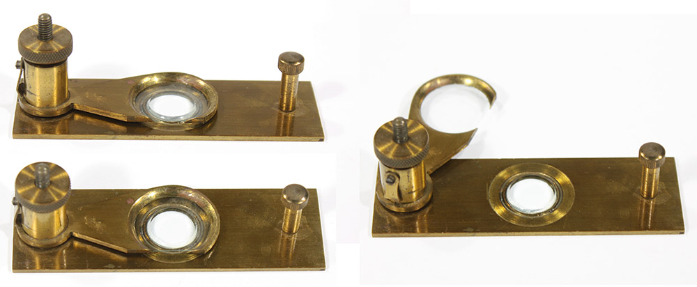
Figure 3B.
Another example of the original design of “Rousselet’s Compressorium”, unsigned.
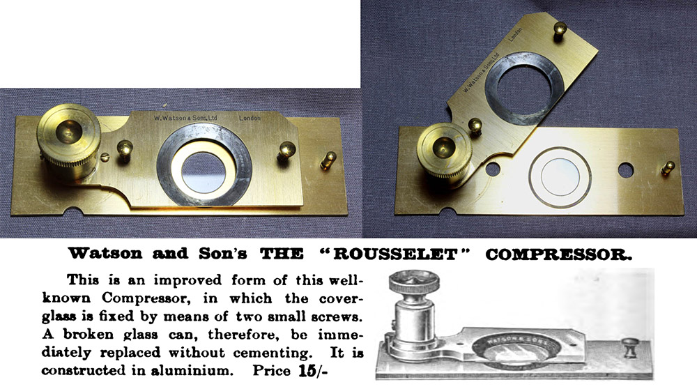
Figure 4.
An “improved” "Rousselet Compressor”, as produced by Watson & Sons, and an 1896 advertisement. This version avoided the difficulties of gluing the coverslip and melting old glue to remove coverslips by attaching a standard round glass slip via a pair of screws. To deal with the height of the screw heads, a pair of holes were drilled through the base to fit them. As Rousselet complained (1905), “These screws have introduced a fresh defect, because the arm has to be raised considerably before it can be turned aside. There is absolutely no reason why any one wanting such a compressor should not have it; but I think I must ask these opticians to be good enough to call their modified model by some other name, for I have always disapproved of it for the reasons stated above, and it is not my model and very much less an ‘improved’ form of it.” Compressorium images adapted by permission from https://microscope-antiques.com/gvhrousselet2.html
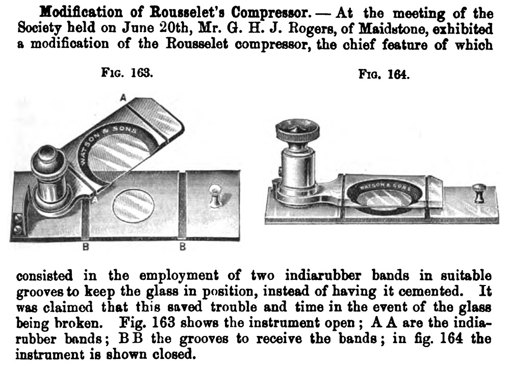
Figure 5.
Another “improved Rousselet’s Compressor”, as designed by George H.J. Rogers, and described in “The Journal of the Royal Microscopical Society”, 1900. Rogers attached the coverslip by two rubber bands, and cut two grooves in the base to fit the bands. Rousselet also objected to this design, “ in the first place, the cover-glass was no longer sufficiently firm and rigid for delicate manipulation; and secondly, the compressorium became quite unsuitable for use with water and oil immersion lenses, as these fluids could run under the brass ring and eventually mix with the water on the slide. The removal of a broken cemented cover is so readily effected by slightly heating it over a lamp, that no modification is required on that account - none at least that cancels two very essential qualities my model.”
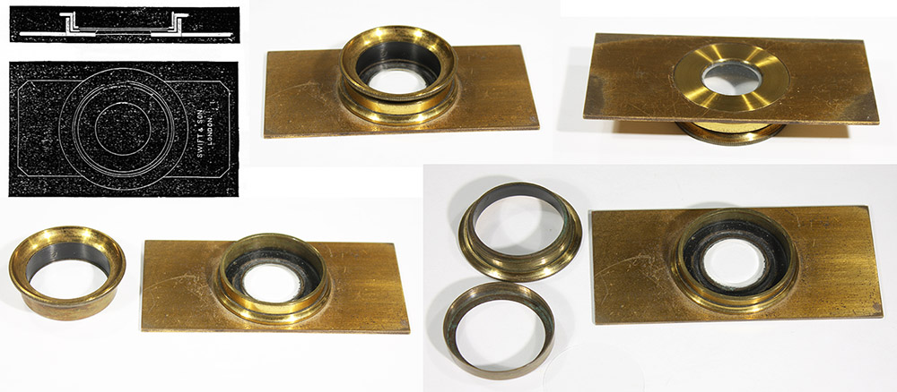
Figure 6.
“Rousselet’s Live-Box”, which he first described in 1887. Important features of his design are placing the bottom glass near the base of the live-box, which allows the microscope condenser to reach as close to the apparatus as desired, and a top that is much wider in diameter than the bottom glass, which provides enough space for a high-power objective lens to be placed close to the top glass and also scan the entire field of view. This example, manufactured and sold by C. Baker, has a two-part top, which screws together to hold a 30mm coverslip between the two parts. The top piece then slips into the base, which presses the coverslip against the bottom glass.
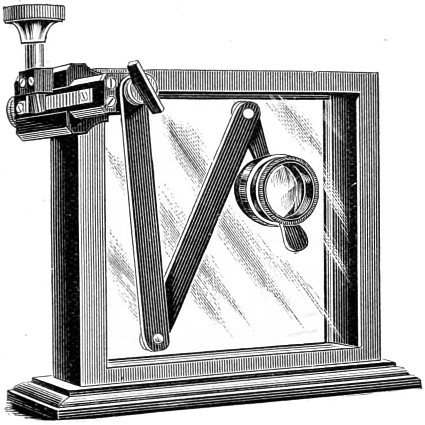
Figure 7.
“Rousselet’s Tank Microscope”, described by him in 1889, and subsequently produced for retail by C. Baker. Rousselet wrote, “I desire to bring to your notice a small tank microscope, designed for the purpose of rapidly looking over pond water and weeds, collected at a day’s excursion and placed in a small parallel-sided window aquarium… One of Zeiss’ aplanatic lenses is carried on a jointed arm, which moves parallel to the side of the tank, and the lens is focused by means of a rack and pinion, the whole being fixed to the upper left-hand corner of the tank by means of a screw clamp… The definition of these aplanatic lenses is excellent; the lowest power has enough working distance to focus through all my tanks, and the magnification (6 diam.) is sufficient to permit of the identification of all ordinary rotifers, and anything uncommon, or new, is at once recognized. Such delicate creatures as the Floecules, which are all but invisible with the ordinary pocket lens, are seen without difficulty, and the whole contents of the tank can be ascertained with a great saving of time.”
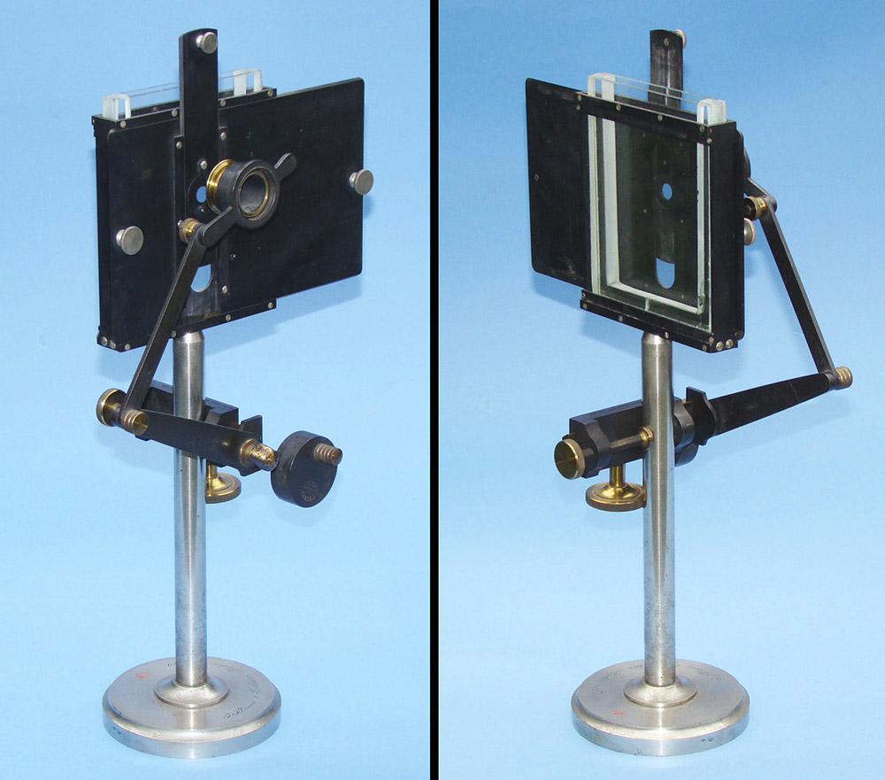
Figure 8.
A surviving “Rousselet’s Tank Microscope”, signed "C. Baker, 244 High Holborn, London", ca. 1900. It has been modified such that it clamps onto a stand, rather than on the tank as originally designed (See Figure 7). This modification may have been done by an owner, as the stand is signed "J. Klönne and G. Müller, Berlin, No. 2110" and the tank more closely resembles a Klönne and Müller observation tank than those sold by Baker. Adapted for educational, nonprofit purposes from an antiques catalogue.
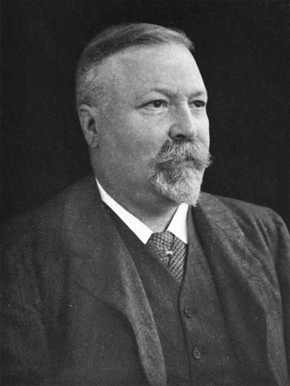
Figure 9.
Charles Frederic Rousselet, adapted from his obituary in “The Journal of the Royal Microscopical Society”, 1921.
Charles Frederic Rousselet was born on October 10, 1854, in Friedrichsdorf, Hesse (Germany). His naturalization papers give his parents’ names as Louis Frederic and Elise Judith Rousselet. His obituary on The Journal of the Royal Microscopical Society stated, “Rousselet came of an old Huguenot family, and his direct ancestor, Esaïe Rousselet, left his home at Perrière, near Soissons, after the revocation of the Edict of Nantes. He and several others, some thirty families in all, founded a French settlement afterwards named Friederichsdorf, from the Landgrave of Hesse-Homburg, Friederich II. Here their descendants have continued to live, speaking French as their mother-tongue, to the present day.”
The JRMS further noted, “Mr. Rousselet came to London in 1873 and was naturalized in 1889. During the years 1881-1902 he was the London representative for the Bordeaux (i.e. wine) firm of A. de Luze et fils.”
He joined the Quekett Microscopical Club on January 26, 1883, and became a Fellow of the Royal Microscopical Society in 1888.
In 1887, Rousselet demonstrated to the QMC two pieces of apparatus that he had made to his order: a new form of live box that (see Figure 6) and a portable binocular microscope. I have not seen any pictures of his microscope, but he did describe it in the Journal of the Quekett Microscopical Club. It had 8 inch body tubes (“which is as short as they could be made without causing a strain to the eyes”), “the diameter of the tubes is only a little over one inch, but the tubes carrying the eye-pieces are wider at the top, and receive the B eye-pieces of my large microscope; this is of importance, because with small eyepieces the field of view is greatly reduced, and the result is not so satisfactory. The stage is simple, and has a broad but very thin sliding-stage, which moves very smoothly. The condenser, also, belonging to my large microscope fits in an adapter, which is screwed on to the stage below, and it is focussed by a spiral motion, which brings it up just above the stage if required. There is no tail-piece, the mirror being carried by a double-jointed arm fixed at one corner of the stage, which allows it to be brought above the stage for the illumination of opaque objects; for this purpose the concave mirror has a very short focus (1 1/2 in.), and acts as a very effective silver side-reflector. The focussing is effected by a good coarse rack-work; a fine adjustment was not required for my purpose. … The whole is packed into a small case, which, when quite complete, weighs just under 6 lbs.”
Rousselet’s other 1887 invention was a new form of live-box (Figure 6). A key feature is that the bottom glass is considerably narrower in diameter than is the upper glass. The purpose of this was explained by Rousselet, “It is very frequently desirable to examine an object with a high power after it has been found with a low one, and we all know how very fond living creatures are of getting to the edge of the drop of water in which they are placed, and to shift them to the centre is frequently a very tedious work, and often fatal to the animal. To remedy this defect, I have had a live box constructed in which the glass tablet is somewhat reduced in diameter, but the outer ring is enlarged sufficiently to allow any high power to focus to the very edge of the glass tablet, and the result is very satisfactory. An object lying anywhere in the live box can be reached by the condenser from below, and by both low and high powers from above; besides which, it acts as a very good compressor, capable of fixing, without hurting, the smallest rotifers, and when you know how to do this, it is also possible to get a rotifer in so small a drop of water that is unable to swim out of the field of view of a quarter inch objective. I have had it in constant use for animals of all sizes, from the smallest infusoria to tadpoles, and I hope it will be found equally useful to my fellow pond-hunters.” This live-box was later manufactured and retailed J. Swift & Son.
Two years later, in 1889, Rousselet unveiled another invention to the QMC. “Rousselet’s Tank Microscope” (see Figures 7 and 8) was a fairly simple device that allowed the user to easily search through a large aquatic sample to identify interesting specimens, which could then be removed with a pipet for closer examination in a live-box. This invention was produced on a large scale and retailed by C. Baker.
Rousselet first exhibited his famous compressorium in 1893, to the Royal Microscopical Society. It was described as being “devised for the purpose of facilitating the examination of minute living and free-swimming animals, such as rotifers, infusoria, &c.” Baker also produced and sold this instrument.
An awkward aspect of Rousselet’s compressorium is that the coverslip is glued to the underside of the arm, so that if the coverslip cracks, one needs to remove the broken glass by scraping off or melting the glue, then gluing a new coverslip in place. At least two variants of were soon developed by other microscopists, which attached the coverslip by other means. Both were retailed by W. Watson & Son (Figures 4 and 5).
Rousselet responded to these “improvements”: “I must ask these opticians to be good enough to call their modified model by some other name, for I have always disapproved of it …, and it is not my model and very much less an ‘improved’ form of it.”
During 1905, Rousselet traveled to South Africa to attend the joint meeting of the British and South African Associations for the Advancement of Science. The trip included collecting excursions to Victoria Falls, Zimbabwe, and Mozambique.
Rousselet’s health began to fail in 1916. During that year, The Journal of the Quekett Microscopical Club wrote, “The Committee would like to give some expression - in which they are sure the members will join - to their sorrow at the serious and long-continued illness of Mr. C.F. Rousselet, and to their sympathy with him." In 1917, the J.Q.M.C. added, “The chairman announced that Mr. C.F. Rousselet had presented to the club several portraits, and also a further collection of photographs of drawings of Rotifera to be added to those he had already given. He said that Mr. Rousselet's health was slightly improved, and expressed the hope that it might in time be sufficiently restored to enable him again to attend the meetings of the Club.”
Despite his poor health, Rousselet prepared a 9-page paper on collecting and mounting Rotifera for The Journal of the Quekett Microscopical Club, published in 1917.
Charles Rousselet died on October 15, 1921.
A.W. Shephard wrote this obituary for The Journal of Royal Microscopical Society:
“It is with great regret we have to record the death of Mr. C.F. Rousselet, who for over a quarter of a century was a familiar figure at our meetings. He was born at Friederichsdorf in 1854, and died on October 25, 1921, after a long illness of several years.
Rousselet came of an old Huguenot family, and his direct ancestor, Esaïe Rousselet, left his home at Perrière, near Soissons, after the revocation of the Edict of Nantes. He and several others, some thirty families in all, founded a French settlement afterwards named Friederichsdorf, from the Landgrave of HesseHomburg-Friederich II. Here their descendants have continued to live, speaking French as their mother-tongue, to the present day. Mr. Rousselet came to London in 1873 and was naturalized in 1889. During the years 1881-1902 he was the London representative for the Bordeaux firm of A. de Luze et fils.
Even before the publication of Hudson and Gosse's ‘Rotifera’, Rousselet's general interest in this group had given place to serious study, and by unremitting research he soon became the recognized authority on the subject. His contributions to the Journal of this Society, as well as of that of the Quekett Microscopical Club, embodying the results of his original observations, were as valuable as they were numerous, and resulted in considerable additions to the number of known species. His work was very methodical and exact, and his descriptions were always accompanied by accurate drawings representing both the species and the details necessary for its determination. In this way Rousselet set an excellent example to the band of workers he gathered round him. His method of preservation and mounting is perhaps more widely known than his descriptive work; and the process of “ narcotizing which he introduced, or some modification of it, is now practised in all laboratories where biological work is in progress. His last paper, ‘Some Further Notes on Collecting and Mounting Rotifera’, was published in Vol. 13, Journal of the Quekett Microscopical Club, p. 321 (October 23, 1917), where a bibliography is given of his various papers in this subject, in which he achieved such marked success.
He was elected to the Fellowship of the Royal Microscopical Society in 1888, and served for many years on the Council. In January 1898 he was appointed Honorary Curator, and in this capacity his knowledge of the history of the microscope enabled him to render valuable service to the Society in the preparation of the Catalogue of the Collection of Instruments. He held office until December 1918, when increasing physical distress compelled him to relinquish the work. In January 1917 Mr. Rousselet presented to the Society an extensive collection of reprints, etc., which he had received from his many correspondents in all parts of the world. These papers, numbering about a thousand, form probably the most valuable collection of papers on the Rotifera available to workers for reference in this country.”
The Journal of the Quekett Microscopical Club reproduced much of the JRMS obituary, adding, “He served the Club for many years in the office of Foreign Correspondent and also as Vice-President, and was a frequent attendant at both our meetings and excursions, where his expert knowledge was always freely available.”
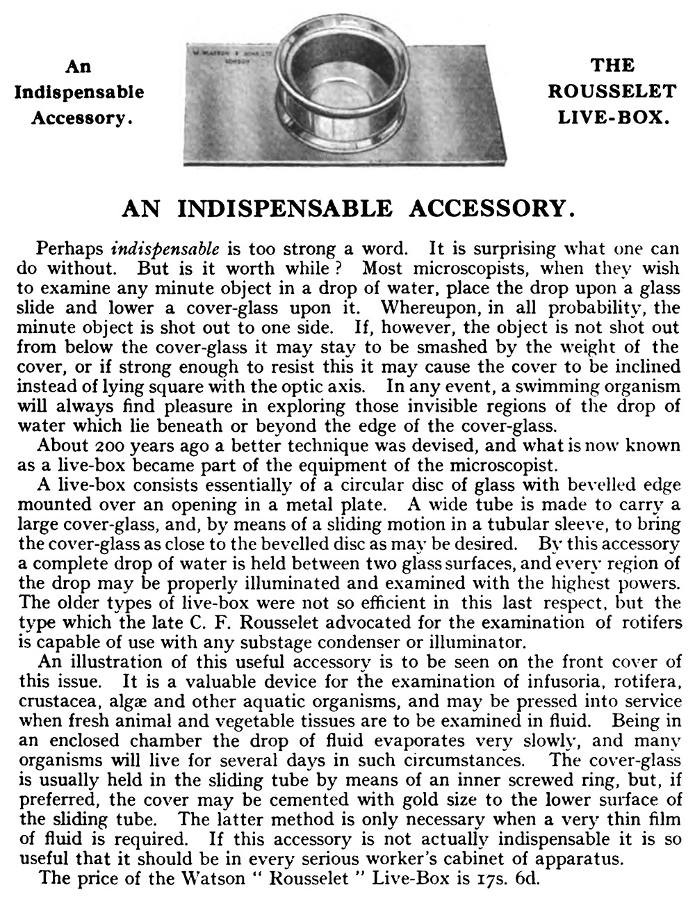
Figure 10.
Description of Rousselet’s live-box, from “Watson’s Microscopical Record”, 1930.
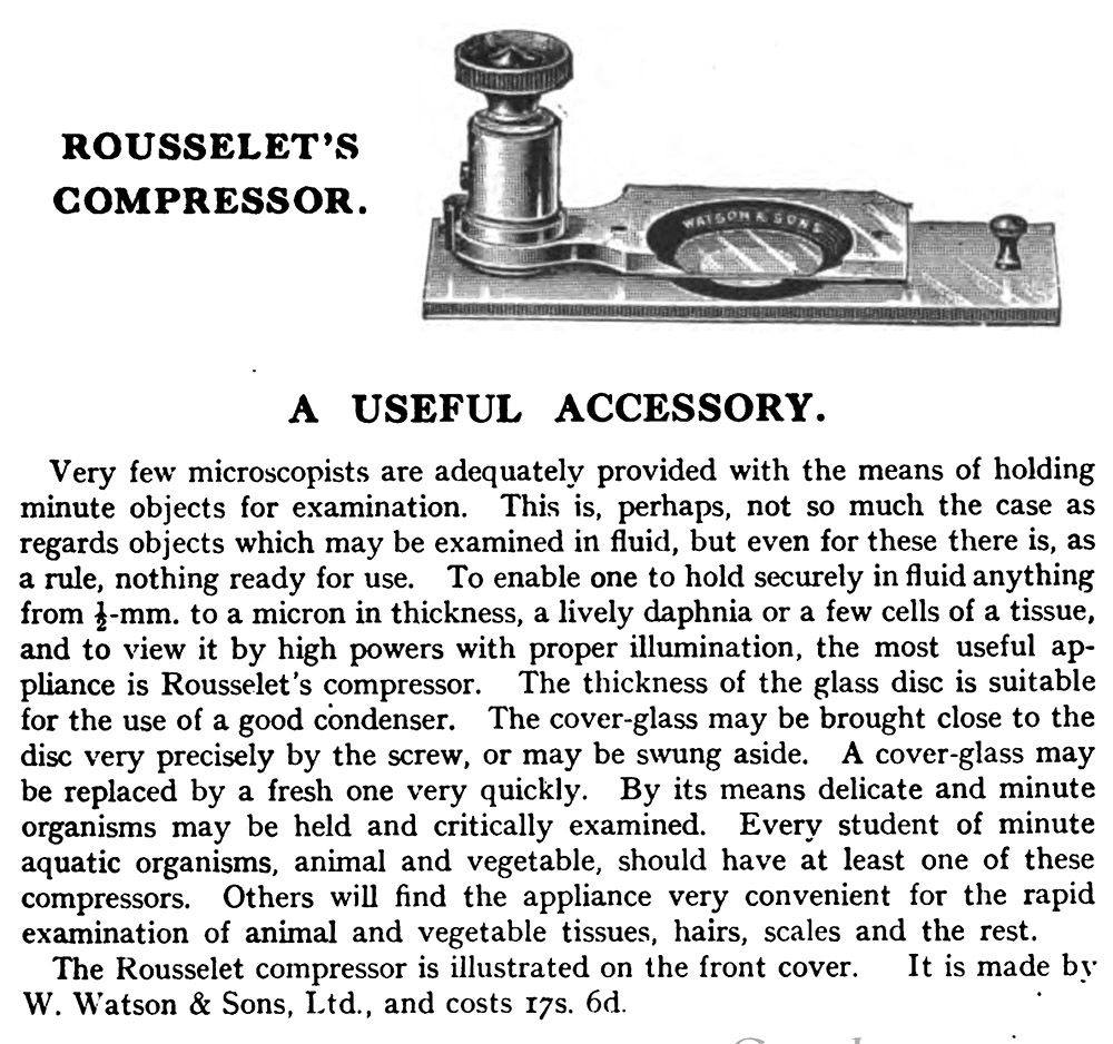
Figure 11.
Description of Watson & Sons’ version of “Rousselet’s compressorium”, from “Watson’s Microscopical Record”, 1930.
Acknowledgements
Thank you to Barry Sobel for permission to use images from https://www.microscope-antiques.com/rouss.html, and to Joe Zeligs for helpful insights.
Resources
Bracegirdle, Brian (1998) Microscopical Mounts and Mounters, Quekett Microscopical Club, London, pages 81 and 166, and Plate 31-N
Cross, M.I. and M.J. Cole (1895) Live cages, in Modern Microscopy, Bailliere, Tindall, and Cox, London, pages 102-103
Eliot Merlin, A.A.C. (1904) Note on a modification of the Rousselet live-box, Journal of the Quekett Microscopical Club, pages 169-170
England census and other records, accessed through ancestry.com
International Journal of Microscopy and Natural Science (1896) Advertisement from Watson and Sons, Vol. 15
Journal of the Quekett Microscopical Club (1883) Members, “Jan. 26, 1883, Rousselet, Charles, 42, Welbeck street, W.”
Journal of the Quekett Microscopical Club (1887) Members, “Jan. 26, 1883, Rousselet, Charles, 308, Regent street, W.”
Journal of the Quekett Microscopical Club (1887) Members, “Jan. 26, 1883, Rousselet, Charles, F.R.M.S., 308, Regent street, W.”
Journal of the Quekett Microscopical Club (1891) Members, “Jan. 26, 1883, Rousselet, Charles, F.R.M.S., 27, Great Castle street, Regent street, W.”
Journal of the Quekett Microscopical Club (1899) Members, “Jan. 26, 1883, Rousselet, Charles F. (Hon. Secretary for Foreign Correspondences), Curator R.M.S., 2, Pembridge Crescent, Bayswater, W.”
Journal of the Quekett Microscopical Club (1916) page 300
Journal of the Quekett Microscopical Club (1917) page 390
Journal of the Quekett Microscopical Club (1921) Charles Frederic Rousselt, F.R.M.S., 1854-1921, pages 379-380
Journal of the Royal Microscopical Society (1893) Rousselet’s new compressorium, pages 386-387
Journal of the Royal Microscopical Society (1900) Modification of Rousselet’s compressor, page 635
Knowledge (1905) The Quekett Microscopical Club, page 69
https://www.microscope-antiques.com/rouss.html (accessed April, 2023) Rousselet compressorium
Naturalization papers of Charles Rousselet (1889) accessed through ancestry.com
Nelson, E.M. (1912) The Rousselet compressor, Journal of the Quekett Microscopical Club, pages 511-513
Probate of the will of Charles Rousselet (1922) “Rousselet Charles Frederic of 15 Cloudesley-road St Leonards-on-Sea died 15 October 1921 Administration (with Will) London 20 January to Cecile Pauline Emilie Rousselet widow. Effects £2802 2s 2d”, accessed through ancestry.com
Rousselet, Charles (1887) On a small portable microscope and a live box, Journal of the Quekett Microscopical Club, pages 175-176
Rousselet, Charles (1889) On a simple tank microscope, Journal of the Quekett Microscopical Club, pages 53-54
Rousselet, Charles (1892) On Conochilus unicornis and Euchlanis parva – two new rotifers, Journal of the Quekett Microscopical Club, page 367
Rousselet, Charles (1905) A description of the Rousselet compressorium, Journal of the Quekett Microscopical Club, pages 137-140
Rousselet, Charles (1908) Pond-life collecting for the microscope, Knowledge, Vol. 26, pages 188-189
Rousselet, Charles (1912) Comments on E.M. Nelson’s paper “The Rousselet compressor”, Journal of the Quekett Microscopical Club, page 513
Rousselet, Charles (1917) Some further notes on collecting and mounting Rotifera, Journal of the Quekett Microscopical Club, pages 320-328
Sheppard, A.W. (1921) Charles Frederic Rousselet, Journal of the Royal Microscopical Society, pages 382-383
Watson’s Microscope Record (1930)