Ernest Henry Shaw, 1867 - 1956
by Brian Stevenson
last updated October, 2019
English surgeon and pathologist Ernest H. Shaw was an early advocate for immediate histological examination of tumor biopsies during operating sessions. He recommended that stopping a surgery for the few minutes required to prepare and examine a tissue under a microscope was an efficient way to determine how to continue. In 1923, he wrote to The Lancet:
“It is now 12 years since I ventured to draw the attention of the medical profession to the value of immediate microscopic diagnosis of tumours at the time of operation. After an experience of a practice of the method which now extends over a period of 23 years, I have no hesitation in again urging its great value, in the hope of making its use more widely known and more appreciated.
The method of examination which I practise and for which I claim great advantages, is very simple … The whole process, including freezing, cutting, mounting, staining, and examining of a section, can be accomplished, when the pathologist is well acquainted with histology and morbid anatomy, in the short space of five minutes or less. It will be seen, therefore, that the delay in the performance of an operation is very short and is amply compensated for by the information gained as to the exact nature of the tumour with which the surgeon has to deal. The accurate diagnosis of a tumour cannot always be made from clinical evidence alone, and the aid of the microscope is often required”.
The microscope slide shown in Figure 1 is dated November 26, 1900, a few years before he competed his medical training. It is finely finished and includes a customized label, suggesting that it was a special slide, possibly for his personal use or as an exchange with another practitioner.
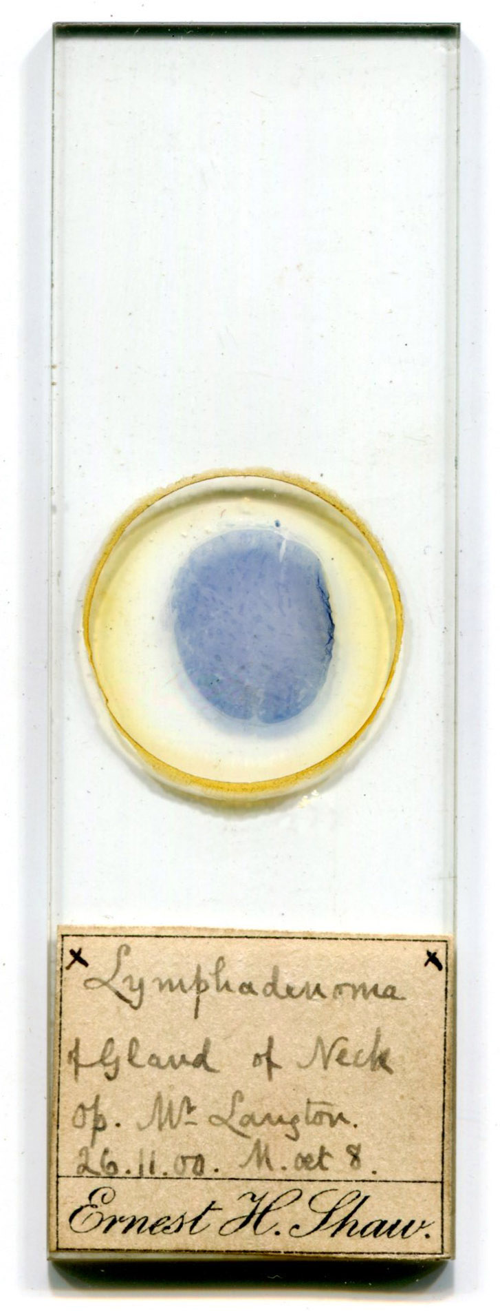
Figure 1.
A microscope slide by Ernest H. Shaw, dated November 26, 1900, labeled “Lymphadenoma of Gland of Neck” (i.e. cervical lymph node). It also notes that the operation was performed by “Mr. Langton”, probably John Langton (1840-1910), surgeon and lecturer at Saint Bartholomew’s Hospital, London, where Ernest Shaw was trained. Shaw completed his medical training a few years afterward, so this would have been one of his very earliest of slides.
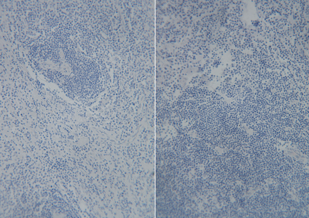
Figure 2.
Magnified views of the specimen on the slide shown in Figure 1. Based on Shaw’s 1907 publication, the stain is probably Loeffler’s methylene blue (see Figure 3). With the caveat that I am not a pathologist or a histologist, and so may have missed photographing key features, an examination of these and other photographs of the specimen by a professional pathologist, Jeffrey Silverman, led him to conclude, “this looks like a benign, hyperplastic lymph node. The normal nodal architecture of cortex and medulla is effaced and replaced by sheets of benign looking lymphocytes. The darker areas are reactive germinal centers and the paler rounded structures are blood vessels cut in transverse plane or, if more elongated, longitudinal plane. The node does not look like it harbors a malignant lymphoma but looks reactive. Of course, the precise diagnosis of lymphoma, which is a huge family of malignancies, requires perfectly fixed and stained sections and even then requires immunohistochemical staining for accurate classification”. Imaged with a C-mounted digital SLR camera and a 25x objective lens.
Ernest’s father, Henry Shaw, was a customs official, and moved several times during his career. Ernest was the eldest child, born on May 30, 1867, while his parents were stationed in Cork, Ireland. The little family soon moved to India: sister Edith was born in August, 1869 in Benares (now Varanasi, Uttar Pradesh). The Shaws were soon reassigned to Murree (now in Pakistan), when son Frederic was born in August, 1871. This was followed by a short move to Nowshera (also now in Pakistan), where Bessie was born in July, 1872, then back to Murree, where Charles was born in January, 1875. The family returned to England and settled in Lambeth, Surrey, where the sixth child was born in 1878.
Ernest was working as an assistant in a museum in 1891, when he married Clara Coughtrey in London. Son Edgar was born there in 1892, while daughter Jessie was born in Cambridge in 1899. They were back in London soon afterward, with Ernest studying medicine at Saint Bartholomew’s Hospital.
The pathology slide shown in Figure 1 is dated November 26, 1900. The 1901 census listed the family in the Saint Paul area of London, and Ernest was recorded as being a “medical student”.
Shaw was receiving training in microscopical analysis of pathological specimens. He coauthored “An analysis of the tumours of the breast removed at St. Bartholomew’s Hospital during the year 1900”, along with H. Morley Fletcher, M.D. (Shaw did not have any initials in suffix). They noted that “A microscopical examination was made in all cases, and in many instances sections were made from several parts of the same tumour; the glands were also cut in all cases in which they were removed”.
Ernest Shaw became a Member of the Royal College of Surgeons in July, 1905. Later records indicate that he went on to become a Fellow of the R.C.S. He became a Member of the Royal College of Physicians in 1910.
He was part of a demonstration to the Hampstead Division of the British Medical Association in 1907. C.B. Lockwood, F.R.C.S., Surgeon to St. Bartholomew’s Hospital, gave a presentation on “The immediate microscopical diagnosis of tumours during the course of operations”. Lockwood announced that “Mr. Ernest Shaw is present to-night to demonstrate to you how easily and quickly a perfect section of a tumour can be placed under the microscope, and how certain that makes the diagnosis”. Shaw’s method is reprinted as Figure 3, below.
Shaw published a sole-authored paper in The Lancet in 1910 on the same topic, “The immediate microscopical diagnosis of tumors at the time of operation”. He wrote another paper with essentially the same title for The Lancet in 1923.
He was appointed Director of Clinical Pathology at Great Northern Central Hospital, London, in 1911.
Ernest Shaw died in 1956. The British Medical Journal his death, “On September 26, 1956, Midhurst Avenue, Fortis Green, London, N., Ernest Henry Shaw, F.R.C.S., M.R.C.P., aged 89”.
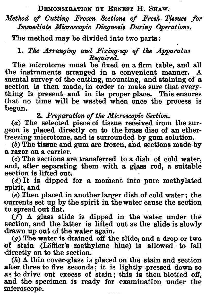
Figure 3.
Ernest Shaw’s method for preparing tissue sections during a surgery. From “The British Medical Journal”, 1907.
Acknowledgement
Thank you to Jeffrey Silverman for his expert opinions on Shaw’s pathology specimen.
Resources
Blackhall-Morison, Alexander, and Ernest Henry Shaw (1920) Cardiac and genito-urinary anomalies in the same subject, Journal of Anatomy, Vol. 54, pages 163-165
The British Medical Journal (1910) Obituary of John Langton, F.R.C.S., Vol. 2, page 918
The British Medical Journal (1956) Obituary of Ernest Henry Shaw, page 950
Burfield, J., and Ernest H. Shaw (1905) A case of bilharzia infection of the vermiform appendix, The Lancet, pages 368-370
England census and other records, accessed through ancestry.com
Fletcher, H. Morley, and Ernest H. Shaw (1902) An analysis of the tumours of the breast removed at St. Bartholomew’s Hospital during the year 1900, Saint Bartholomew’s Hospital Reports, Vol. 37, pages 295-308
The Lancet (1901) Examining board in England by the Royal Colleges of Physicians and Surgeons, pages 497-498
The Lancet (1901) “Shaw, Ernest H., M.R.C.P. Lond., has been appointed Director of Clinical Pathology at the Great Northern Central Hospital,. Holloway-road”, pages 1109-1112
A List of the Fellows, Members, Extra-Licentiates and Licentiates of the Royal College of Physicians of London (1913) Members, 1910, “Ernest Henry Shaw, 355, Camden-road, Holloway, N.”, page 52
Lockwood, C.B. (1907) The immediate microscopical diagnosis of tumours during the course of operations, The British Medical Journal, Vol. 1, pages 127-129
London Medical Press and Circular (1905) Royal College of Surgeons, England - New Members, Vol. 131, page 129
Marriage record of Ernest Henry Shaw and Clara Coughtrey (1891) Parish records of St. James, Bermondsey, accessed through ancestry.com
Shaw, Ernest H. (1910) The immediate microscopical diagnosis of tumors at the time of operation, The Lancet, page 939
Shaw, Ernest Henry (1917) Types of micro-organisms met with in the female genito-urinary tract, in The New System of Gynaecology, Thomas W. Eden and Cuthbert H.J. Lockyer, eds, Macmillan, Toronto, pages 97-117
Shaw, Ernest H. (1923) The immediate microscopical diagnosis of tumors at the time of operation, The Lancet, Vol. 201, pages 218-223
Shaw, Ernest H. (1926) Discussion on “pre-cancerous states”, Proceedings of the Royal Society of Medicine, Vol. 19, pages 6-7