Percy Evershed Wallis, 1852 - 1921
by Brian Stevenson
last updated June, 2016
P.E. Wallis was a physician, surgeon, and amateur microscopist who lived in East Grimstead, Sussex, England. For many years, he was Medical Officer of Health for East Grinstead, Surgeon for the East Sussex Police, and served on the staff of the East Grinstead Cottage Hospital. As might be expected, Wallis’ microscope slides are of medical or anatomical subjects (Figure 1). His surviving slides probably date from the 1880s, when he is known to have been an active hobby microscopist.
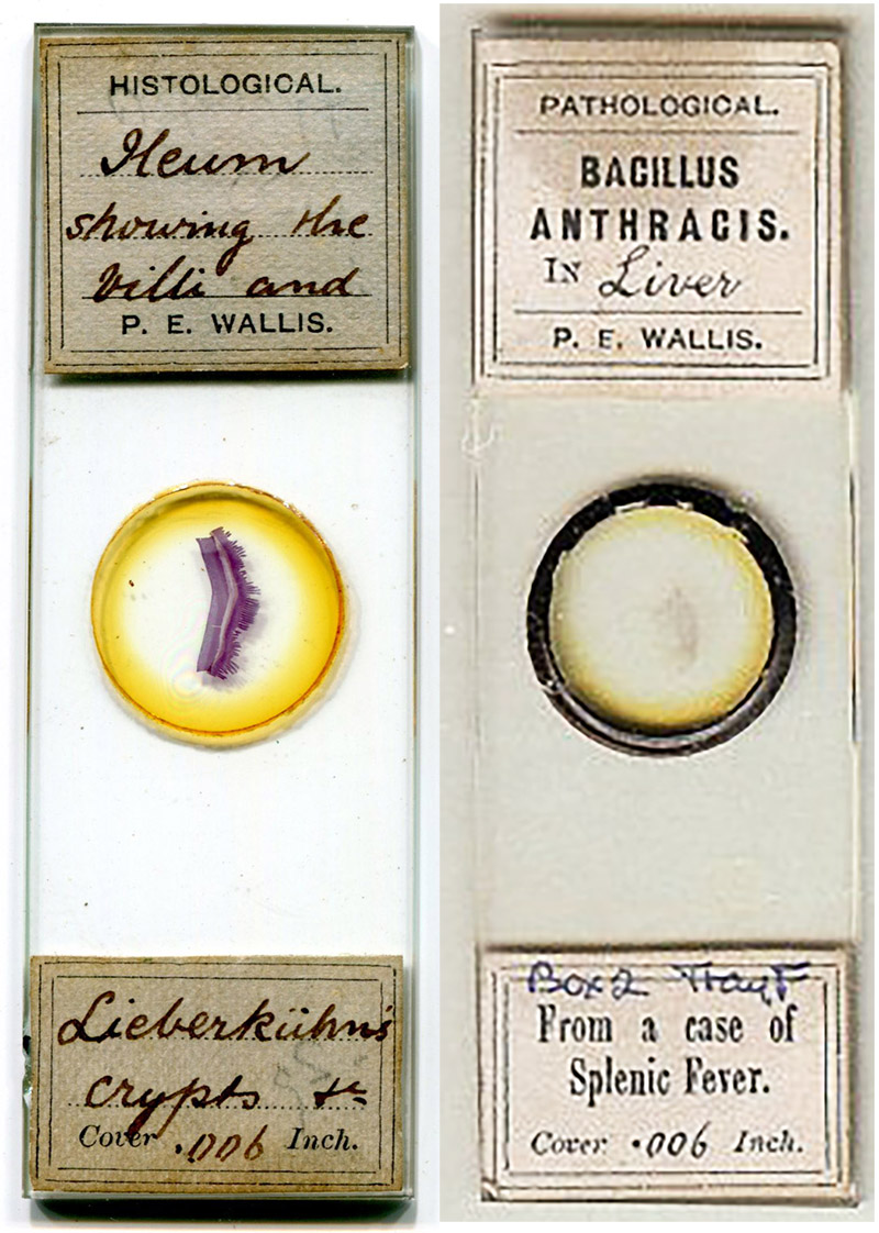
Figure 1.
Two circa 1880s microscope slides that were prepared by P.E. Wallis. As a surgeon and public health official, Wallis undoubtedly had ready access to tissues from human patients. Records indicate that Wallis encountered several anthrax patients, both human and quadruped, and the type-set labels of the Bacillus anthracis specimen on the right suggest that Wallis prepared and distributed slides of specimens from many of those patients. In 1883, he offered to exchange slides of Bacillus anthracis in lung (Figure 2). Bracegirdle’s “Microscopical Mounts and Mounters, Plate 37-D, illustrates a Wallis slide of the bacteria responsible for cholera. The image of the B. anthracis slide was adapted from an internet auction site for nonprofit, educational purposes.
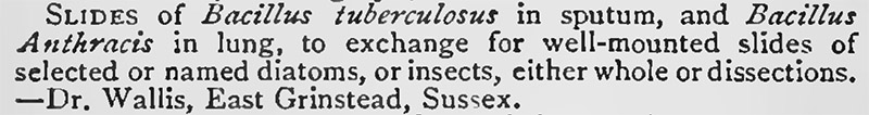
Figure 2.
1883 exchange advertisement from “Hardwicke’s Science-Gossip”.
Percy Wallis’ father and paternal grandfather were also surgeons/physicians. He was the second eldest of four children, and the only son. Life in the family home of "Oak Lea House", in Hartfield, Sussex, appears to have been quite comfortable, with cooks, maids, and other servants.
Percy entered Epsom College, Surrey, in 1864, and left in 1868. He completed his medical training in 1877, at Guy’s Hospital, London. In addition to earning his Licentiate of the Royal College of Physicians (L.R.C.P.) and Member of the Royal College of Surgeons (M.R.C.S.), Wallis studied in Edinburgh and qualified as a Licentiate in Midwifery (L.M. Edin.).
After training, Wallis set up a medical practice in East Grinstead, about 8-9 miles from Hartfield. The 1881 census found him living there as a lodger.
An interest in general microscopy was evident by 1883, when he offered slides of tuberculosis and anthrax specimens from patients in return for slides of diatoms or insects (Figure 2).
Wallis is noted as having mounted pathology specimens for other purposes. In 1885, local veterinarian H. Leeney wrote, “The prevalence of Anthrax in the northern part of Sussex has been, for generations, a great source of loss to stock-owners …. By post I send a specimen of splenic blood, mounted by Dr. P.E. Wallis”. An 1888 paper on hermaphroditism by C. Stonham thanked, “Dr. Wallis, of East Grinstead, for making the microscopical sections of the testis and epididymis”.
To the best of my knowledge, the only surviving slides by Wallis are the pathological/anatomical mounts that were likely produced for exchange. He is known to have prepared other subjects, although they were temporary mounts. For example, his 1887 report on “The development of the tadpole”: “Having seen Mr. C. Rousselet's note in the last number of Science-gossip, I am happy to be able to confirm his observations on the existence of the ciliated epidermal cells of the young tadpole. I have been aware of the existence of the cilia in question for three or four years, and had no idea that the fact was unknown to biologists. I first noticed the currents produced by the cilia while watching the circulation of the blood with a 1/2-in. objective. With this glass the cilia are not easily seen, but the currents produced by them are very distinct. In order to see the cilia well, the tail should be gently scraped and the free cells examined in a little water, with a 1/8-in. objective; the cilia are then plainly visible. A very weak solution of osmic acid brings them still better into view, but quickly destroys the vitality of the cell; the cilia become stationary, and after a short time seem to be attracted to the body of the cell and are lost to view. I fear Mr. Rousselet will meet with no success in his attempt to preserve stained sections, owing to this capillarity between the cilia and the cell, which comes into play as soon as the protoplasm of the cell ceases to live”.
He also wrote that year of means to observe features of test diatoms. However, his exchange offer of 1883 suggests that the diatom mounts that he owned were prepared by other microscopists (Figure 2).
Wallis’ name appears occasionally in medical publications after 1890, although none that I found refer specifically to microscopy. It is likely that at his growing family and medical practice led Wallis to curtail his amateur microscope hobby.
In 1891, 38 year-old Percy was a bachelor, living with his elder sister, Ellen, in East Grinstead. They were served by a cook, a parlour maid, and a housemaid. Percy married Mabel Louisa Penelope Bovill in 1893. Ellen moved out, but was recorded in the 1901 census as “living on her own means” with a surgeon and his family (and three servants) in Eastbourne, Sussex. Percy and Mabel had three children, and continued to live in “Old Stone House” on Judge’s Terrace, East Grinstead, along with several servants.
Although not directly related to microscopy, a short paper by Percy Wallis, from 1901, reveals some insight into his life: “To The Editor Of The ‘Spectator’, Sir, Your article in last week's Spectator on ‘The Rat Plague’ is of very general interest. If sanitary authorities and others would first read it, and then act upon it, you would have conferred a benefit upon the community at large. May I add another indictment to those you bring against rats, - viz., the pollution of the shallow wells by them in rural districts? A few years ago I had to investigate the origin of an outbreak of septic pneumonia at a farmhouse, where five persons were seriously ill of the disease, two of the cases ending fatally. Of course, the water supply was, as usual, declared to be above suspicion. Certainly a glass of water drawn from the pump was clear and sparkling; but on removing the cover of the well, there were the bodies of three rats, in varying stages of decomposition, floating on the surface of the water. The well was only 12 ft. deep, and the water-level was within 6 in. or 8 in. of the top of the brick lining. It was obvious that through one of several holes in the rickety well-head the rats - who are extremely thirsty animals - came to the well to drink. If the water-level was lowered a few inches, the rats, in trying to reach down to the water, fell in and were drowned. On having the well emptied, there was found at the bottom a horrible mass of putridity consisting entirely of decomposed rats. The men who cleaned out the well told me the stench was so awful that it was as much as they could do to finish the work. I could quote many other instances of a similar nature. Let me add a word to what you say about rat-catching as a sport. Many a happy and exciting day I spent as a boy with an old rat-catcher. It was a delight to watch the old man with his five or six terriers. There was a perfect understanding between master and dogs; a whispered order from the old man was instantly obeyed by 'Teaser,' 'Jock,' or 'Toby.' The dogs were quivering with excitement, but as still as mice till a rat was bolted by the ferret. The best day's sport I remember to have had was at a large farm at threshing time. The bag amounted to about three hundred and sixty rats. I have since enjoyed nearly all forms of British sport, including deerstalking in the Highlands, salmon-fishing, and grouse-driving, but I doubt if anything ever much surpassed the pleasure and sustained excitement of that day's rat-hunt among the old barns and rick-yard. - I am, Sir, &c, P.E. Wallis, Medical Officer of Health, &c. East Grinstead”.
P.E. Wallis died on June 26, 1921, at his "Old Stone House" home in East Grinstead.
Figure 3.
Human “ileum showing villi and Lieberkühn’s crypts, &c”., prepared ca. 1885 by Percy Wallis (see Figure 1). Photographed using a C-mounted SLR digital camera on a Leitz Ortholux II microscope, with a 10x objective lens.
Resources
Bracegirdle, Brian (1998) Microscopical Mounts and Mounters, Quekett Microscopical Club, London, pages 97 and 178, plate 37-D
England birth, census, marriage, death, and probate records, accessed through ancestry.com
Epsom College Register from October, 1855, to July, 1905 (1905) “Wallis, Percy Evershed [William Wallis, Esq., Hartfield, Sussex]; b. 1852, 1. 1868. (entered 1864) M.O.H. East Grinstead, Surg. E. Sussex Police, E. Grinstead Cott. Hosp., and Brighton Ry. Prov. Soc., L.R.C.P. (Licentiate of the Royal College of Physicians) and L.M.Edin. (Licentiate in Midwifery, Edinburgh), M.R.C.S.Eng. 1877 (Guy's). Old Stone House, East Grinstead”, page 42
Guy’s Hospital Reports (1877) “Gentlemen admitted to practice &c., in the year 1877”, page 407
Hardwicke’s Science-Gossip (1883) Exchange offer from P.E. Wallis, Vol. 19, page 144
Leeney, H. (1885) Splenic fever, British Veterinary Journal, Vol. 20, page 415
Stonham, C. (1888) Case of perfect uterus masculinus with perfect Fallopian tubes and testes in the broad ligament. Complex or vertical hermaphroditism, Transactions of the Pathological Society of London, Vol. 39, pages 219-226
Wallis, P.E. (1887) Amphipleura pellucida, Hardwicke’s Science-Gossip, Vol. 23, page 44
Wallis, P.E. (1887) The development of the tadpole, Hardwicke’s Science-Gossip, Vol. 23, page 44
Wallis, P.E. (1887) The rat plague, The Spectator, Vol. 93, page 426