“Larva of Anthomyia Caniculata, from Stomach of Girl - Microscopical Transaction, 1844”
by Brian Stevenson
last updated October, 2012
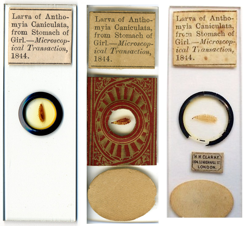
Figure 1.
Three slides of “Anthomyia caniculata’, maggots that
were passed in the feces of a 5 year-old girl in London, 1837. The original
description of the case and the maggots was published in 1844 by Arthur Farre,
M.D. The leftmost slide is in my collection, the other two images are internet
screenshots from eBay (used herein for non-profit, educational purposes).
Microscope
slides of a “Larva … from Stomach of Girl” pop up occasionally in internet
auctions (Figure 1). The specimens are well-prepared maggots of an uncertain
species of Diptera (fly).
The child
had suffered from Intestinal Myiasis, an
infestation of her intestine with fly larvae (maggots). According to the US
Centers for Disease Control and Prevention, “Myiasis is the infestation of live human and vertebrate animals with
fly (dipterous) larvae, which, at least for a certain period, feed on dead or
living tissue or ingested food of the host. Intestinal myiasis occurs when fly
eggs or larvae previously deposited in food are ingested and survive in the
gastrointestinal tract. Some infested patients have been asymptomatic; others
have had abdominal pain, vomiting, and diarrhea. . . Many fly species are
capable of producing intestinal myiasis”.
The original description of this girl’s case of intestinal maggots is presented below, following an description of these antique microscope slides.
The
printed descriptive labels indicate large-scale production. The center slide in
Figure 1 bears patterned paper that dates from circa 1870. Noting that the
paper was trimmed to fit within the other labels, it is likely the paper was
added after purchase, probably to help hold on the cover slip. Likewise the
black ringing on the other two illustrated slides, a style which dates to after
the 1880s. The early paper on the middle slide suggests that the “H.H. Clarke”
trade label on the rightmost slide was probably added by Clarke for re-sale
when he was active in the early 1900s.
The glass
of the slide in my possession is thicker than modern, and the edges finely
ground, suggestive of production during the 19th Century. The finish
is very fine, and professional in quality.
The
citation “Microscopical Transaction”
is a corruption of the journal’s actual name, Transactions of the Microscopical Society of London. Farre referred
to the maggots as being larvae of Anthomyia
canicularis, whereas the slides’ labels name the insect Anthomyia caniculata. That information
suggests that the slides were not made by Farre or anyone associated with the
Microscopical Society of London (later, the Royal Microscopical Society), since
such people would have used Farre’s name for the insect and/or the correct name of the journal.
Dr. Farre noted in his presentation, “For
a specimen of this larva consult the Museum of the Royal College of Surgeons,
London”. Although Farre made his presentation in 1841, the report
was not published until 1844. Widespread demand for specimens would probably
not have arisen until after that date.
The maker of these slides has not been identified. A strong possibility is that they were produced
on order of the Museum of the Royal College of Surgeons, to fulfill demand
after publication of Farre’s report.
Other than
these slides, the only place I located the use of the spelling “Anthomyia caniculata” was in The Microscopic Journal and Structural
Record for 1841, the forerunner of Transactions
of the Microscopical Society of London. Page 63 reported minutes of the
April 25, 1841 meeting of the Microscopical Society of London, stating, “A paper was then read by Dr. Arthur Farre,
entitled ‘On the minute Anatomy of the Larva of Anthomyia caniculata of Meyen’ ".
A summary of Farre’s talk, presented on pages 129-130 of that journal, used the
other spelling, Anthomyia canicularis.
The
species of fly responsible for the girl’s myiasis is not known. Neither Anthomyia canicularis nor Anthomyia caniculata are currently
accepted names. In The Animal Parasites
of Man (1906) Braun wrote “Anthomyia canicularis, Meig. Syn.: Scalaris, Fabr., and manicata, Meig. The larvae live in vegetables, cabbage, &c.), and are beset
with feathered bristles. They frequently invade the intestine of persons, and
produce alarming symptoms until vomited up or passed with the faeces. . . This
species is known as Homalomyia
canicularis, Linn. It is common to Europe and North America, and is an
abundant house-fly. It is the small house fly so often seen on windows. Besides
living on vegetable matter, they have also been found in the nests of the
humble bee”. Blankenmeyer (1914) reported that “it is now positively known that the Anthomyia
canicularis is the larva of the small black flower-fly known, according to Stitt, as
Anthomyia pluvialis”. It is probable that Anthomyia canicularis was a catch-all name for any dipteran larvae associated
with intestinal myiasis. Any information readers can provide on the true
identity of these 180 year-old maggots will be greatly appreciated.
From the Transactions
of the Microscopical Society of London, Volume 1, pages 51-57:
On
the Minute Anatomy of the Larva of Anthomyia canicularis, Meigen. By Arthur
Farre, M.D., F.R.S.
Read April 28, 1841.
The
subject of the present memoir has come under my notice as a parasite of the
human body, of which, however, it appears to be a rare inhabitant, as I have
met with but a single instance of the kind, and I believe there are only two or
three similar cases on record.
The
mere circumstance however of this insect in its larva state being found in the
human intestine, it is not now so much my object to record, as it is to bring
before the Society a brief description of the minute anatomy of this singular
parasite, with a view of showing the peculiar adaptation of its organs,
particularly those of the digestive system, to the circumstances in which it is
thus occasionally placed.
The
insect considered as a parasite appears to have its parallel in the oestrus or
bot of the horse and sheep, and may perhaps be considered as constituting the
bot of the human subject, though it does not appear to be altogether limited to
man, but has been also observed to occur in the Boa constrictor.* (*footnote: See Lancet, vol. ii. 1839-40, p.
638.).
The
case which afforded me the opportunity of making the following observations was
that of a rather sickly child, a girl 5 years of age, who was brought as an
out-patient to St. Bartholomew's Hospital, in the month of June, 1837, having
the ordinary symptoms of irritation produced by worms, for which a brisk
purgative was prescribed. This had the effect of bringing away a vast quantity
of the parasites, which were stated to be alive at the time they were passed,
and were described by the parent of the child as coming away by handfuls at a
time, and which continued to be passed at intervals for three weeks, when the
case was lost sight of.
A
similar case occurred to Dr. Haviland of Cambridge, in the year 1836, in the
person of a clergyman 70 years of age, who, after suffering disagreeable
sensations about the epigastrium, which he described as a tremulous motion,
accompanied by loss of appetite and general weakness, passed in the summer and
autumn of the same year very large quantities of the larvae, and, according to
his own statement, the chamber-vessel was sometimes half full, and he thinks
that altogether he must have passed several quarts : they were alive, and
continued to be passed for several months. This case is recorded by the Rev.
Leonard Jenyns, in the 'Transactions of the Entomological Society,' vol. ii.
part 3, and is accompanied by a very accurate figure of the insect. A rather
rude drawing of evidently the same insect also accompanies a paper by Dr.
Bateman in the seventh volume of the ' Edinburgh Medical and Surgical Journal,'
p. 48, on the subject of larvae found in the human body ; while a much older,
though more accurate one, will be found in Swammerdam's ‘Bibl. Nat.' tab. 38,
figs. 3 & 4. And lastly may be mentioned a case published in the 2nd volume
of the ‘Memoirs of the Medical Society of London,' which appears to be of a
similar kind. These are the only cases that I find recorded of the occurrence
of the larva in the human subject, but it has also been observed in the Boa constrictor, as appears from an
instance recorded by Mr. Iliff, to which I have just alluded, and where the
larvae were passed along with the masses of urate of ammonia which constitute
the excrement of that animal.
There
appears to be little doubt that in all these cases the insect is the same, and
that it is the larva of the Anthomyia
canicularis of Meigen, or Musca
canicularis of Linnaeus.
Its
minute anatomy does not appear to have been investigated, and it is this
deficiency which I shall attempt to supply from my notes of the dissection of
the specimens obtained from the first case to which I alluded.* (*Footnote: For
a specimen of this larva consult the Museum of the Royal College of Surgeons,
London ; Cat. Nat. Hist. Series, pt. iv. No. 609, D.).
The
larva (PI. v. fig. 1, d) is five lines in length by one and a half in breadth.
It is of a dull brown or blackish brown colour, soft and flexible, but having a
tough integument, which however is sufficiently transparent to allow of the
alimentary canal being seen through it. The body consists of eleven segments,
but the last is apparently formed of three blended into one, (figs. 2 & 3).
Each segment carries a pair of feathery branchial appendages, which project at
right angles from the body, constituting a double row on either side. There is
also a double row of small eminences extending down the dorsal surface, but the
abdominal surface is nearly smooth. The lateral appendages, (fig. 4), of which
the upper series is much larger than the lower, are pinnate. The central shaft
of these, which is long and pointed, is hollow, and communicates apparently
with the tracheae. The lateral pinnae are again pinnated on their outer margin.
The integument, (fig. 5), which appears smooth to the naked eye, is found when
examined under the microscope, to be granulated all over with minute dentiform
or pointed processes, which appear to be of a harder nature than the rest of
the tegument, and resemble on a small scale the spinous prominences in the
tegument of certain cartilaginous fishes, as the sturgeon ; and it appears to
be only an extraordinary development of these latter processes which
constitutes the long feathery lateral appendages already described.
The
mouth of this larva is perhaps the most interesting part of its anatomy. The
head (fig 6) is furnished with two broad fleshy lips, (a) which together
constitute a broad disk, having in its centre a minute aperture (b) leading to
the oesophagus, and flanked on either side by the hook-shaped mandibles, the
sharp points of which are directed downwards and somewhat outwards, (c) and are
nearly retracted each within a separate sheath, the aperture in the extremity
of which just allows their points to protrude. Each of these broad fleshy lips
is crossed by transverse parallel plaits or folds of membrane, about
twenty-five in number, which in their free margin exhibit a delicately notched
appearance, and in fact in every particular resemble a similar structure which
is seen on a larger scale in the sucking disk situated upon the dorsum of the
head of the Remora, by which that
fish is enabled to attach itself firmly to various objects. In the present
instance, however, the structure, though precisely similar, is exceedingly
delicate, and so minute as to be invisible to the naked eye, though there can
be no doubt that it is intended to answer the same purpose in both. For by the
aid of this sucker the larva is enabled to fix the head, so as the more readily
to insert its sharp hook-shaped mandibles into the soft mucous membrane of the
intestine which it inhabits, and draw therefrom its nutrient juices, which
would thus readily flow into the little aperture of the mouth, situated in the
centre between the mandibles, assisted also by the partial vacuum which would
thus be produced during the act of adhesion.
The
mandibles are sunk so deep between the two fleshy lips, having only the points
projecting from the aperture of their investing sheaths, that it is necessary
to disconnect them entirely from the soft parts before they can be accurately
examined. They are placed parallel to each other, with their hooked points
directed downwards, so as the more readily to be inserted. The mandibular
apparatus (fig. 7, a, and fig. 8) on each side consists of three portions. The
first portion is hooked and sharp pointed, (fig. 8, a) and is only the 1/60 of
an inch in length. It is nevertheless furnished at its base with a delicately
shaped ball, adapted accurately to a socket in the second joint, and has
projecting from either side of its base a sharp spine or trochanter, for the
insertion of the tendons of the abductor and adductor muscles by which its
movements are effected. The second joint (fig. 8, b) bears the socket to which
the ball of the first is adapted, and at its opposite extremity is united to
the third and principal portion of the jaws, (fig. 8 c), which consists of a
broad expanded corneous plate, of less density than the preceding, and
resembling in form and office a similar structure well known as occupying the
centre of the large claw of the lobster, being for the attachment of the muscles
by which the mandibular hooks are moved to and fro. The whole mandibular
apparatus measures about the ^ of an inch in length, and, being very firm and
solid, presents a remarkable contrast in texture to the surrounding soft parts
with which it is connected, and from which it is easily detached.
>If
the body of the insect be laid open the alimentary canal is seen to be of
considerable length, and much convoluted, (figs. 9 & 10). It commences by
an exceedingly delicate hair-like oesophagus, (fig. 7, b), so narrow that it
would appear to be specially destined to transmit fluid nourishment, and
nothing else. This terminates about the third segment of the body in a minute
globular cavity or proventriculus, (fig. 7, 6-), which is of the same diameter
as the rest of the alimentary canal, and immediately below which four very
short salivary vessels enter, (fig. 7, d). From this point commences the large
intestiniform stomach, (fig. 10, a), which after contracting in its first third
to the finest thread, (fig. 10, 5), again dilates and proceeds of uniform
diameter to the point where the four slender biliary vessels enter, (fig. 10,
c), where it again contracts and forms a short intestine. The whole alimentary
canal is about six times the length of the body, and of this length the stomach
forms about five-sixths. Its greatest diameter does not exceed one-third of a
line, and its least is that of a mere thread. The whole structure appears to be
that of an animal adapted to live on fluid nourishment.
The
principal external openings to the tracheae appear to be two apertures situated
on the dorsum of the last segment of the body, and which constitute the last
pair of the series of dorsal eminences formerly noticed. These apertures
correspond with the very remarkable and conspicuous pair of organs occupying a
similar situation in the last segment of the oestrus of the sheep, and which
are also the external openings of the respiratory apparatus in that insect.
None
of the insects were alive when they came into my possession, and they were
placed in spirit of wine for the purpose of preserving them previously to their
being dissected. Several days after my attention had thus been directed to the
subject, I happened to observe at the bottom of a jug of New-River water a small
living object, which appeared very much to resemble the larvae which I had
recently been examining ; and upon placing this under the microscope I found
the resemblance to be complete, except that the animal was only about
two-thirds the size of the former. It was deficient also in the pinnae upon the
lateral spines, which were simple, but the mandibular apparatus was perfectly
formed. On making further search two other individuals of the same species were
found in different stages of growth; the smallest, however, not exceeding
one-third of a line in length, though still possessing some of the characters
of the larger ones. One of these specimens was very lively, moving freely at
the bottom of the water, and frequently protruding and retracting its proboscis,
by which it dragged itself along.
This
fact is a matter of some interest, as furnishing a clew to the source of these
parasites, since it is evident that the larvae can pass along the water-pipes
which supply the metropolis, and may thus be swallowed in the water used for
food : and in the present case the larvae, or the ova, must have traversed a
distance of at least a mile. At the same time it is evident that this cannot
always afford an explanation of their mode of entrance into the body, because
in the case of the clergyman at Cambridge it is expressly stated that " he
never drank water unmixed, but generally beer, tea," and the like; at the
same time the water used for these beverages was entirely supplied from a pond
on a stiff clay. If therefore the ova found entrance with the fluid aliments,
they must have withstood the action of heat, as in making the beer, tea,
&c.; while on the other hand it is difficult to suppose that they passed
in with the solid food, because the larvae are evidently aquatic. Perhaps the
most inexplicable part of the case is, the fact of their occurrence in such
immense numbers. In the Cambridge case several quarts were passed in a few
months, and in the instance which I have just recorded they were described as
coming away by handfuls. It is extremely difficult to account for this fact,
because a number of larvae, or their ova, must have been swallowed equal to
those which were evacuated, since they could not multiply by generation in the
alimentary canal, they being in the larva state, and having, as the dissection
showed, and as is well known in the case of larvae, the generative organs
undeveloped ; indeed no trace of generative organs was visible : while it is
difficult to suppose that the parent animal could have been accidentally
swallowed, and its ova, previously impregnated, have become developed in the
bowels; though this is perhaps the least objectionable supposition. At any rate
the parent animal could not live in the alimentary canal, since the larva has
been recognised by several entomologists as being that of a well-known fly,
(the Anthomyia canicularis). The latter supposition however which I have
advanced, namely, that the fly, having its eggs previously impregnated, may
have been swallowed, and thus, perishing in the digestive canal, have left the
ova unencumbered, and in a possible situation for development, derives some
countenance from the circumstance of the extreme rarity of the occurrence of
these larva? as parasites, there being, as I have mentioned, very few cases on
record ; which would give to the circumstance the air of an accidental
occurrence, of which however it is again immediately robbed when we contemplate
the singular and very obvious adaptation of its organization to the peculiar
circumstances in which it is thus placed. The anatomy being clearly that of an
animal destined, or at least adapted, to live by adhesion and suction on fluid
nourishment, though it is clear from the fact of some being found nearly
two-thirds grown in simple river water, that the larva is also capable of life
and growth in other elements than the contents of the alimentary canal, and in
other capacities apparently than that of a parasite.
Much,
it appears, may be advanced on either side, and indeed the whole subject appears
to me to be calculated to afford interesting points for discussion; and it is
chiefly with this view that I have brought it before the notice of the Society.
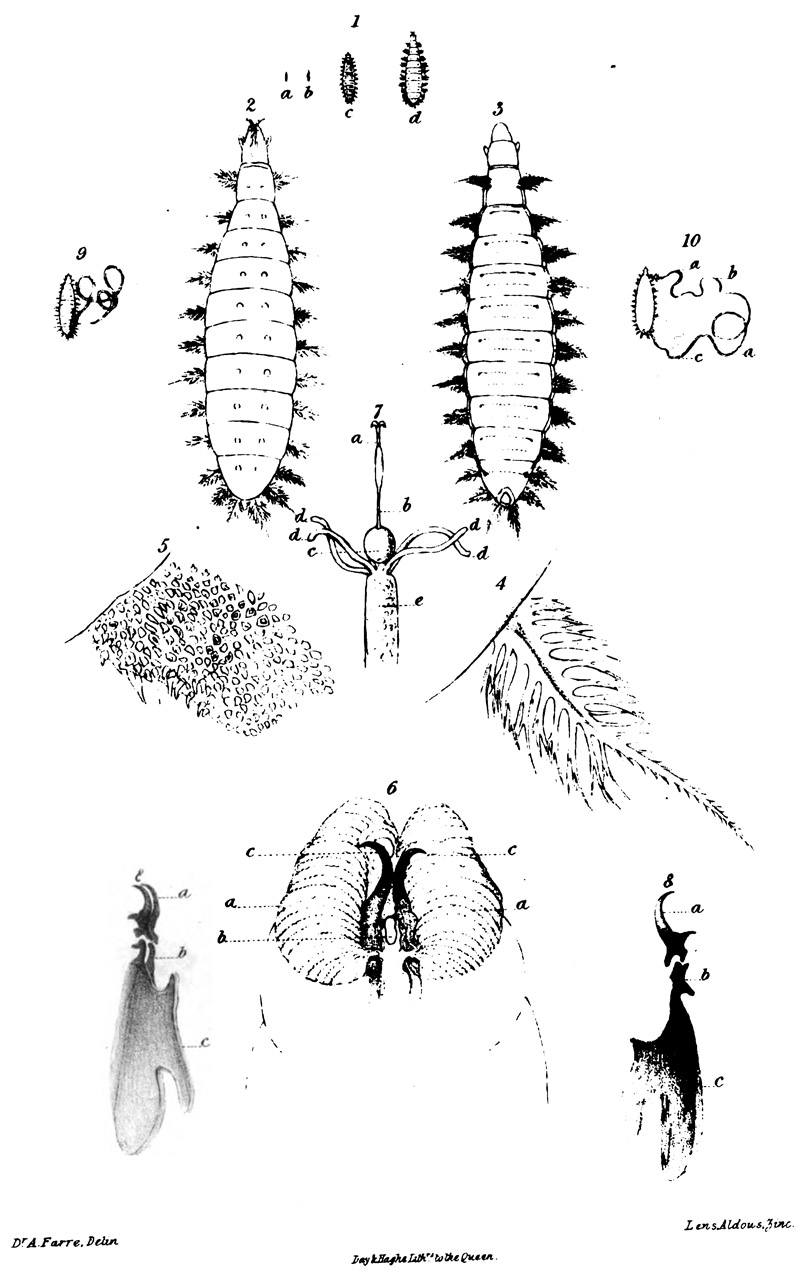
EXPLANATION OF PLATE V.
Fig. 1. a, b, c, d. Larvae of natural size, in different stages. The three first from the New-River water; the fourth from the intestine.
Figs. 2 & 3. The same amplified five diameters ; 2 dorsal, 3 ventral aspect.
Fig. 4. One of the upper series of branchial appendages.
Fig. 5. Portion of tegument of the back.
Fig. 6. Head. a. Two plaited lobes forming the suctorious disk. b. Mouth. c. Points of the mandibles just exserted from their sheaths. The remaining portion of mandibles seen indistinctly through the semitransparent flesh.
Fig. 7. Mandibular apparatus and upper portion of alimentary canal, a. Mandibles. b. Oesophagus. c. Proventriculus. d. Salivary tubes. e. Commencement of stomach.
Fig.
8. Two views of the mandibles, a. First portion, sharp pointed at its
extremity, and showing two trochanters at its base for insertion of adductor
and abductor muscles, and bearing a ball adapted to socket in b, the second
joint, c. Third portion for origin of muscles.
Fig.9. Larva (natural size) opened to show alimentary canal.
Fig. 10. Ditto, a. Intestiniform stomach, b. Contracted portion, c. Point where biliary vessels enter.
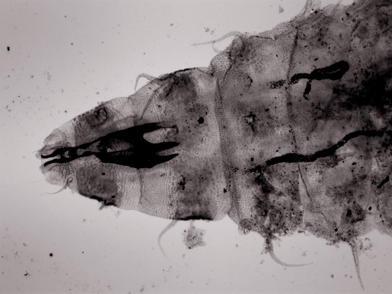
Figure 3.
Photomicrograph of the mouthparts of the maggot in the left slide shown in Figure 1. Photographed using an Olympus BX51 microscope, 4x objective lens, brightfield illumination and a Retiga 2000R Fast
1394 with QCapture Pro software.
Resources
Blankenmeyer,
H.C. (1914) Intestinal myiasis, Journal
of the American Medical Association, 43:321.
Bracegirdle,
B. (1998) H.H. Clarke / Clarke & Page, Microscopical
Mounts and Mounters, Quekett Microscopical Club, London, page 22.
Braun,
M.G.C.C. (1906) The Animal Parasites of
Man, Translated by P. Falcke, Edited by L.W. Sambon and F.V. Theobald, J.
Bale, Sons and Danielsson, London, pages 435-436.
Farre, A.
(1841) On the minute anatomy of the larva of Anthomyia canicularis, The
Microscopic Journal and Structural Record (1841) pages 129-131.
Farre, A.
(1844) On the minute anatomy of the larva of Anthomyia canicularis, Meigen, Transactions
of the Microscopical Society of London, pages 51-57 and Plate V.
The Microscopic Journal and Structural Record (1841)
page 63.
Morbidity and Mortality Weekly Reports (1985)
Intestinal myiasis – Washington, Vol. 34: 141-142.