Frederick H. Evans, 1853 – 1943
by Brian Stevenson
last updated July, 2017
The four slides
shown in Figure 1 contain small, oval labels with the handwritten signature “F H Evans”. The specimen labels of the two center slides, “Pulex Iritans (sic) – Flea” and “Spine of Echinus”, are written in the same handwriting, suggesting that this slide was made by Evans. The writing on the specimen label of the left slide is that of John Barnett, a professional slide-maker, and so was evidently purchased by Evans. The slide on the right carries a descriptive label with handwriting that indicates production by amateur Frederick Marshall. Brian Bracegirdle’s Microscopical Mounts and Mounters illustrates a slide with an Evans label on plate 15D, with a cover paper indicating that it was made by professionals J. & T. Jones. Bracegirdle interpreted the scrawled handwriting to be “F N Evans”. Confirmation that these slides were signed by Frederick H. Evans can be seen at a surprising source: a signed photograph by Evans, of Aubrey Beardsley, is on display at the Musee d’Orsay in Paris, France. A copy-protected image can be seen on the museum’s web site. Clicking on the image, then on the magnifying glass symbol, will provide a clear view of Evans’ signature. A screen-capture of the signature is included in Figure 2, below. Other examples of his signature and handwriting can be found on the web by searching databases such as Google Images.
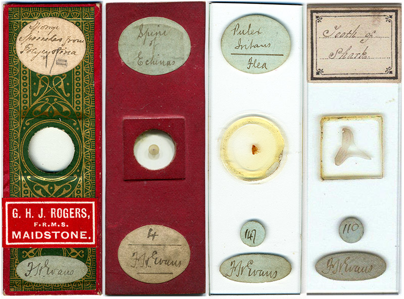
Figure 1. Four microscope slides bearing labels with Frederick Evans’ signature. The two slides in the center, of an echinus spine and a flea, also carry descriptive labels in the same hand, and were presumably made by Evans. The left slide, a papered arrangement of sponge spicules, bears a specimen label with the handwriting of professional slide-maker John Barnett. It is not known whether Evans bought the slide through G. Rogers, or if Rogers disposed of it after Evans had owned the slide. The slide on the right, a thin-section of a shark tooth, was carries a descriptive label from Frederick Marshall, an amateur microscopist who was well-known for his thin-section mounts of echinus spines.
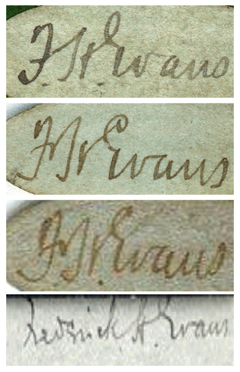
Figure 2. F.H. Evans’ signature, from the three microscope slides shown in Figure 1, and from Evans’ photograph of Aubrey Beardsley (lower).
Frederick
Henry Evans was born June 26, 1853 in Whitechapel, London. He was the fourth of John and Sarah
Evans’ five children. His father,
John, was a school master, and on the 1881 census was recorded as being a music
teacher. Frederick was recorded as
working as a clerk on both the 1871 and 1881 censuses, with the ’81 census
elaborating his job as being with a “preserved provision merchant”.
Like many
other people of his time, Frederick acquired an interested in microscopy. He also developed skills in
photographic techniques. He
obviously blended both interests very well, as he proved to both the Royal
Microscopical Society and the Quekett Microscopical Club in May, 1886. From an account of the RMS
presentation: “Mr. F. H. Evans exhibited
some photo-micrographs, produced by the Woodbury-type process, from negatives
taken by himself, and transferred to glass for the purposes of lantern
illustration, and so that in many cases the objects could be seen on the screen
more perfectly than under the microscope. To show what an advance had been made
in this direction, 60 of the slides were shown upon a portable screen by Mr.
George Smith (of the Sciopticon Company), who had printed the slides from the
original negatives. The objects illustrated comprised diatoms and desmids,
foraminifera, polycistina, star fishes, sections of Echinus spines, insect
preparations, animal parasites, and anatomical and vegetable sections, the
remarkable clearness of most of the photographs calling forth frequent
favourable comments from the Fellows present…The President said that the
meeting were very much obliged to Mr. Evans and his colleague for this exhibition,
which was certainly the most interesting which he had yet seen. He had been
much struck by the beauty of many of the pictures...The photographs were all
taken with an A eyepiece, and by the light from an ordinary paraffin microscope
lamp. The slides were prepared by what was known as the Woodbury process—that
is, they were printed from metal plates; the result being that they got very
much greater transparency and better detail, with a uniform colour. When once
the proper tone had been obtained, any number of prints could afterwards be
produced of exactly the same depth. The process was undoubtedly the finest
possible for the purpose…Mr. Smith said that there was nothing unusual about
the process in any way; it was simply a question of manipulative skill. Some of
the transparent objects were illuminated by the spot lens, and ordinary
objectives were used…Mr. Crisp said that for the next number of the Journal he
had written a note on the question whether photographs of microscopic objects
were better for purposes of class illustration than the objects themselves
thrown on the screen, and had expressed himself in favour of the natural
objects. What, however, he had seen that evening certainly required him to
alter his opinion…Mr. Smith said that in the first instance a photographic
negative was taken in the usual way. This negative was upon a glass plate, and
was so called because all the lights and shadows were reversed from what they
were in the natural picture. From this negative an ordinary photograph was
produced by printing from it in the usual way. The Woodbury-type process made
use of the property acquired by gelatine when mixed with bichromate of potash,
in virtue of which it became insoluble after being exposed to the action of
light. A film of gelatine so prepared had the photograph placed upon it, and
after being exposed to light was washed in hot water, which dissolved away
those parts which the light had not affected. In this way a very delicate film
was obtained not exceeding the 1/500in. in thickness, but containing every line
of the picture in relief. This film was put upon a steel plate, and a piece of
lead having been placed upon it, they were subjected to a pressure of many tons
weight, by which means an intaglio mould was formed upon the lead. The plates
were practically casts from this mould made in gelatine and darkened with
lampblack.”

Figure 3. An example of Evans’ cellebrated photomicrographs of natural objects, this being a transverse section of an echinus (sea urchin) spine.
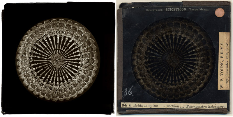
Figure 4. A lantern slide of a cross-sectioned echinus spine, produced by the Sciopticon Company. The subject matter, style and distributor all suggest that the original photograph was taken by Frederick Evans. Views were taken with transmitted light (left) and reflected light (right).
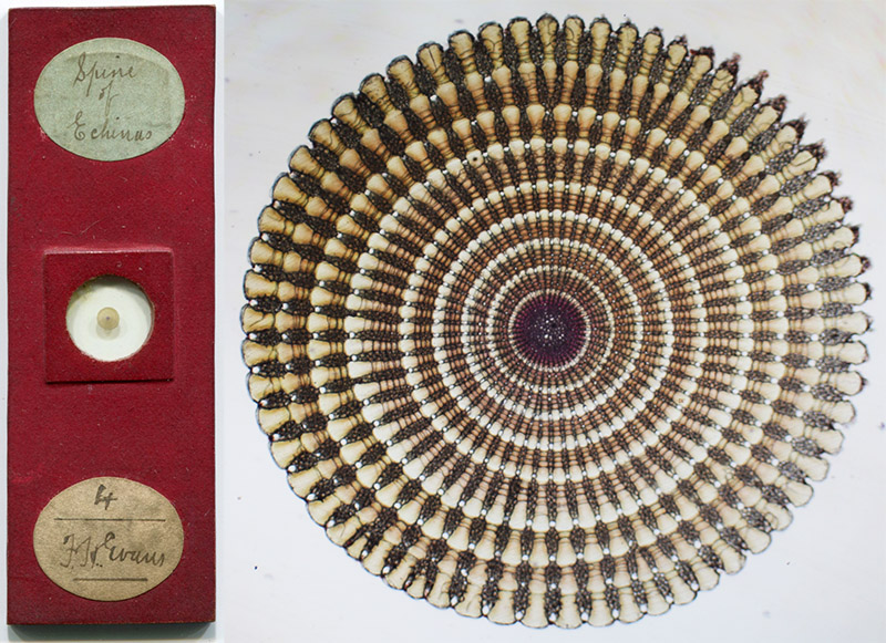
Figure 5. The echinus spine slide shown in Figure 1, and a photomicrograph of the specimen, taken with a C-mounted digital SLR camera and a 3.4x objective lens.
Also in 1886,
Evans developed a screen to aid in focusing a microscope for use with a camera
(Fig. 4). The Journal of the Royal Microscopical Society described it as follows:
“Evans's Focusing Screen for
Photomicrography.—Mr. F. H. Evans refers to the difficulty which exists in
focusing, by means of an ordinary focusing lens, the microscopic image
projected on a screen of patent plate glass. This is due to the power of
accommodation of the eye, in consequence of which the focal plane of the image
is frequently assumed to be on the outer instead of the inner surface of the
screen. He suggests that this difficulty may be readily overcome by ruling on
the inner surface of the glass screen (i.e. the surface towards the Microscope)
a series of fine lines similar to those shown in fig. 83 (reproduced here as
Fig. 6); the eye has then before it a definite object in the focal plane upon
which the focusing lens is adjusted, so that the almost involuntary movement of
accommodation is practically arrested thereon, and the focusing of the
microscopic image on that plane is thus greatly facilitated.”
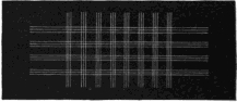
Figure 6. Evans’ Focusing Screen for Photomicrography, as illustrated in the Journal of the Royal Microscopical Society, 1887.
The Royal
Photographic Society awarded Evans a medal in 1887 for his photomicrographs of
shells. The 1890 book, Practical photo-micrography: by the latest
methods, described Evans as having produced “useful photographs of physiological preparations.”
On Dec. 18,
1891, Frederick Evans was elected to be a member of the Quekett Microscopical
Club. At the time he lived at 77
Queen St., London. He resigned his
membership by 1897.
By the late
1880s, Evans and a partner had opened a book shop, Jones and Evans, near
Cheapside. Among the clientele
were George Bernard Shaw, Aubrey Beardsley and J.M. Dent. Evans introduced Beardsley to Dent, who
then hired Beardsley to produce his famous illustrations for Le Morte d’Arthur in 1893. In 1894, Evans joined “The Linked
Ring”, a group who promoted the artistic values of photography over its
technical matters. Evans gave up
the book shop and turned full time to photography in 1898. He photographed his
friends, including Beardsley, as noted above. Evans also made celebrated photographs of buildings,
including English and French cathedrals. Alfred Stieglitz published several of Evans’ interior views of
cathedrals in a 1903 issue of Camera Work. Evans used the platinotype technique to
produce his stunning photographs. However, the escalating cost of the platinum required for that technique
led to him abandon photography by 1915.
He died June 24, 1943.
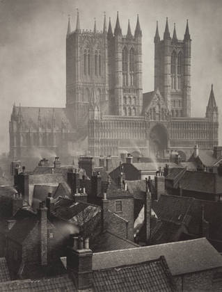
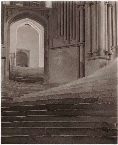
Figure 7. Photographs by Frederick Evans. (right) Lincoln Cathedral: From the Castle, 1898, and (left) Wells Cathedral: A Sea of Steps, 1903.
Acknowledgments
Thanks to Peter Paisley for sharing pictures of slides.
Resources
Bracegirdle, Brian, 1998, Microscopical
Mounts and Mounters, Quekett Microscopical Club, London.
England vital statistics, accessed through www.ancestry.co.uk
English mechanic and
world of science, 1888, Scientific Societies - Royal Microscopical Society,
vol. 43, page 275
Frederick H. Evans,
Photographer of the Majesty, Light and Space of the Medieval Cathedrals of
England and France, 1975, Aperture, vol. 18, no. 1. Contains many photographs by Evans, and several photos of him at various times in his life.
Hannavy, John, 2008, Encyclopedia of
Nineteenth-century Photography, vol. 1, page 504, CRC Press.
Journal of the Royal
Microscopical Society, 1887, Evans’s Focusing Screen for Photomicrography,
page 320
The Lancet, 1886,
Medical Diary for the Ensuing week, vol. 1, page 907
Luminous Lint, http://www.luminous-lint.com/app/photographer/Frederick_Henry__Evans/A/
Nature, 1886, Diary
of Societies, vol. 34, page xiii
Pringle, Andrew, 1890, Practical
photo-micrography: by the latest methods, Scovill and Adams, New York
Quekett Journal of
Microscopy, 1892, Second series, vol. 5, pages 92 and 287
Quekett Journal of
Microscopy, 1892, Second series, vol. 6, page vi
Warren, Lynne, 2006, Encyclopedia
of twentieth-century photography, vol. 1, page 461, CRC Press.