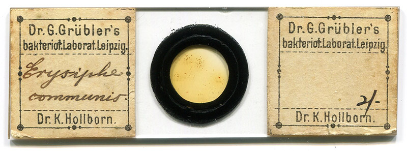
Figure 1. A circa 1900 microscope slide from “Dr. G. Grübler’s Bakteriologisches Laboratorium (Dr. K. Hollborn)”, with specimens of Erysiphe communis (now known as Erysiphe cruciferarum, a fungus that causes “powdery mildew” on brassica).
George Grübler, 1850 - 1915
Karl Hollborn, 1862 - 1942
by Brian Stevenson
last updated February, 2024
The development of chemical dyes boomed in the mid-1800s, as offshoots from the Industrial Revolution. This included analine compounds that were derivatived from byproducts of coal gas production. Germany became a major center of dye production, reported to have developed and tested thousands of new dye chemicals during those years. Biologists quickly noted that certain dyes were particularly good at staining of tissues for microscopical examination, since some chemicals had particular affinities for specific substances.
George Grübler developed a business during 1880 in Leipzig, wherein he acquired samples of commercial dye lots, tested them for efficacy as biological stains, and then purchased large amounts of suitable batches. He then packaged and sold them to microscopists. Since Grübler had tested each lot, and possessed large quantities, he was ale to provide consistent products of very high quality. As a result, his business quickly became the preferred source for microscopists throughout the world.
Grübler sold his business to Karl Hollborn in 1897, who continued it under Grübler’s name. Around that same time, Hollorn commissioned and sold microscope slides under the name of “Dr. G. Grübler’s Bakteriologisches Laboratorium (Dr. K. Hollborn)” (Figure 1). Those slides are not very common nowadays, and are generally of very good quality.
Figure 1.
A circa 1900 microscope slide from “Dr. G. Grübler’s Bakteriologisches Laboratorium (Dr. K. Hollborn)”, with specimens of Erysiphe communis (now known as Erysiphe cruciferarum, a fungus that causes “powdery mildew” on brassica).
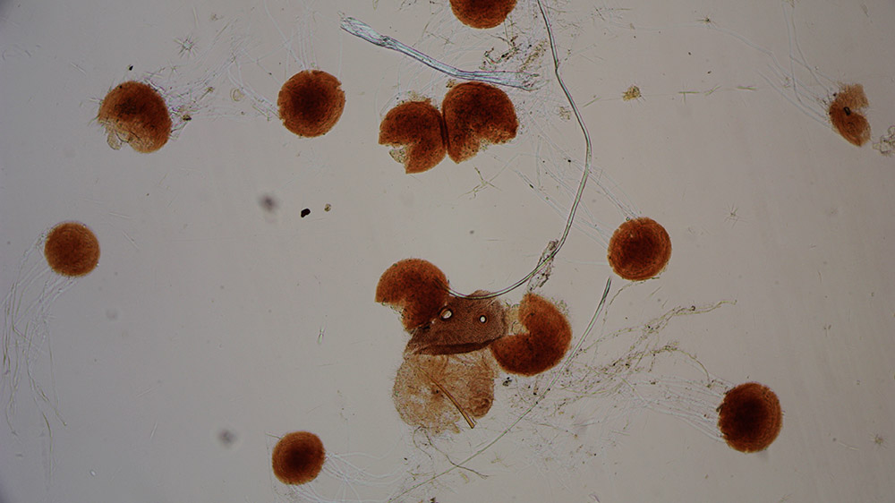
Figure 2.
Magnified details of Erysiphe communis / Erysiphe cruciferarum, from a slide retailed by Dr. G. Grübler’s Bakteriologisches Laboratorium, Dr. K. Hollborn. Clear filaments can be seen emerging from several of the brownish chasmothecia (fruiting bodies). A thick, colorless organic filament can also be seen in the center, probably a fiber from the host plant. Imaged with a 10x objective and C-mounted digital SLR camera on a Leitz Ortholux II microscope.
George Grübler born in 1850 in Meerane, Saxony (Germany). The son of a pharmacist, George followed in his father’s footsteps, graduating from the University of Leipzig in 1874. He then worked as a pharmacist while attending graduate school. He earned his Ph.D. in 1880.
Also in 1880, Grübler started a private chemistry lab, and began experimenting with dyes. He was soon acquiring large batches of dyes that he found to be biologically useful, then marketed them across Europe and North America (Figures 3 and 4).
>p>Arthur Bolles Lee’s 1890 The Microtomist's Vade-mecum: A Handbook of the Methods of Microscopic Anatomy specifically recommended Grübler‘s dyes, “A word of caution to beginners and others:You are not likely to succeed in staining, especially in the beautiful processes of staining with coal-tar colours, unless you see to it that you are working with chemicals of the proper quality. You cannot ensure this by going to a generally trustworthy house for chemical products-at all events, not in the case of coal-tar colours. It is not sufficient that these should be analytically what they are described to be, they may be pure, and yet not give good stains. I have a collection of coal-tar colours obtained from one of the best London houses, of the purity of which I have no doubt; a large proportion of them are useless for staining purposes. I therefore feel constrained to advise everybody to get his reagents - at all events his anilins - from the well-known chemists Grübler or Münder. Grübler has all the tried reagents in stock, and supplies only such as have been found by experiment with tissues to furnish the desired stain. He also makes up fixing and staining solutions, injection and imbedding masses, &c., according to the classical formulae, and sends them out neatly packed and ready for use. From experience I can most highly recommend these preparations, which are, in nine cases out of ten, better than those the observer is likely to make for himself. They may be ordered from the price-list, or by quoting the numbers of the formulae in this work. The address is-Herr Dr. G. Grübler, Chemiker, Baiersche Strasse, 12, Leipzig. Grübler can correspond in English”. Lee also recommended that microscopists should acquire Grübler’s “Zoologist’s Travelling Case” full of stains (Figure 5).In 1896, Karl Hollborn began working for Grübler. Hollborn was born in 1862 in Hildesheim, Saxony. As had his employer, Hollborn earned a degree in pharmacy from the University of Leipzig, in 1889. He then took a PhD from University of Rostock in 1894.
During 1898, Grübler‘s “Labor für Physiologische Chemie und Mikroskopische Chemie“ (“Physiological Chemistry and Microscopical Chemistry Laboratory”) was divided to form a business that was dedicated to the mail-order distribution of his dyes, "Dr. G Grübler & Co., Farben, Chemische Präparate für Mikroskopie und Bakteriologie” (“Dr. G Grübler & Company, Dyes, Chemical Preparations for Microscopy and Bacteriology”).
Grübler sold his business to Hollborn in 1897, then moved to Dresden. In that city, he opened a new business, providing pepsin, trypsin, and other proteins for physiological and biochemical studies (Figure 6). He died in Jena in 1915.
Sale of the Leipzig business was announced in Chemiker-Zeitung, “Die Firma Dr. G. Grübler's mikroskopisch-chemisches und bakteriologisches Laboratorium ist auf Dr. phil. Karl Theodor Immanuel Hollborn übergegangen; künftige Firmirung Dr. G. Grübler's mikroskopisch-chemisches und bakteriologisches Laboratorium [Dr. K. Hollborn]”, (“The company Dr. G. Grübler's microscopic-chemical and bacteriological laboratory is located on Dr. phil. Karl Theodor Immanuel Hollborn passed over; future name Dr. G. Grübler's microscopic-chemical and bacteriological laboratory [Dr. K. Hollborn])“.
Around that same time, Hollborn’s company began to sell prepared specimens for the microscope (Figures 1, 2, and 7).
Hollborn wrote a brief “History and Theory of Staining” for the 1900 edition of The Illustrated Annual of Microscopy. He co-authored the article with Herbert F. Angus, one of his English distributors. The brief essay is still worth reading:
"For how many sciences can it be said that the seed was sown a full century before the young shoot, destined to become a great tree, showed itself above ground? Certainly it is true of a very great number, and the science of demonstrating histological, and cytological details by means of stains is one of them."
"To Prof. Reichet, of Leipzig, is due the credit of having sown the seed; others came after, who, by grafting, so altered the fruit of the tree that the learned professor would now, perhaps, hardly recognise it as of his planting, but he it was, who, in the year 1758, used a decoction of Pernambuco wood for studying the histology of plants. Thus was the seed sown, but it was nearly a century before any further advance was made, until in the year 1849 the botanists, Messrs. Göppert and Cohn, used carmine solution to study the rotation of the cell contents of Nitella flexilis; five years later Hartig made exhaustive experiments as to the possibility of fixing the carmine in relation to the capacity of the different elements of the plant cell; that is to say, of stopping the staining process when, owing to their varying affinity for carmine, the elements were each stained to a different degree, and thus differentiated; he was so far satisfied with his results as to prophecy a great future for the new process."
"Already, the knowledge of these experiments had spread widely, and two years later, A.D. 1856, we find Lord Sydney Godolphin Osborne, who was deeply interested in botany, growing plants in carmine solution, and in the next year the chemist, Maschke, of Breslau, endeavoured to further his botanical researches with the same stain. So far all the recorded experiments had dealt only with plant histology, and it was reserved for Gerlach, anatomist, of Erlangen, to first describe the results obtained by the new method in the field of human morphology; he it was who urged on the histologists of his day the necessity of further experiment, both with this and with other stains; and, if to Prof. Reichet is due the credit of planting the seed, to Gerlach is certainly due the credit of so grafting the resulting shoot as to give it its present-day character."
"In answer to his appeal there arose a school of histologists devoted to mastering the technique of staining, and in 1871 Weigert succeeded in staining the Cocci-Zooglea, as well as the nucleus of the cell, by means of an ammoniated solution of carmine followed by treatment with muriatic acidglycerin; in the following year Messrs. Eberth and Wagner succeeded in staining Cocci, but not Bacilli, with Haematoxylin, and subsequently Weigert discovered that Cocci, especially in Zooglœa, could be stained by various nuclear stains, for this purpose, and for the first time he used an anilin dye, viz., Methyl Violet."
"These anilin dyes, destined, in a great measure, to displace carmine and haematoxylin, were first put on the market in 1856, and, although recommended by Waldeyer in 1863, were still very little used. Fischer, however, in 1875 introduced Eosin, much used at present as an ingredient in multiple stains, and also for blood work; and, about the same time, Ehrlich threw the weight of his authority into the scale; in 1878 we find Weigert recommending the use of Bismarck Brown, and in the next year Ehrlich published his far-reaching classification of the coal tar colours, dividing them into three groups, the Basic, the Acid, and the Neutral; the first class being, in general, sharp nuclear stains, the second, plasma stains, that is to say, stains with a special affinity for cytoplasm, and intercellular substances; and the last, stains with special affinity for certain cell contents; this classification, with one or two reservations, is accepted as correct at the present day."
"In the year 1881 Ehrlich conclusively demonstrated that only the basic colours were suitable for bacteriological investigation: Bismarck Brown, Fuchsin, Methyl, and Gentian Violet, etc., but especially Methylene Blue."
"The tide had now turned completely in favour of the anilins, and the next few years are particularly rich in the application of these dyes to the various branches of microscopic research."
"Weigert applied Acid Fuchsin to the study of the nervous system; Strasburger used Methyl Green, combined with acetic acid, to demonstrate the mitotic figures of cell division; and in the same year, 1882, Koch demonstrated the Bacillus Tuberculosis by staining first with alkaline Methylene Blue, and subsequently with Vesuvin, whereby the Bacilli were stained blue, the remainder, including other micro-organisms, brown. This method was modified by Ehrlich, who, instead of an alkaline solution of the primary stain, used one of the basic dyes, Gentian Violet, or Fuchsin, in a solution of anilin. Ziehl Neelsen still further modified the process by using a carbolized solution of basic Fuchsin, and, it is this last method which is in general use to-day. It will be noticed that the dyes, in that they all belong to the basic group, are the same, the only difference being in the vehicle in which they are dissolved; the importance attached to the choice of vehicle will best be appreciated by reference to the process of ordinary industrial dyeing, in which a great number of colours directly stain the material immersed, while others require the presence of some substance, generally a metallic salt, or hydrate, technically known as a mordant, before they can be made to give a satisfactory result; the same holds good in microscopic staining, and the Tubercle Bacillus, although stained slowly by an aqueous, or alcoholic solution of one of the basic colours, stains much more rapidly when an alkaline, carbolized, or anilin solution is used; these substances serving as mordants."
"These bacilli, being thus difficult to stain, are equally difficult to decolorize, and, while immersion for a short time in acid or alcohol will effectually decolorize the other elements of the preparation, they still retain their colour, and remain. unaffected, even when the decolorized elements are again stained with some contrast stain, as in Koch's original demonstration, in which the bacilli were stained blue, the epithelial cells, etc., brown."
"This discovery, opening up as it did the science of Bacteriology, is one of the greatest triumphs of latter-day staining methods. The country folk in many districts, especially Naples, considered Tuberculosis an infectious disease; already Willemin, of Paris, had proved the same by the vaccination of experimental animals, but efforts hitherto made to demonstrate microscopically the germ of the disease, had failed, until Koch succeeded with the above-mentioned stain."
"Now, scarcely any accurate observations are made, in either Histology or Bacteriology, without the aid of stains; and the literature of the subject has increased so largely that it would be impossible, in the space at our command, to give more than a very general outline of the theory, underlying the hundreds of formulæ recommended; perhaps, under the circumstances, it will be best to merely draw together the threads of what has already been said."
"The chief reason for staining microscopic objects is to differentiate them, that is, to exhibit one group of cells in a different colour or shade from another group (a histological stain); or to demonstrate the nucleus, or granules, of a cell by these same colour differences (a cytological stain)."
"Stains may be divided into two great groups (1) General, and (2) Specific; the latter of which may be again divided into three sub-divisions, after the classification of Ehrlich, already mentioned, and into the details of which we need not now enter. Those comprised in the first group, the general stains, colour the whole of the preparation, although all the elements are not equally affected; the specific stains on the other hand, as their name implies, colour only certain groups of cells, or elements of cells; but there is scarcely any stain so specific that it does not need careful attention as to strength, time it is allowed to act, etc., and these details necessarily bring us to a consideration of the methods employed."
"There are two distinct methods, all the various modifications being grouped under one or the other:-(1) The Progressive and (2) The Regressive. The progressive method consists in staining just so long as to bring out the elements required, and stopping its action before the other elements are affected sufficiently to destroy the differentiation."
"The regressive consists in allowing the whole of the preparation to become stained, and then washing out by means of alcohol or acid the stain from those elements, which give it up most readily; of this method we have already given an example, when speaking of the discovery of the Bacillus Tuberculosis; the elements thus freed from stain can be restrained with another colour, those which have resisted the washing out process being unaffected by this second staining, thus we have what is called a double stain."
"These double stains are not confined to just that branch of research from which our example is taken, but are equally applicable to animal and vegetable histology; nor is it always necessary to wash out one stain before applying the next, as some stains have the power, when applied subsequently, of replacing the original stain in those elements for which they have a special affinity; others, again, may be mixed in one solution, and thus applied simultaneously, each picking out its own especial elements; there are also some valuable triple stains belonging to this 'one solution' class, by means of which our knowledge of the leucocytes of the blood has been greatly increased."
"The last point needing mention, the theory of staining with mordants, we have already explained at some length; we cannot here enter into the question of the preliminary treatment of the object to be examined, which, according to the various reagents used, favourably or unfavourably affects the subsequent staining."
"Here then we have the salient points, at least, of that science, without which the microscope, despite the recent improvements in both brass and glass, would still be little more than a scientific plaything, useful, no doubt, for exhibiting the beautiful and the curious, but incapable of supporting by its evidence any of those numerous sciences, which to-day look to it as their main stay."
"For our own part, we are convinced, that in every one of these sciences the advance of the future will depend more on the mastery of the technique of staining, than upon any optical improvement of whatever kind.”
The mail order side of the business was sold to Johannes Schmid in 1905, although he continued to acquire stains from Hollborn’s firm. However, in 1921, Schmid began distributing stains from other sources. As a result, there were then two separate “Grübler” businesses that sold stains from different sources and which had undergone different tests.
After World War I, German dye companies formed a cartel, named “I.G. Farben”, where each company produced certain amounts of specific products. Hollborn’s business became the sole distributor for I.G. Farben by the 1930s.
In 1932, Karl Hollborn’s two sons joined the business, which became “Dr. Karl Hollborn und Söhne”. It is still in business in Leipzig under that name.
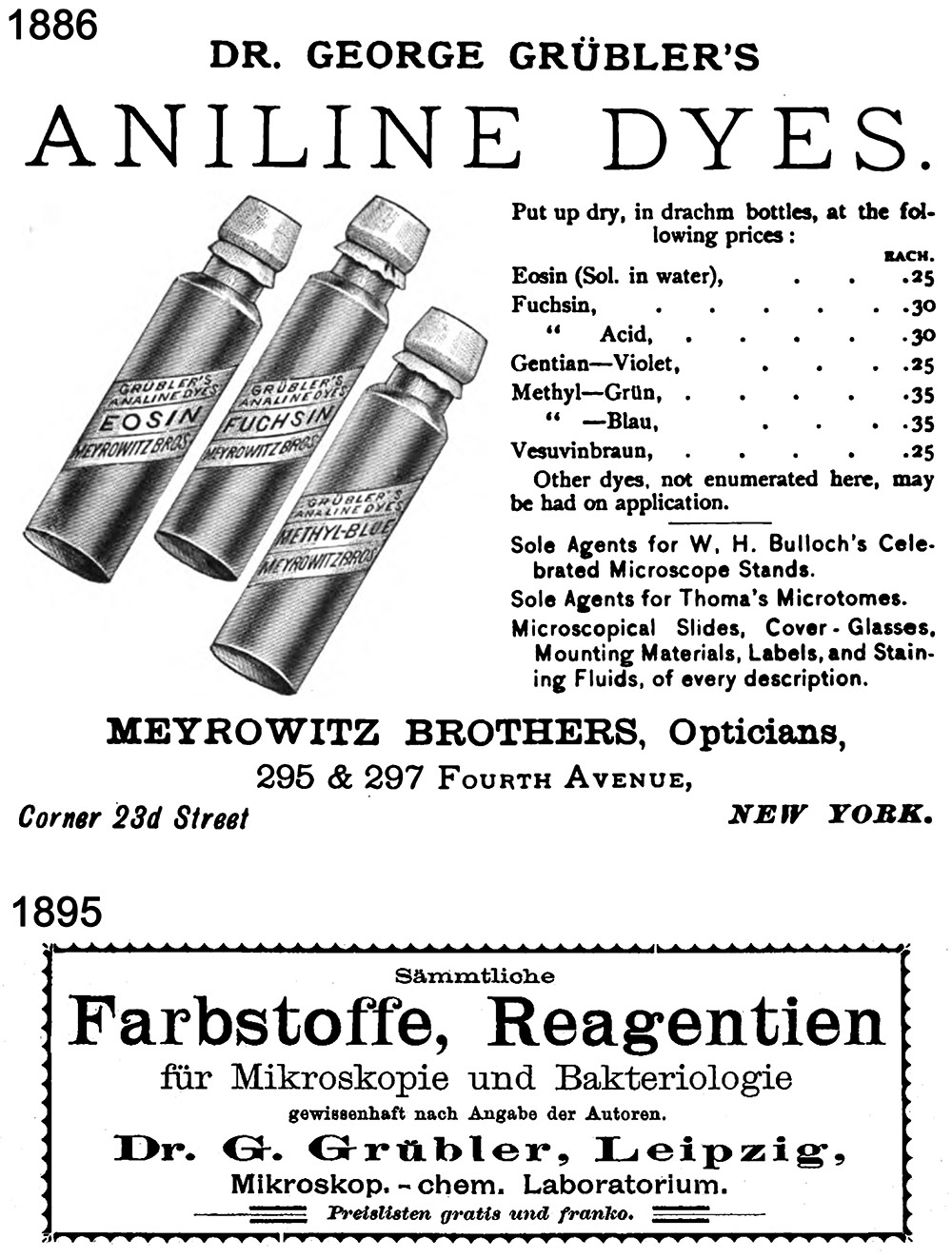
Figure 3.
Advertisements for George Grübler’s biological dyes, from “The Journal of the New York Microscopical Society” (USA, 1886) and “Dr. Paul Börner's Reichs-Medicinal-Kalender für Deutschland” (Germany, 1895).
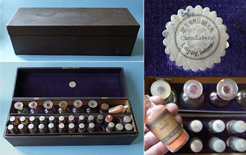
Figure 4.
A ca. 1890 stain kit from George Grübler. Adapted for nonprofit, educational purposes from https://www.kugener.com/de/laboratoires-d-analyses/67-artikel/1476-mikroskopie-faerbemittel-von-gruebler.html
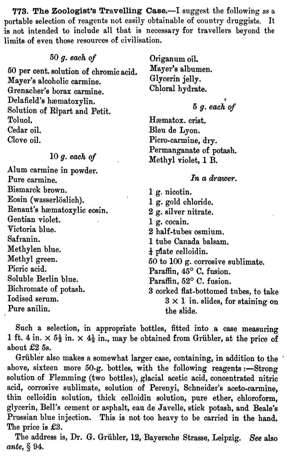
Figure 5.
Description of Grübler’s “Zoologist’s Travelling Case”, as described in Arthure Bolles Lee’s 1890 “The Microtomist’s Vade-mecum”.
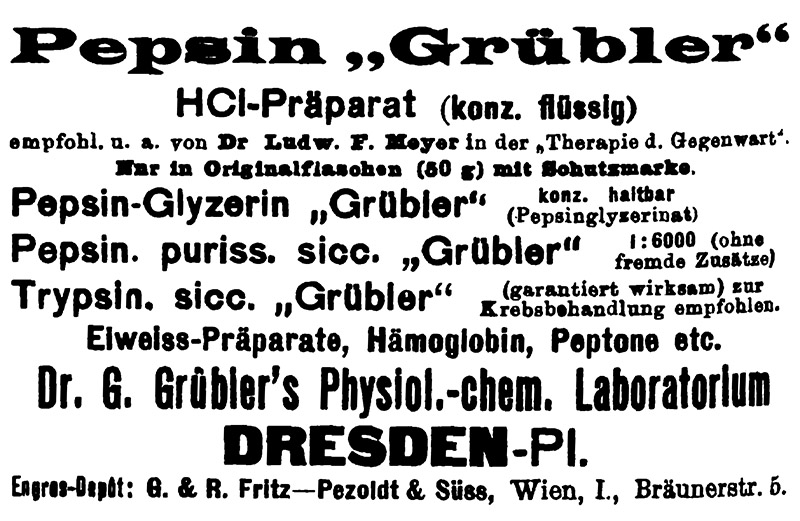
Figure 6.
After selling his dye business to Karl Hollborn in 1897, George Grübler opened business in Dresden that provided enzymes and other proteins for physiological and biochemical analyses. From “Pharmazeutische Post”, 1909.
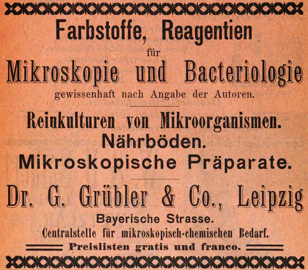
Figure 7.
1898 advertisement from Karl Hollborn’s business, “Dr. G. Grübler & Company”, including notice that the firm provided preparations for the microscope (“Mikroscopische Präparate”).
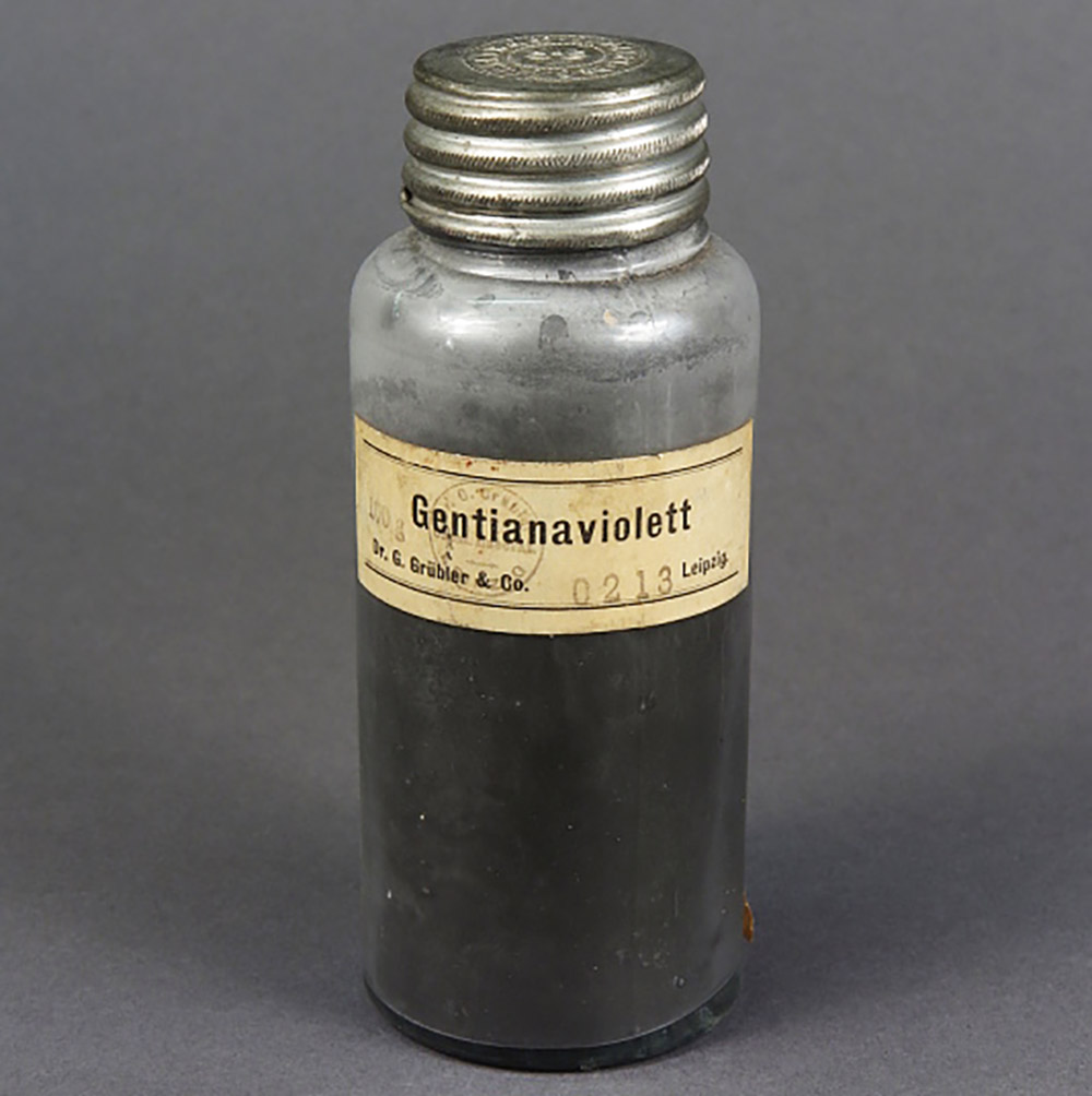
Figure 8.
A vial of gentian violet stain from “Dr. G. Grübler & Co.”. Adapted for nonprofit, educational purposes.
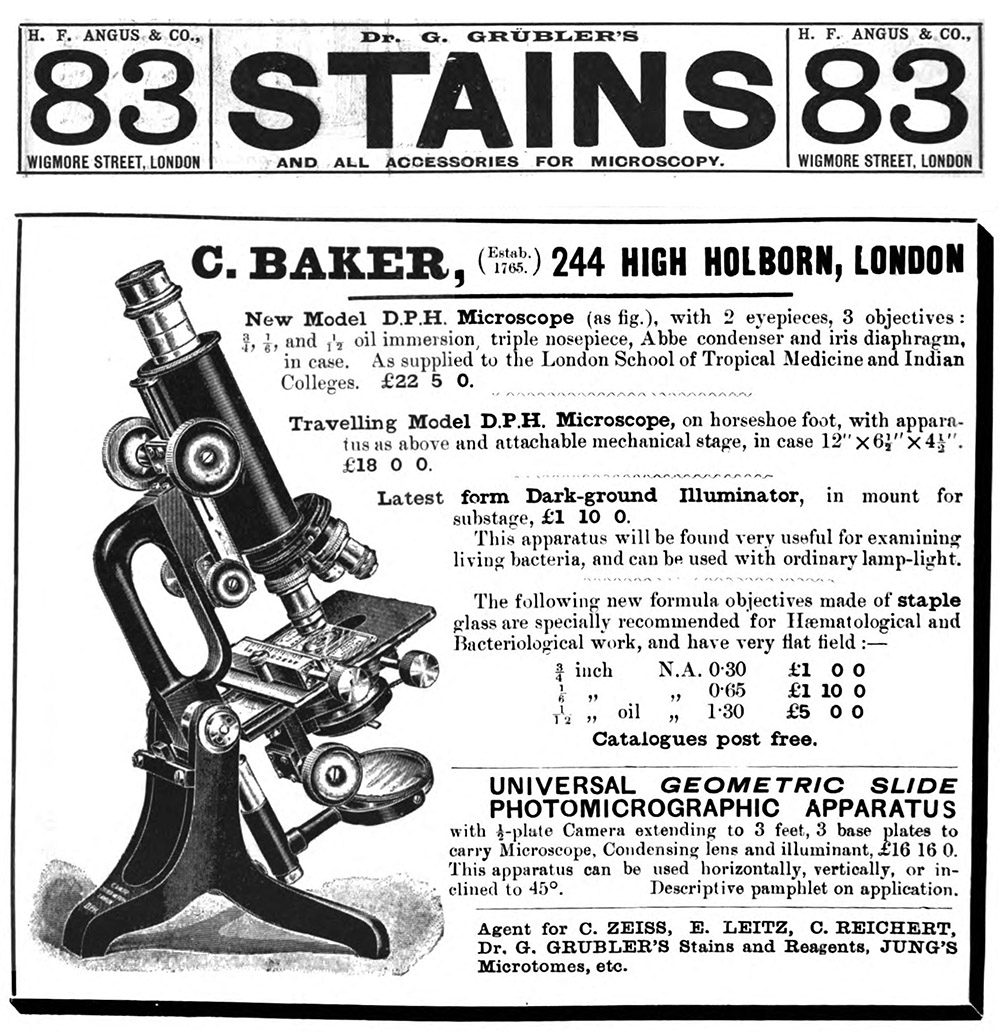
Figure 9.
Advertisements from English dealers for stains from Hollborn’s “Dr. G. Grübler”. From (top) “The British Medical Journal”, 1899, and (bottom) “The Journal of the London School of Tropical Medicine”, 1912.
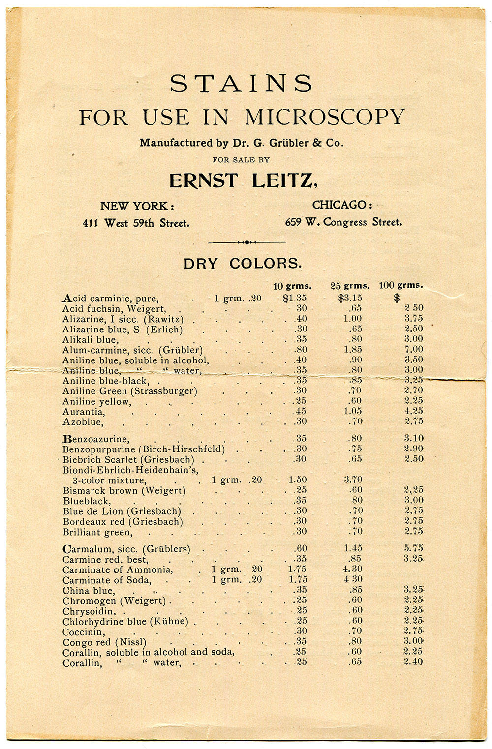
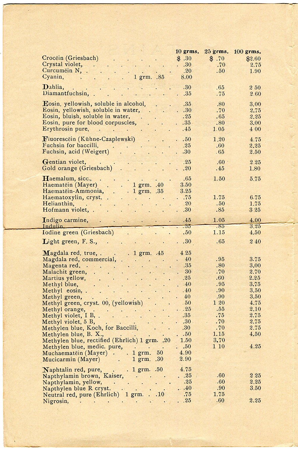
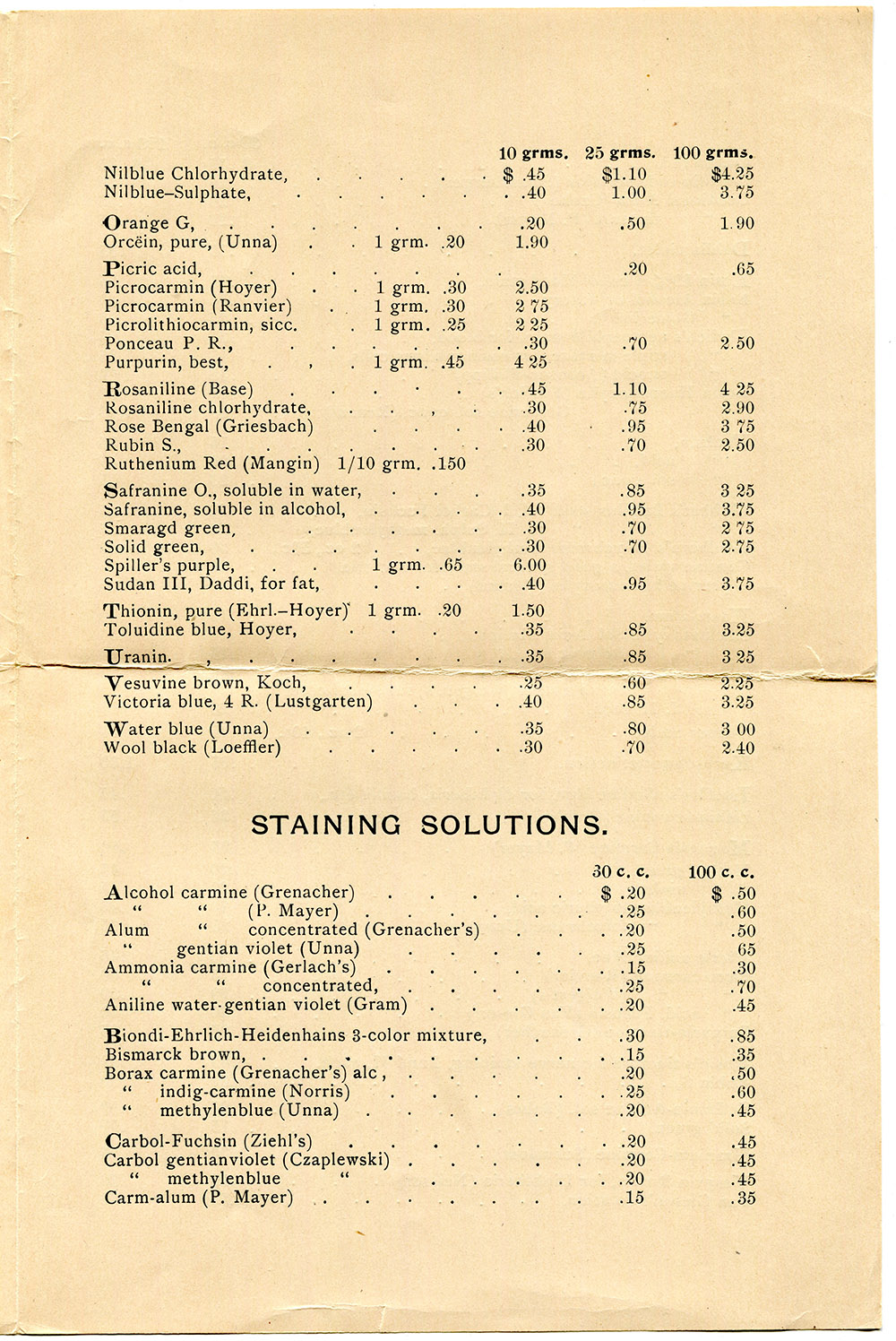
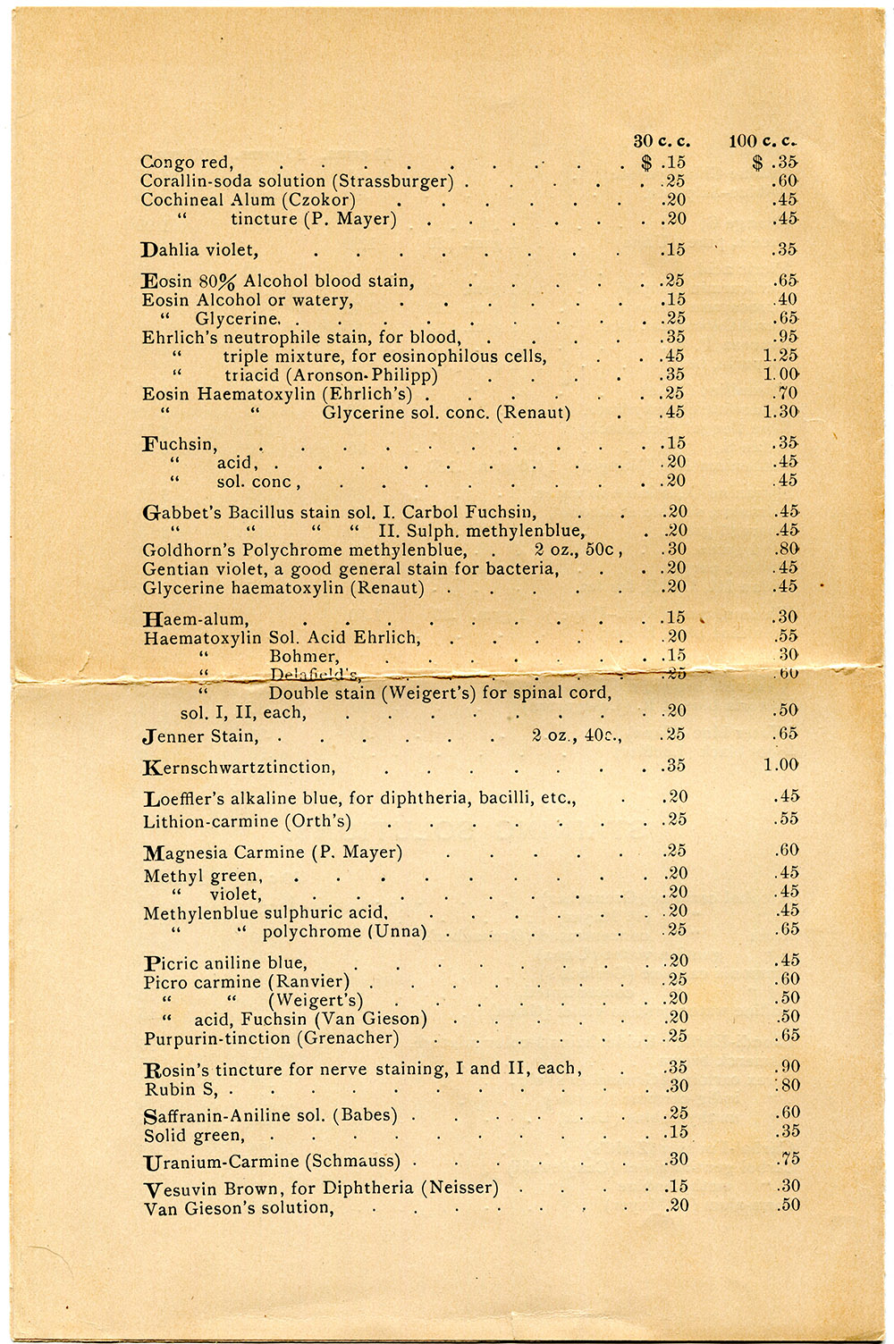
Figure 10.
A 1903 list of stains by “Dr. G. Grübler & Co.” that were distributed in the USA by Ernst Leitz.
Resources
Arbeiten aus dem Bacteriologischen Institut der Technischen (1898) Advertisements from Dr. G. Grübler & Co., multiple issues
Biologisches Centralblatt (1898) Advertisements from Dr. G. Grübler & Co., multiple issues
The British Medical Journal (1899) Advertisement from H.F. Angus for G. Grübler’s stains, page 38
Chemiker-Zeitung (1897) “Leipzig. Die Firma Dr. G. Grübler's mikroskopisch-chemisches und bakteriologisches Laboratorium ist auf Dr. phil. Karl Theodor Immanuel Hollborn übergegangen; künftige Firmirung Dr. G. Grübler's mikroskopisch-chemisches und bakteriologisches Laboratorium (Dr. K. Hollborn)”, page 452
Chemiker-Zeitung (1897) “Neue Firmen: … Dresden, Dr. G. Grübler's physiologisch-chemisches Laboratorium”, page 483
Dr. K. Hollborn & Söhne GmbH & Co. KG (website accessed February, 2024) http://www.hollborn.de
Dr. Paul Börner's Reichs-Medicinal-Kalender für Deutschland (1895) Advertisement from George Grübler
The Journal of the London School of Tropical Medicine (1912) Advertisement from C. Baker
Journal of the New-York Microscopical Society (1886) Advertisement from Meyrowitz Brothers
Hollborn, Karl, and Herbert F. Angus (1900) The history and theory of staining, The Illustrated Annual of Microscopy, pages 25-27
Lee, Arthur Bolles (1890) The Microtomist's Vade-mecum: A Handbook of the Methods of Microscopic Anatomy, second edition, P. Blakiston, Son & Co., Philadelphia, pages 54 and 388-389
Pharmazeutische Post (1909) Advertisement from G. Grübler, Dresden, Vol. 4, page 288
Schafer, E.A., C.S. Sherrington, R.W. Boyce, W.H. Thompson (1897) The physiological effects of peptone and its precursors when introduced into the circulation, Report of the Meeting of the British Association for the Advancement of Science, pages 531-537 (used products from George Grübler's Dresden business)
Titford, Michael (1993) George Grubler and Karl Hollborn: Two founders of the biological stain industry, Journal of Histotechnology, Vol. 16, pages 155-158
Titford, Michael (2007) Comparison of historic Grübler dyes with modern counterparts using thin layer chromatography, Biotechnic and Histochemistry, Vol. 82, pages 227-234