
Figure 1. Examples of microscope slides that were prepared by Henry Underhill. He generally wrote the date and method of preparation, and occasionally signed slides with his initials or surname.
Henry Michael John Underhill (H.M.J.U.), 1855-1920
by Brian Stevenson
last updated October, 2015
Henry M.J. Underhill’s microscope slides can generally be recognized by the tidy handwritten labels that list the date and describe the method by which he prepared the specimen. They are usually neatly made, and are often artistically arranged.
Underhill, who was familiarly called Harry, was a relatively well-to-do grocer in Oxford, England. He began studies with a microscope when in his teens, and wrote extensively on a variety of related topics. He was also one of the first to use a microtome to produce serial sections of insects, spiders, flowers, etc., thereby permitting detailed examinations of internal structures.
Figure 1.
Examples of microscope slides that were prepared by Henry Underhill. He generally wrote the date and method of preparation, and occasionally signed slides with his initials or surname.
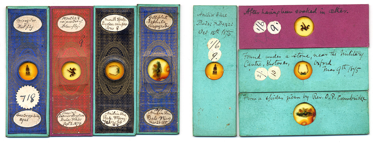
Figure 2.
Additional slides by Underhill. It was not uncommon for him to write additional information on the reverse, giving mounting technique or specimen sources.
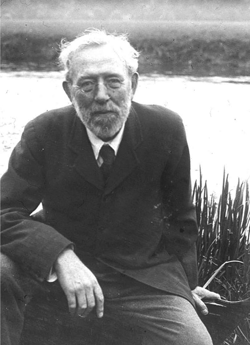
Figure 3.
Undated photograph of Henry Michael John Underhill. Courtesy of Dr. Megan Price.
H.M.J. Underhill was born January 14, 1855, the eldest son of Henry Scrivener and Ann Underhill. The father was a grocer, as was his own father – the Underhills having been merchants in Oxford for several centuries. Harry’s uncle, Charles Underhill, served as Mayor of Oxford from 1887-88.
Harry exhibited interests in art and science as a child. He was taught oil and watercolor painting by Adrian Reviére. A painting he produced when only 12 years old was shown in a local gallery. His artistic skills show in many of Underhill’s microscope slides, and in the illustrations he made to accompany many of his writings. He was also known for his skills of painting magic lantern slides – an excellent on-line article on that subject, by Deborah Harlan and Megan Price, can be seen at http://archaeologydataservice.ac.uk/archives/view/underhill_na_2004
Evidently, Harry developed microscopy skills as a young teenager. He published two exchange offers in Hardwicke’s Science-Gossip when only 16. Both probably refer to objects for the microscope: “Batrachospermum Moniliforme - For this alga, send stamp and object (other algae preferred) to H.M.J. Underhill, 7, High-street, Oxford”, and “For Palates of Neritina fluviatilis and Paludina vivipara (unmounted) send stamp and objects of interest to H.M.J Underhill, 7, High Street, Oxford”.
He was definitely making slides by the following year, 1872, as indicated by this request, “For exchange, slides of Entomological objects, &c. Send lists to H.M.J. Underhill, 7, High Street, Oxford”. Two of the slides illustrated in Figure 1 were produced in 1872, when Harry was 17 years old.
These two exchange advertisements appeared in 1873, “Wanted, specimens of Sabanus and Gasterophilus preserved in spirit, or mounted or unmounted tongues and other parts of the same. Good mounted or unmounted objects (mostly entomological) offered in exchange - H.M.J. Underhill, 7, High-street, Oxford”, and “Wanted, Specimens of Breeze-flies (Tabani) and Gad-flies (Gasterophili) preserved in spirit, or mounted or unmounted parts of the same. Slides or unmounted objects offered - H.M.J. Underhill, 7 High-street, Oxford”.
A query published by Underhill in the December, 1873, issue of Hardwicke’s Science-Gossip indicates that Underhill was still learning slide-making techniques, “Palates of Spiders: Will F. Bernard, who wrote a short article on ‘palates of spiders’ in the May number of this magazine, mind inserting a notice in next month’s Science-Gossip, stating his method of preparing insect specimens with carbolic acid and chloroform? I have tried it, and found it a very expeditious way of mounting, but I totally failed in making good slides, because I could not by any means get rid of the ‘interior arrangements’, which on being pressed squeezed out. - H.M.J. Underhill”.
Underhill’s skills at microscopic mounting, dissection, and illustrating developed to excellent levels by the time he turned 20. In 1875, he published an authoritative, two-part article on “Spider’s webs and spinnerets”. It was illustrated by several drawings from Underhill (Figure 4). A diagram of a cross-sectioned spider abdomen indicates that he had also become proficient with use of the microtome.
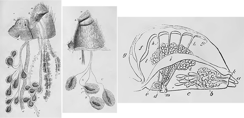
Figure 4.
Drawings of spider silk-spinning organs and a cross-section of a spider abdomen by Underhill, from his 1875 articles on ‘Spider’s webs and spinnerets’.
During 1875 and 1876, Underhill and his cousin, Frank J. Allen (1854-1942), wrote a 6-part series on various dipteran flies. Allen was the son of Alfred Allen, Secretary of The Postal Microscopical Society and Editor of the Society’s journal. Underhill’s artistic skills highlighted the diptera papers (Figure 5).
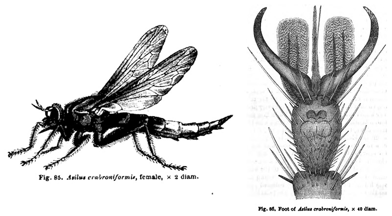
Figure 5.
Drawings of a fly, and that species’ foot, by Underhill, from the 1875-76 series of papers on diptera written by him and Frank Allen.
In 1879, Underhill wrote an authoritative two-part series on “The preparation of insects for microscopical examination”.
Harry undertook a lengthy investigation of the internal structures of spiders, and published two papers on the topic in 1886. Both were reprinted in the Journal of the Postal Microscopical Society in 1889. His studies were aided by cutting fine, serial sections of the spiders, which were then laid out alongside each other on microscope slides. One can then move from section to section and mentally assemble a three-dimensional image.
At the October 22, 1886, meeting of the Quekett Microscopical Club, Henry Freeman presented a talk on Underhill’s work, “Mr. Freeman said he had brought a series of sections of Spiders, cut by Mr. H.M.J. Underhill, of Oxford, who, a few years ago, wrote some articles on Spiders, and Diptera in ‘Science Gossip’. Mr. Underhill had recently been cutting sections of the Garden spider, Epeira Diadema, and from a careful study of these sections had prepared a drawing, giving, what he believed to be, a correct representation of the internal organs in their natural position. Some of the results of his investigations had been given to the world in an article which appeared in ‘The Welcome’ for the present month, a copy of which periodical was on the table. The principal drawing illustrating that article had been built up, as it were, from various sections taken in different directions through the Spider, it being evident that a single section would not convey a very comprehensive view of the interior economy of the creature. He (Mr. Freeman) had also brought the original drawings, which had been kindly lent by the editor of ‘The Welcome’. The article was written in a free and simple style, in order to interest the general public, and especially the young, but he thought it would be found interesting to many of the members. Mr. Underhill's chatty, though terse, style made the dryest details readable. In his next article he proposed to deal with the web-making, and web-making apparatus, of the spider. The sections were cut with an ordinary machine, and were remarkable not only on account of their anatomical value, but, like all Mr. Underhill's work, for their careful and methodical preparation. He (Mr. Freeman) remembered hearing Dr. B.T. Lowne, some years ago, suggest that a consecutive series of sections of the human spinal cord, from the top to the bottom, would be most instructive and valuable, and thought it could be done in a few hundreds of slides. Mr. Underhill had gone a good way in that direction, for some of his slides had as many as 60 distinct sections of a species of slug on a 3 x 1 slip. Of the Spider series some have as many as 32 sections on a slide, grouped with the greatest regularity. On seeing the article it occurred to him that some of the members of the Quekett Club would like to have an opportunity of examining the preparations themselves, and he therefore communicated with Mr. Underhill, who most kindly, perhaps somewhat rashly, sent some slides up for exhibition. He had asked Mr. Underhill to send one that would best illustrate the drawings. He said, in reply, ‘The fact is I have not one single perfect section. It is only by diligently comparing some number of slides that you can see all that I have put into the drawings. I think they are quite accurate’."
Underhill visited the Quekett Microscopical Club for their May 27, 1887, meeting. He presented further information on sectioning spiders and discussed their anatomy. He also exhibited a number of microscope slides for the Club, including “Section of Lycosa, showing brain, heart, gills, liver, silk glands, &c.; Small spider, unnamed; Eyes of Lycosa; Section of eye of Lycosa, showing lens, vitreous body, and retina; Spinnerets of Hair Curler Spider, Ciniflo similis; Threads of Ciniflo; Spinnerets of Clubiona, showing silk tubes; Nest of spider, Theridion; Calamistrum or ‘comb’ on hind leg of Ciniflo; Foot of Web-walking Spider, Epeira; and Foot of Ground-walking Spider, Dysdera”.
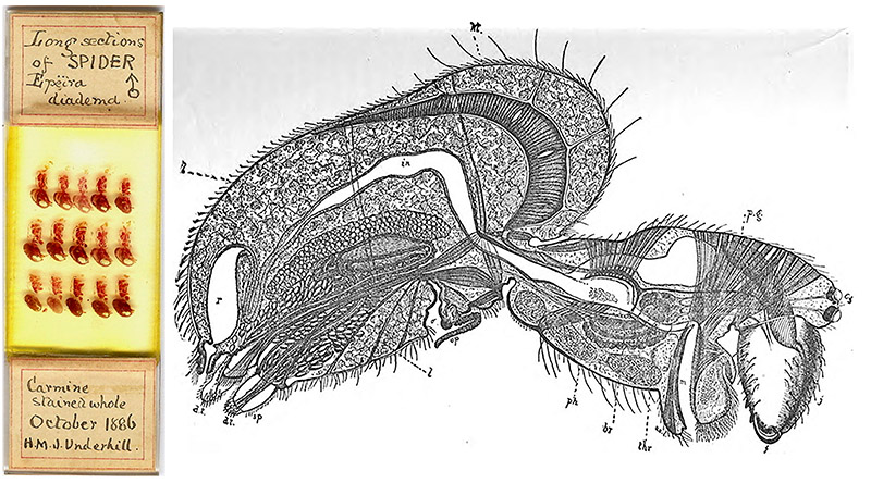
Figure 6.
An 1886 Underhill slide with 15 longitudinal sections of an Epeira diadema spider, and his drawing from that year’s paper on this spider. Slide picture courtesy of Trevor Gillingwater.
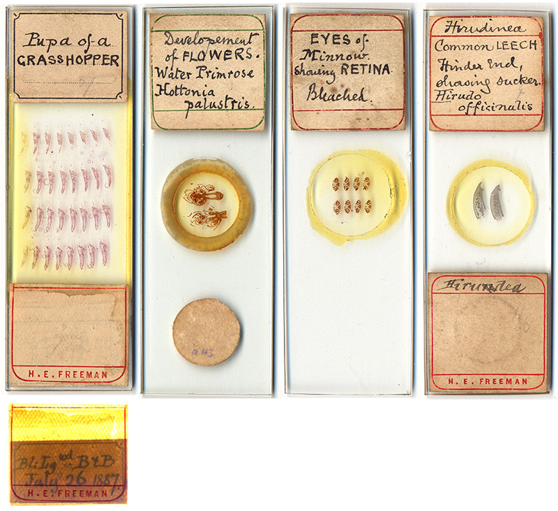
Figure 7.
Additional section slides by Underhill, two of which were additionally labeled by Henry Freeman. The slide on the left, of a grasshopper pupa, has Underhill’s label underneath Freeman’s, visible with strong back-lighting. An unsigned (and unidentified) slide of a sectioned Limax agrestis slug is shown in B. Bracegirdle’s ‘Microscopical Mounts and Mounters”, plate 43-K. Although Bracegirdle suggested that serial section slides would have been possible only after the invention of the automatic microtome in 1884, Underhill wrote that he made these slides with a standard microtome and a hand-held blade (see below). A detailed drawing of the internal organs of a cross-sectioned spider suggests that Underhill was using this method in the mid-1870s (see Figure 4).
Underhill’s sectioning method, excerpted from his 1888 instructional paper on the topic, was “Soak the insects in (common methylated spirits) for a week or two, or as much longer as you like. … Then let them have three or four days in absolute alcohol, changing the alcohol once. Transfer them to oil of cloves, in which let them stay for a day; and, after the oil of cloves, let them be in turpentine another day. … Now melt some paraffin wax before a gas-stove or over a water bath. Be very careful to have it only just above its melting-point. Warm the bottle of turpentine containing the specimens, pick them out, and drop them into the melted wax. This must be maintained at the same temperature for about ten hours. … The vessel in which the wax is allowed to get cold must be flat bottomed; and the objects should be arranged in it at intervals in such positions that the bottom of the vessel is at right angles to the plane of the sections which you intend to cut. When the wax is quite hard, the specimens may be carefully cut out in little cubes of wax, and kept for any length of time until wanted. … If you have a microtome of the common sort, with a well, fill the well with melted wax, and let it get quite cold. Then screw it up until about half an-inch of wax appears above the cutting plate of the instrument. Remove this with the razor ... Take now one of the cubes of wax containing an insect to be cut; square it roughly with a pen-knife; fasten it to the wax in the well by means of the heated blade of an old knife. When it is cold and hard again, finish off the squaring process accurately, taking care that the opposite sides are parallel. Screw down the machine until the top of the little cube is level with the surface of the cutting plate, and the specimen is ready for cutting. Take off a few preliminary slices, and when you have cut down to the object, place the razor with its edge parallel to and almost touching one of the sides of the cube. Draw it sharply towards you without any motion sideways, but in a direction exactly at right angles to that side of the block with which its edge is parallel. Let the section remain on the razor blade, and after again turning up the screw of the microtome, repeat the cut, using exactly the same portion of the razor edge to cut with by placing the section already cut precisely behind the little squire block of wax. The new section will stick to the edge of the first one and push it across the razor blade. The same process, indefinitely repeated, will produce a ribbon of sections almost as neatly as a rocking microtome will do it. … After a little practice, I could readily get unbroken ribbons of 20 to 40 sections. After this, they became too long to be manageable. The advantages of ribbons over single sections I consider to be two: they are much easier to manipulate; and they facilitate getting a series of sections in their proper order. This is an important point when you are cutting up an insect. Besides, having all the sections cut before any are put upon the slide, you are able to count them, and so calculate the space they will occupy. You can, therefore, arrange them with proper regard to the middle of the slide - a great thing to all who love neatness”.
Underhill was a frequent contributor to the Postal Microscopical Society’s Journal of Microscopy and Natural Science in the late 1880s and throughout the 1890s. He offered mounting and collecting tips, as well as descriptive articles on various topics.
He was a charter member of the Oxfordshire Natural History Society, and served as its President in 1893. He became an avid photographer, and joined the Oxford Camera Club. It was said that Underhill’s photographs “were as near perfection as artistic feeling, technical skill, and good instruments could make them”.
Harry Underhill never married. The 1911 census shows him living with his unmarried sister, Maud, and a domestic servant. He died of cancer on October 2, 1920, at the age of 65. An obituary wrote, “Reticent and unassuming, Harry Underhill passed his life within a narrow circle, yet was a well-known man in the city and universally respected. He was beloved and faithfully served by a long succession of employees, many of whom had begun as boys in the old-established firm of M. Underhill and Sons, and grown grey in the service of his father and himself. All who met him could not but feel that he possessed great force of character and reserve of strength, and could a kindred spirit induce him to unbend, he would find Harry Underhill one of the most delightful and entertaining of companions”.
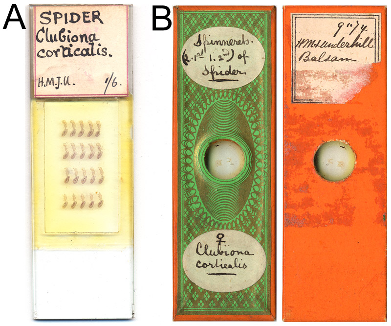
Figure 8.
Two microscope slides that were produced by Harry Underhill, and re-labeled by later owners.
A. Longitudinal sections of the spider Clubiona corticalis.
B. Spinnerets of C. corticalis, dated September, 1874, and re-papered by Edward Ward.
Acknowledgements
Many thanks to Dr. Megan Price of Oxford University for enthusiastically sharing information on Harry Underhill, and to Trevor Gillingwater for generously providing images of Underhill’s slides.
Resources
Allen, Frank J., and Henry M.J. Underhill (1875) Notes on the Diptera, Hardwicke’s Science-Gossip , Vol. 11, pages 79-81 and 147-150
Allen, Frank J., and Henry M.J. Underhill (1876) Notes on the Diptera, Hardwicke’s Science-Gossip , Vol. 12, pages 60-62, 103-106, and 155-159
Bracegirdle, Brian (1998) Microscopical Mounts and Mounters, Quekett Microscopical Club, London, pages 96 and 190, plate 43-K
England census, birth, marriage, and death records, accessed through ancestry.co.uk
Gunther, Robert T. (1937) Early Science in Oxford, Vo. 11, self-published, Oxford, pages 321-322
Hardwicke’s Science-Gossip (1871) Exchange offers from H.M.J. Underhill, Vol. 7, pages 144 and 168
Hardwicke’s Science-Gossip (1872) Exchange offer from H.M.J. Underhill, Vol. 7, page 284
Hardwicke’s Science-Gossip (1873) Exchange offers from H.M.J. Underhill, Vol. 9, pages 168 and 192
Harlan, Deborah, and Megan Price (2004, updated 2013, accessed September, 2015) H.M.J. Underhill Archive, http://archaeologydataservice.ac.uk/archives/view/underhill_na_2004
Journal of the Royal Microscopical Society (1887) Notes on Freeman’s presentation to Quekett, page 698
Oxford Chronicle (1920) Obituary: “Mr. H.M. Underhill”, October 8
Price, Megan (2008) Amateurs and professionals in nineteenth-century archaeology; The case of Oxford ‘Antiquarian and Grocer’ H.M.J. Underhill (1855-1920), in Archives, Ancestors, Practices: Archaeology in the Light of its History, ed. by N. Schlanger and J. Nordbladh, Berghahn Books, Oxford, pages 109-119
Price, Megan (2008) H.M.J. Underhill (1855–1921); Oxford antiquarian, artist, and ‘provision merchant’, Oxoniensia, Vol. 73, pages 99-120
Probate of H.M.J. Underhill (1920) “Underhill Henry Michael John of 231 Woodstock-road Oxford grocer died 2 October 1920 Administration Oxford 10 January to George Edward Underhill, esquire. Effects £6435 18s 7d”, accessed through ancestry.co.uk
Oxford History: Mayors & Lord Mayors (accessed September, 2015) Charles Underhill, http://www.oxfordhistory.org.uk/mayors/1836_1962/underhill_charles_1887.html
Quekett Journal of Microscopy (1886) Ordinary meeting of October 22, 1886, Series 2, Vol. 3, pages 24-25
Quekett Journal of Microscopy (1887) Ordinary meeting of January 28, 1887, “The following objects were exhibited : serial sections of slug, Limax agrestis, cut by Mr. J Underhill, (exhibited by) Mr. H.E. Freeman”, Series 2, Vol. 3, page 88
Quekett Journal of Microscopy (1887) Ordinary meeting of May 27, 1887, Series 2, Vol. 3, pages 157-159
Underhill, Henry M.J. (1873) Palates of spiders, Hardwicke’s Science-Gossip Vol. 9, page 283
Underhill, Henry M.J. (1875) Spiders’ webs and spinnerets, Hardwicke’s Science-Gossip, Vol. 11, pages 88, 132-135, and 195-198
Underhill, Henry M.J. (1875) The preparation of insects for microscopical examination, Hardwicke’s Science-Gossip, Vol. 15, pages 101-103 and 121-122
Underhill, Henry M.J. (1888) Section-cutting applied to insects, Hardwicke’s Science-Gossip, Vol. 24, pages 1-4
Underhill, Henry M.J. (1875) Spider gossip, part 1, Journal of the Postal Microscopical Society, Vol. 8, pages 16-25
Underhill, Henry M.J. (1875) Spider gossip, part 2: How the spider makes her web, Journal of the Postal Microscopical Society, Vol. 8, pages 86-95