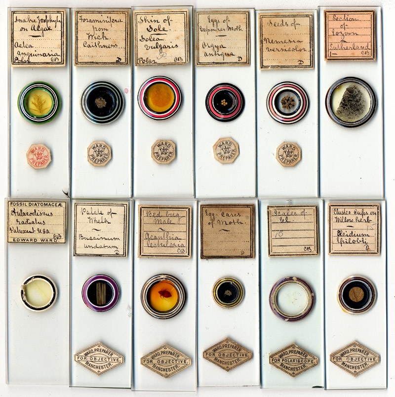
Figure 1. Examples of Edward Ward’s commercially-prepared microscope slides.
Edward Ward, 1844 – 1901
by Brian Stevenson
last updated November 2015
From the late 1870s to the turn of the 20th century, Edward Ward was a major producer of microscope slides, material for slide-making, and other supplies in Manchester, England. His slides are encountered fairly often in auctions nowadays, and are generally still in excellent condition, attesting to Ward’s great skill and his significant output. Ward’s later slides, which frequently carry his commercial label, tend to be artistically ringed with alternating light and dark paints (Figure 1). His handwriting is easy to recognize. In addition, Ward usually noted the mounting method used in his slides: “CB” for Canada balsam, “G” for glycerine jelly, and “D” for dry. Numerous records indicate that Ward was an active amateur (possibly semi-professional) microscopist for at least a decade before he began his business in Manchester (see below). Handwriting comparisons indicate that Ward’s earlier productions most often used off-the-shelf green front papers, with bright orange (occasionally red) back and side wrapping paper (Figures 2 and 3). He used the same combination to re-paper slides he acquired from other microscopists (Figure 3).
Figure 1. Examples of Edward Ward’s commercially-prepared
microscope slides.
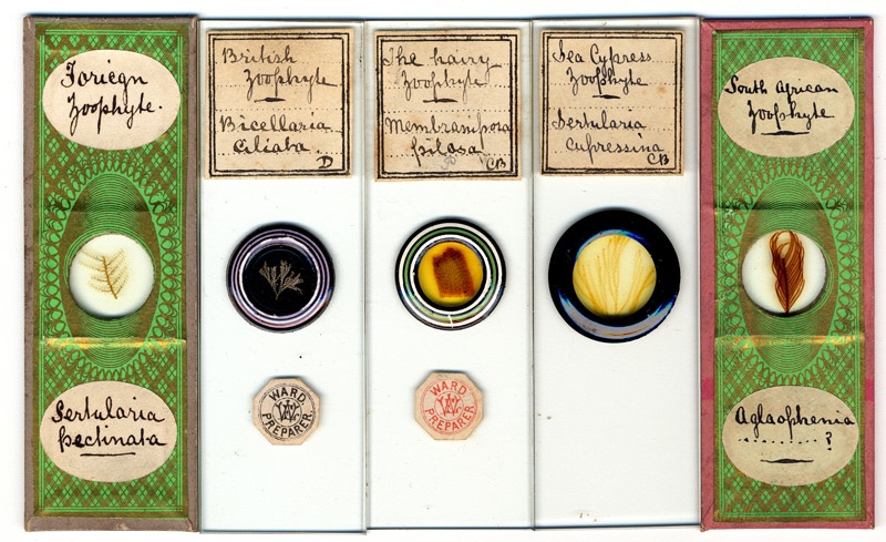
Figure 2. Three of Ward’s post-1880 commercial preparations,
flanked by earlier, paper-covered slides bearing the same handwriting. Neither
of the papered slides bear source notations (see Figure 3), implying that Ward
produced these slides himself. They may have been made for Ward’s private
collection, for exchange with other microscopists, or, possibly, for sale.
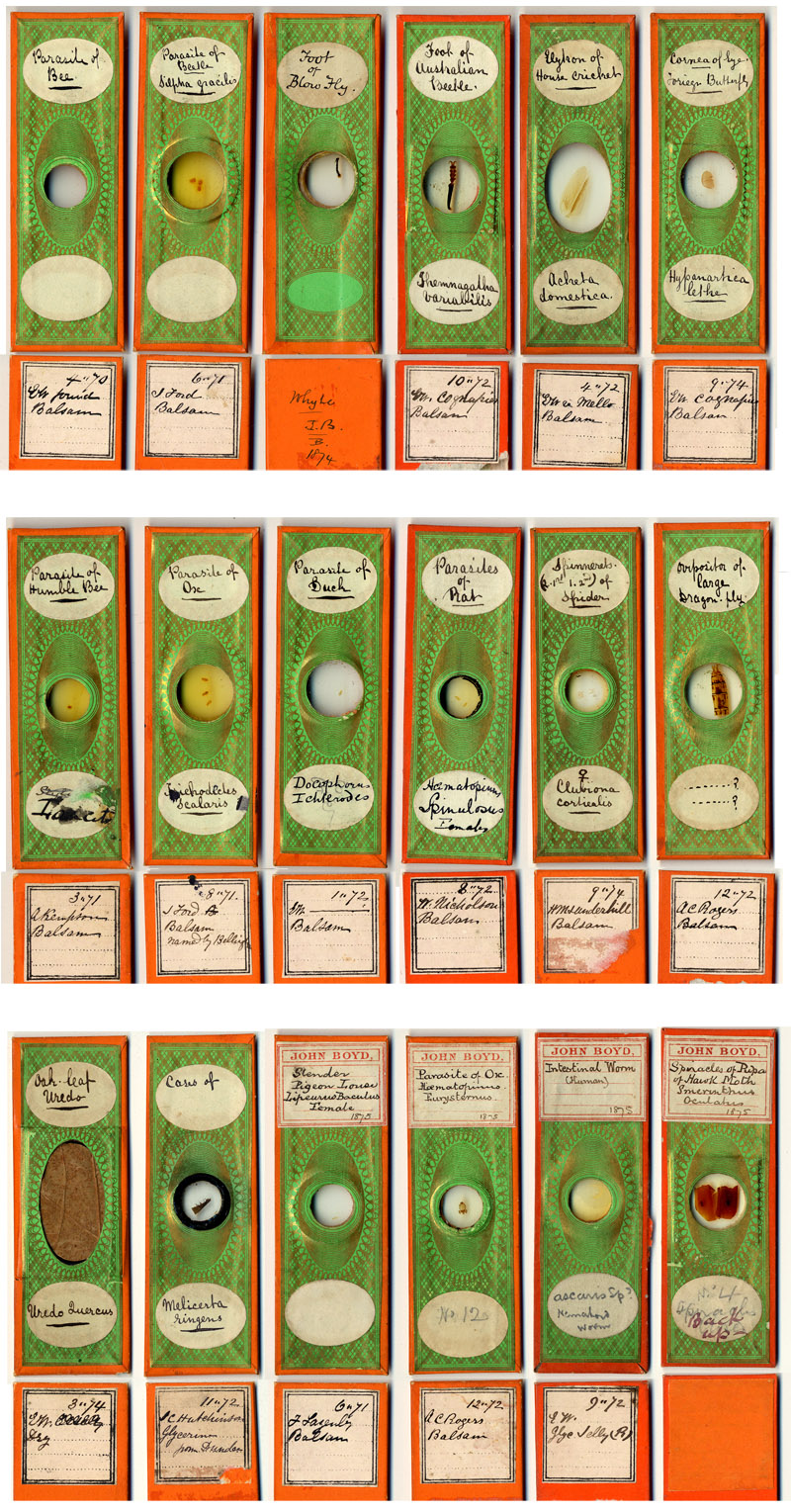
Figure 3. Early, papered microscope slides bearing Edward
Ward’s handwriting, all dated from the early 1870s. Each slide carries another
person’s name on its reverse, apparently the source. Ward frequently advertised
to exchange slides and material during the dates listed on these slides. Notes
on the slides’ sources are included at the end of this essay. The rightmost
slide in the lower row has a square of orange paper covering the source label;
the color is similar to that used by Ward when covering these slides, but I
have no idea why he decided to cover this label. The bottom row shows four Ward
slides that have labels from John Boyd over Ward’s upper oval label. Boyd and
Ward were both members of the Manchester Microscopical Society. Presumably,
these slides were once in Edward Ward’s private collection, and were later acquired by Boyd. An illustrated biography of John Boyd is also presented on this web site.
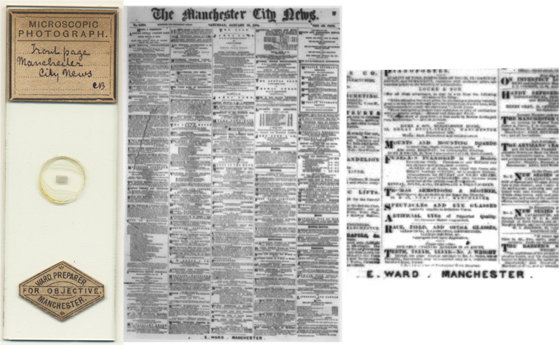
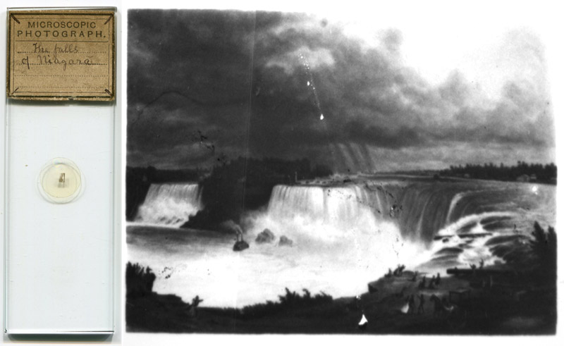
Figure 4A.
Two microphotograph slides retailed by Edward Ward. He was a skilled photographer, and produced the top slide, being the Janauary 19, 1884 issue of the Manchester City News. Note that Ward’s name is incorporated into the photograph, evidence that he made this himself. The lower slide is probably a relabeled mount from John B. Dancer or his successors, being microphotograph number 264 on Dancer’s 1877 list of microphotographs.
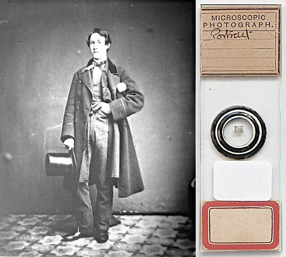
Figure 4B.
Microphotograph slide of an unidentified man. The handwriting and label suggest that this was produced by Edward Ward, presumably as a custom-made novelty for this person.
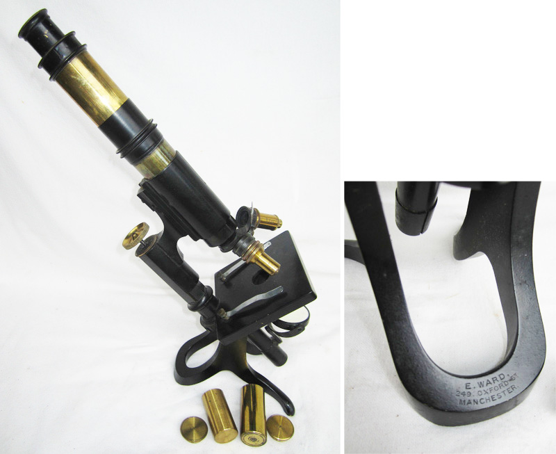
Figure 5.
A microscope, marked as having been sold by Edward Ward. There is nothing in Ward’s background to suggest that he possessed skills in either lens grinding or brass working, so it is very likely that Ward bought the entire microscope from a wholesale manufacturer. Images adapted from an internet auction site for nonprofit, educational purposes.
Edward Ward died in August, 1901. Brian Bracegirdle’s Microscopical Mounts and Mounters noted that magazine advertisements from “Edward Ward” appeared in 1912, and several photographs are known that bear the name “Ward” and address, but date from the 1910s and 1920s. Those photographs and the advertisements referred to by Dr. Bracegirdle were from Ward’s son, Edward Vincent Ward (1872-1921). The son operated from the same location as had his father (249 Oxford Street, Manchester), until his death in 1921 (see Figures 19 and 20 at the very end of this article for examples of the son’s work).
The life of Edward Ward (senior) was summarized in the following 1901 Science-Gossip obituary:
“Edward Ward.—A well-known figure in Manchester scientific society was recently removed by the death of Mr. Edward Ward at the age of 57 years. He was born at Coventry, where in his early life he worked as a ribbon weaver. Having a natural taste for scientific investigation, he soon became possessed of a microscope and later of a primitive camera. His tastes quickly brought him into association with others, which led to his leaving the loom for the vocation of commercial traveller. Notwithstanding the difficulties incident upon the constant change of locality when thus occupied, he contrived while on his journeys to study, dissect, stain, and mount thousands of objects. Mr. Ward eventually settled in Oxford Street, Manchester, in the neighbourhood of Owens College, where he established a ‘University Science Depot’ which afterwards became much patronised by the professors and students at the college. In 1887 he issued his first list of purely scientific lantern slides, which gave an impetus to science work in the district. He was one of the founders of the Manchester Microscopical Society, and for several years one of its presidents (sic, actually, it appears that Ward was a Vice President, not a President), and a lecturer in its Extension Section. It will, however, be on account of his photographic work that he will be best remembered. It is said that he took no less than ten thousand photographs of Geological Sections during the construction of the Manchester Ship Canal, and also of its chief engineering features. To attain such a remarkable pictorial history of that gigantic undertaking, Mr. Ward used at least once a month to walk along the whole course of the works between Manchester and the Mersey above Liverpool during the period of construction, which lasted beyond five years. In addition to these photographs he has left a large collection of other negatives of biological, botanical, and general scientific subjects. The remains of Mr. Ward were cremated at the Manchester Crematorium on August 28th last”.
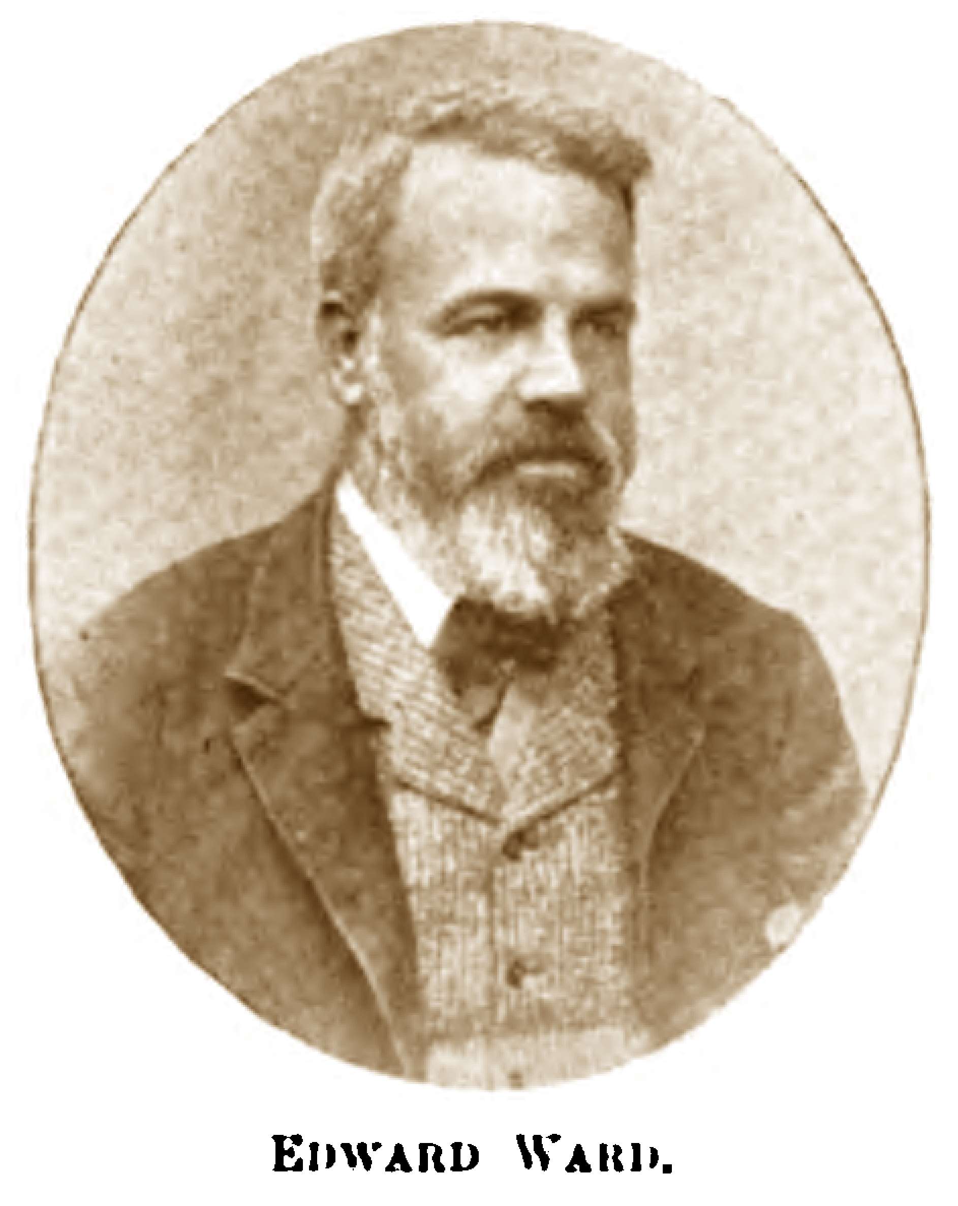
Figure 6. Photograph of Edward Ward, from his 1901 obituary in Science-Gossip.
Edward Ward was born in mid-1844, and christened June 20, 1844 at Holy Trinity Church, Coventry, Warwickshire. He was the second of two children (both sons) of Edward and Mary Innocent Ward. Edward, senior, was a ribbon weaver, as were a good many of the Ward’s neighbors. The 1861 census records that both 16 year-old Edward and his elder brother, William, were then “ribbon weaver apprentices”.
During the spring of 1869, Edward married Frances Goode, in Coventry. Their first child, Elizabeth was born in late summer, 1870, followed by son Edward Vincent in 1872.
Edward Ward’s interest in microscopical studies appears to pre-date 1870. In August of that year, he advertised to exchange a disparate variety of “well mounted” specimens (Figure 6). Thus, Ward believed that he made reasonable microscope slides by 1870, implying that he had been practicing for some time previous.
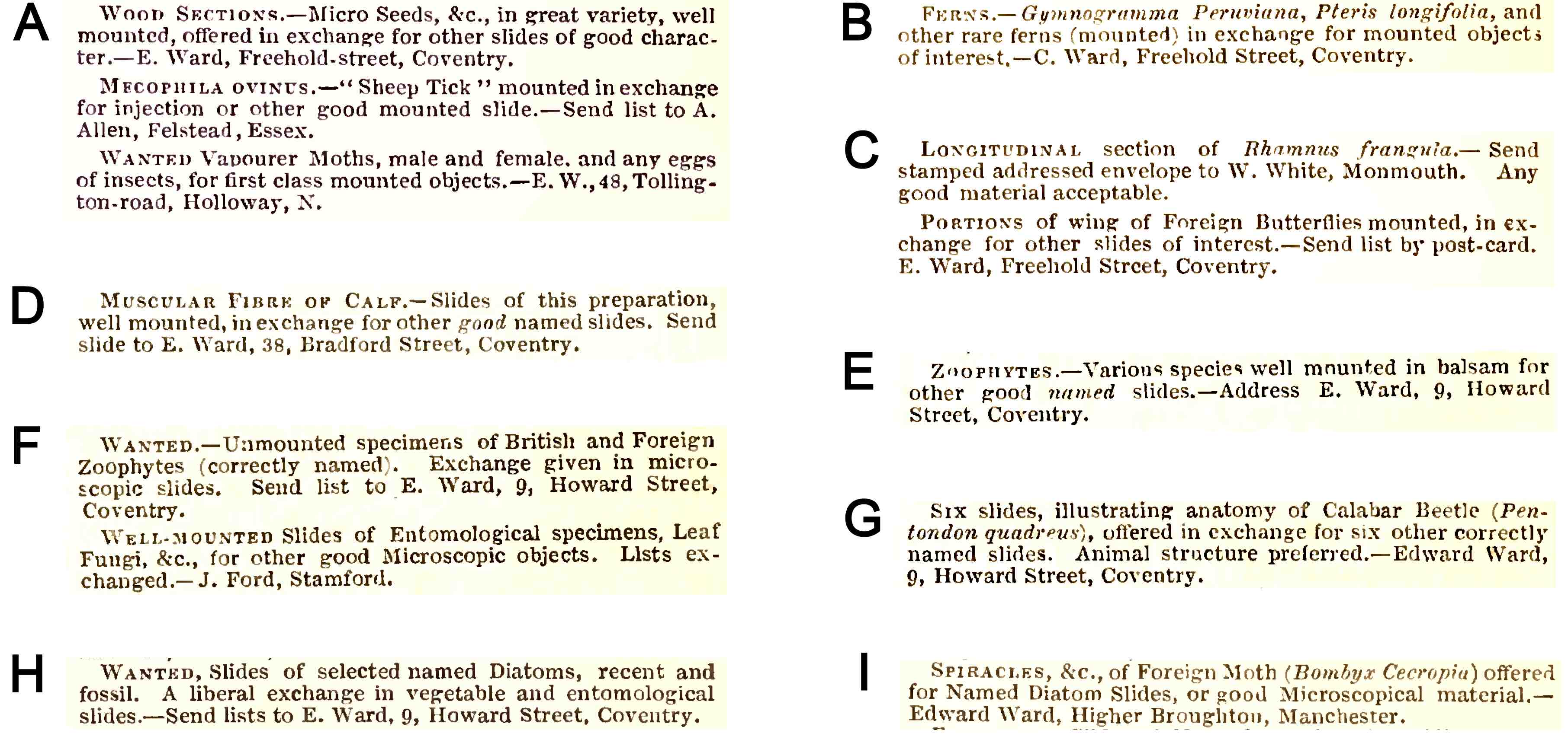
Figure 7. Early exchange offers published by Edward Ward in
Hardwicke’s Science-Gossip. Some exchange offers from other notable
microscopists that coincidentally ran alongside Ward’s ads are also shown.
(A) August, 1870. This is the earliest
known direct evidence of Edward Ward’s interest in making slides. Also shown
are exchange ads from Alfred Allen, of the Postal Microscopical Club, and
professional slide maker Edmund Wheeler. Ward owned several slides that were
made by Wheeler, it is possible that he acquired some of them through direct exchanges
with that maker.
(B) September, 1870.
(C) November, 1870. Also shown is an exchange offer from professional microscopist Walter White.
(D) March, 1871.
(E) July, 1871. Note Ward’s emphasis on obtaining named
specimens, implying frustration in receiving unidentified specimens. See also F.
(F) February, 1872. Ward is known to have exchanged at least one slide with amateur microscopist John Ford (see Figure 3).
(G) September, 1872.
(H) January, 1873.
(I) October, 1874. The ads shown in panels H and I indicate that
Ward moved from Coventry to Manchester during 1873-74.
The 1871 census recorded Edward Ward as being a “traveller” (i.e. traveling salesman), on that day boarding in Hull, Yorkshire. His wife and 9 month-old daughter were back home in Coventry, at 38 Bradford St. Exactly what Ward did while traveling is not known. His obituary indicates that it was connected with his interests in microscopy and photography, so he may have been selling equipment. That source also states that Ward prepared and mounted “thousands of objects” during this time, a volume that suggests commercial production.
At some point between January, 1873, and October, 1874, the Ward family moved to Higher Broughton, Manchester.
By 1879, Ward had opened a shop that specialized in microscope slides, unmounted specimens, and associated apparatus (Figures 6-9). He was also a distributor for Carl Zeiss’ microscopes and lenses. An 1882 advertisement in the Journal of the Postal Microscopical Club indicated that Ward retailed slides of selected and arranged foraminifera produced by Charles Elcock. He was also instrumental in the formation of the Manchester Microscopical Society, as reminisced by J.L.W. Miles in 1905:
“It has been thought desirable that an official record should be made of the origin of the Society, and for this purpose I have been asked to state, shortly, how the Society began its existence. Twenty-seven years ago, I made the acquaintance of the late Mr. Edward Ward, Microscopist (for some time one of our Vice Presidents), who at that time occupied premises in Burlington Street, and from him I purchased my first microscope and acquired a knowledge of microscopical work. In looking back, I cannot help but express my surprise at the energy and perseverance I then displayed in overcoming all the initial difficulties which I encountered. I was not long, however, in finding out how disappointing and disadvantageous it was to lack the acquaintance and companionship of others similarly occupied, and it naturally occurred to me that if only a dozen amateur microscopists could be found in the neighbourhood of Manchester willing to combine for mutual help and intercourse, a club or society could be formed on lines beneficial to every member. After several conversations with Mr. Ward, who offered to assist me as far as he could, I wrote a letter (in the autumn of 1879) which was published in the ‘City News’, advocating the formation of a society for the furtherance of microscopical study. In the correspondence which followed, my idea was somewhat deprecated. I was urged to join the Lower Mosley Street Natural History Society, or become a member of what, if I remember rightly, was called the Manchester Science Association, both of which were stated to utilise the microscope in scientific work. However. I was also the recipient of letters offering support and encouragement, and in the month of November, 1879, I wrote to about fifteen gentlemen stating my intention to call a meeting. This letter caused me to make the acquaintance of Mr. R. Bastow (afterwards the Society's first Secretary), who joined with me in calling a meeting and helping in the formation of the Society. This meeting was held on the 18th of December, 1879, at the old Mechanics' Institution, Princess Street, and was attended by some twenty-seven gentlemen. Dr. John Tatham was in the chair, and after some little deliberation the Society then and there started on its career. Thirty-three names were enrolled, Dr. Tatham was elected President, and a Provisional Committee was appointed (of which I was one) to draw up rules, &c. Amongst those who attended this meeting were several well known names, viz.: Messrs. E. Ward, Rd. Bastow, R. Graham, Thos. Brittain, W. Chaffers, T. W. Lofthouse, E. P. Quinn, W. W. Dawson and J. H. Furnival; all of whom afterwards did their utmost to make the Society a success.”
The 1881 census indicates that Ward’s shop location, 29 Burlington Street, was also the family home. On the night of that census, Edward Ward was staying at the Queen Square Hotel, London. He described himself as a “microscopist” for the census. It is not known whether he was still an active traveling salesman, or if he was in London for only a short trip from home. Ward joined the Royal Microscopical Society in October, 1881, so it may have been that this trip to London was associated with the RMS.
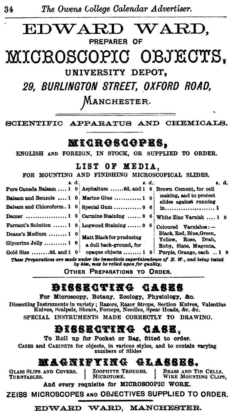
Figure 8. Edward Ward operated a commercial microscopy
enterprise by 1880. Shown is an advertisement from the 1880-81 University
Calendar of Owens College, Manchester. Ward’s business was located adjacent to
the college, now the University of Manchester. In addition to his own slides
and slide-making media, Ward was a distributor for Zeiss microscopes and lenses.
Figure 9. An 1880 Ward advertisement for unmounted objects,
for use in preparing microscope slides. From the Journal of the Royal
Microscopical Society.
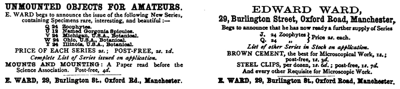
Figure 10. Advertisements from 1881 issues of Hardwicke’s
Science-Gossip. By that time, Ward was selling numerous pre-packaged sets of
unmounted objects, for customers to produce their own microscope slides (see
Figure 9).
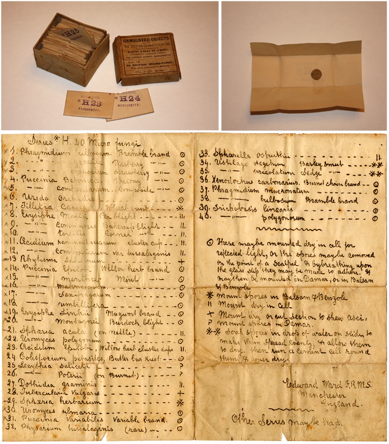
Figure 11. Edward Ward’s ‘Series H’ of unmounted specimens for
production of microscope slides, being British micro-fungi. Each envelope
contains a circular or rectangular section of a plant that is infested with a
particular fungus. Ward supplied a list describing each of the 40 specimens,
with recommendations for appropriate mounting media for each. Although the list
appears to be handwritten, it was actually machine printed, copied from an
original by Ward. This makes sense, since he probably moved hundreds or
thousands of each specimen set. The address of 249 Oxford Street dates this set to after 1882.
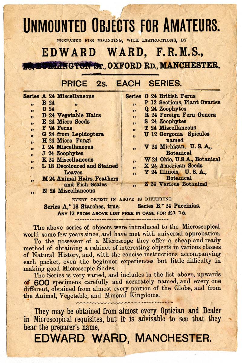
Figure 12. A flier advertising Edward Ward’s various series of unmounted micoscopical specimens. The crossed out Burlington Street address implies that the paper was printed prior to Ward’s ca. 1882-84 move to Oxford Street.
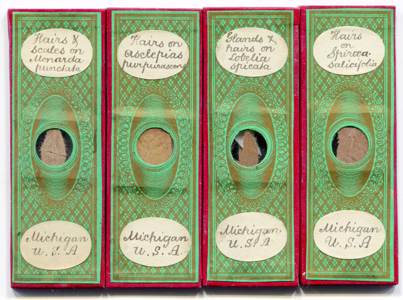
Figure 13. Four slides from England, with descriptions matching Ward’s Series V, "Michigan, U.S.A., Botanical". The sale of material from around the world by Ward and other dealers is one of the main reasons that foreign materials appear on so many English slides.
Figure 14. A slide-making guide by Ward. A standard, 1x3 inch slide would fit into the groove which is lined with light-colored paper. The concentric circles indicate the center of the slide, helping the mounter to accurately place a specimen and cover slip on the slide. This would ensure precise placement of ringing cement/paint when using a spinning ringing table.
A personal view of Edward Ward and his business was provided by this 1883 article in the American Monthly Microscopical Journal:
“Spending a short time in Manchester we were fortunate in discovering, quite by accident, attracted by the display of microscopes and apparatus in the shop-window, Mr. E. Ward, who is well known to most of our readers as a preparer of series of unmounted objects for microscopists. Entering, for a few moments' chat with Mr. Ward, we found him doing a thriving business in selling microscopes, material, and slides of his own mounting. After looking over a large collection of slides, and selecting a few choice ones for ourselves, Mr. Ward proposed that we should go out and make some collections. This was an opportunity not to be neglected; so, in a few moments we were on the tram-car bound for Withington, a suburb of Manchester. The road passes through the finest residence portion of Manchester. On either side, seated as we were on top of the car, we could look over high hedgerows or stone walls into the private grounds of the wealthy. The well kept shaded lawns and winding carriage ways, with here and there a porter's lodge at the gateway, reminded us of descriptions of English homesteads we had read in books. The hawthorn was in blossom, white or tinged with pink, and the laburnum trees were loaded with golden chains of flowers.
Finally we alighted and began our tramp for the open country. One really needs a guide to go collecting in England, for it is so difficult to find the right localities. Mr. Ward is an enthusiastic and experienced collector. He led the way to a place known by the curious name Gatley Carrs, where we found a path along a railroad. By turning over the leaves of the goutweed, and looking at the under surface, we found some good specimens of the fungus Puccinia aegopodia, which looked like black shining spots on the leaf. This was found in considerable abundance. Passing on we came to a turnstile, through which we passed into a path along a brook. This we followed a considerable distance, picking a few blue bells and buttercups to send home, but finding very little to collect. In the brook we found some beautiful Draparnaldia, in abundance, and some Vaucheria, but not much else of value. Still following the path which led away from the brookside, along green and cultivated fields, we at last reached a village, where we entered a public house to wait for the stage to take us back.
Ward does a large business in mounting slides for the trade, and in this work he has become very expert. We are pleased to learn, therefore, that his experience has fully confirmed what we have so frequently stated in these columns, that shellac makes the best cement for general use. Mr. Ward, however, has succeeded in making a mixture which he calls brown cement, shellac being the principal constituent. The brown cement is now to be obtained from the dealers, and has met with a ready sale
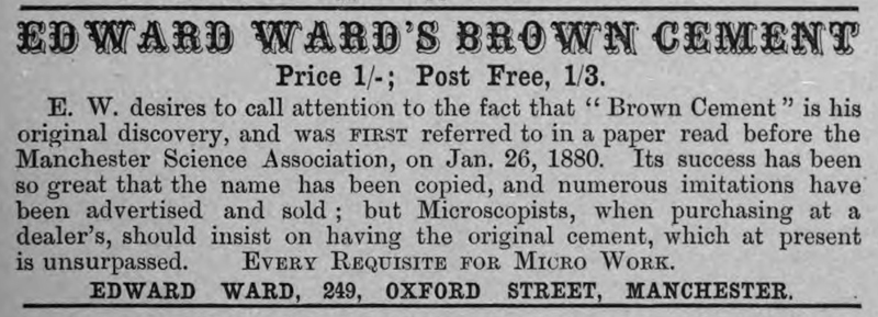
Figure 15. An 1884 advertisement for Brown Cement, from ‘The Journal of the Postal Microscopical Club’. Ward moved from Burlington St. to nearby 249 Oxford Road at some point between 1882 and 1884. This location was on a busier street, and still adjacent to Owens College (by then part of Victoria University).
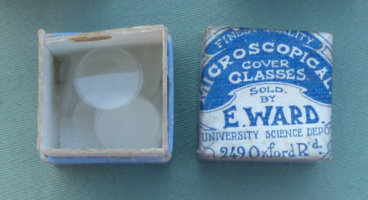
Figure 16. A box of Ward’s cover slips. Adapted from an internet auction site for educational, nonprofit purposes.
At around the same time, Ward wrote, and sold copies, of essays on “Mounts and Mounting” and “Micro-crystallization”. An abstract of the former, from the American Monthly Microscopical Journal, is presented at the end of this essay.
During the 1887 Manchester Royal Jubilee Exhibition, “Edward Ward F.R.M.S., 249, Oxford Street, Manchester, exhibits microscopes and appliances for microscopic investigation. Mounted slides and unmounted specimens, and photographic apparatus and lantern slides of scientific apparatus. An interesting feature of this exhibit is a special row of microscopes ready mounted and furnished with objects to be viewed at any time by those interested in Entomology and Natural History”.
Construction of the Manchester Ship Canal began in 1887, creating navigable water between Manchester and the Mersey Estuary. Edward Ward chronicled its construction with his camera, as reported in his obituary (see above) and the following report, from an 1890 issue of Research, The Monthly Illustrated Journal of Science:
“The progress of the great undertaking which is now engaging the interest and attention of Lancashire men, has afforded some excellent opportunities for the study of the geological characteristics of the country through which the canal bed is being excavated. Sections have been exposed in the post-Pliocene and recent deposits, drift sands and boulder clay, and in various strata of the Trias. Whilst many details have been corrected or verified, upon the whole, little has been discovered that was not already known. Many of the sections in the drift, and in the Red Marl and Bunter formations exhibit features, however, of great geological interest, and it is fortunate that, through the ardour and perserverence of one of the most active members of the Manchester Photographic and Microscopical Societies, photographic records of many of these features have been preserved. Mr. Edward Ward, of Oxford Street, Manchester, has occupied himself with the gigantic task of photographing the canal works in scientific, no less than in some of their social aspects, and has thus rendered a distinct service in recording all that is worthy of note in the excavations so far as they proceed. We are enabled, through Mr. Ward’s courtesy, to reproduce two of his geological photographs, the first of which shows drift sands and river gravel at Barton, with one of the curious ‘potholes’ which occur there. The other photograph exhibits a section of the waterstones at Norton, exposing the ‘slickenside’ face of a fault. These ‘slickensides’ or polished surfaces of faults, are very numerous in the Triassic rocks, and are deserving of study, as throwing light on the direction and character of the movements which have affected the local strata.”
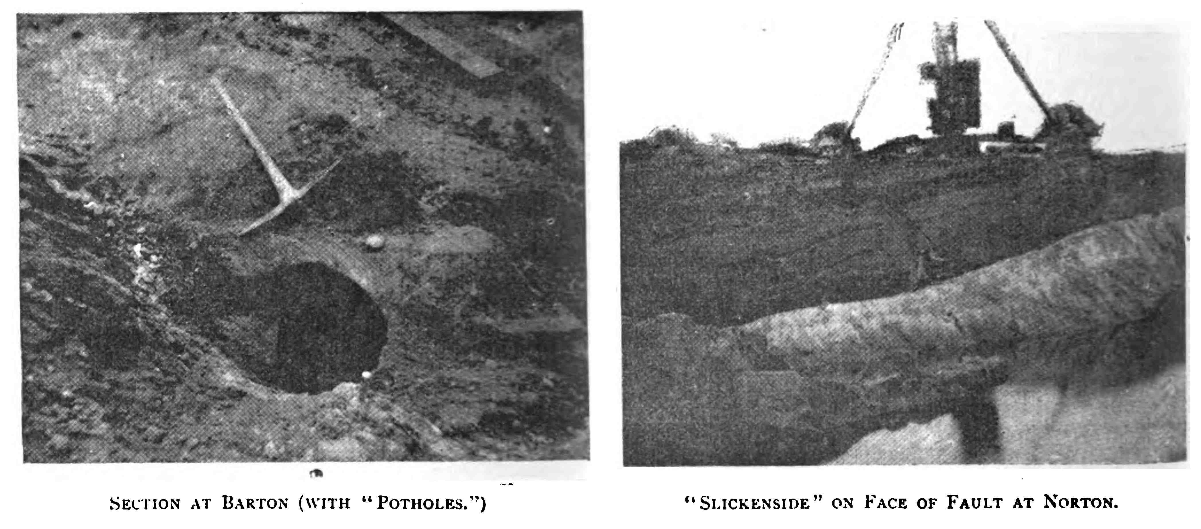
Figure 17. Two photographs taken by Edward Ward of geological features unearthed during construction of the Manchester Ship Canal. These pictures accompanied an article on the canal that appeared in an 1890 issue of Research, The Monthly Illustrated Journal of Science.
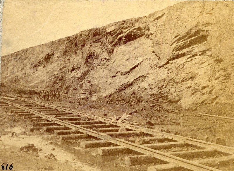
Figure 18. Another Edward Ward photograph of slickenside along the Manchester Ship Canal. From the British Geological Survey.
The 1891 census recorded that Ward’s shop 249 Oxford Street was also the family home. Along with Edward and Frances lived their 20 year-old daughter, Elizabeth (“photographic assistant”), 18 year-old son Edward (“shop assistant – scientific”) and 24 year-old nephew Harry Jones (“photographic operator”). The 1901 census found the family at the same location, with Edward recorded as being an “optician & shopkeeper” and an “employer”.
Edward Ward died in August, 1901, at the age of 57.
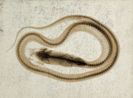
Figure 19. A snake in the process of swallowing a small mammal, probably a mouse, viewed through x-ray. The back of the mount is stamped “E. Ward, 249, Oxford st., Manchester”. Made in 1896 under the direction of the British physicist, Sir Arthur Schuster (1851-1934). Schuster was then a professor at Owen’s College. He was one of the first to receive the offprint of an article entitled “Uber eine neue Art von Strahlen”, along with a set of prints from W.K. Roentgen (1845-1923), the German physicist who discovered x-rays in 1895. From the Wellcome Library (see details in Resources, below).
__________________________________________________
An abbreviated version of Edward Ward’s “Mounts and Mounting”, as it appeared in the American Monthly Microscopical Journal, 1883. Ward sold full-length copies of this essay, along with “Micro-crystallization” in a booklet.
“In the early days of the microscope, mounting must have been a very different matter from what it is now, and as I use (and sometimes break) circle after circle of covering glass, I like to think that it was before my time that mica or talc was used for the same purpose.
I believe that one of the earliest methods of so-called ‘permanent mounting’ was by placing the objects (thin enough to be examined by transmitted light, without preparation) between two thicknesses of mica, and putting these between two card-board slips having a hole punched through them. A little later, so far as I can learn, the plan was adopted of placing the two mica flakes, enclosing objects prepared in the same simple manner, in slips of wood, bone, or ivory, each of these slips sometimes containing several objects.
I wish that I could follow, chronologically, the many improvements made in microscopical mounts - one of the greatest was the introduction of Canada Balsam.
It had long been felt to be one of the greatest needs in the preparation of objects, that some medium should be discovered in which the more opaque ones could be well displayed. That such a medium had been found was shown at a meeting of Micromaniacs, held at the residence of one of their members, where, on an evening, some forty years ago, some objects were placed under the microscope, which, from the clearness and distinctness of the view, caused great delight to the beholders, who naturally wanted to learn how these (to them) most magnificent results were obtained.
The disappointment must have been intense when they were told that this was a profound secret, known only to three persons, each one of whom was pledged to the others not to divulge it; but a gentleman present, while holding up one of the slides for examination, pressed the glasses together between his fingers, and when warned of the danger of thus handling newly-mounted specimens, changed its position and held it by its opposite edges. But he had already pressed out a very small portion of the mounting medium, which, smearing his fingers, disclosed, by its now wellknown scent, the secret of mounting in Canada Balsam, and we enthusiasts may imagine the delight of this worthy worker, when, without any underhand action, he had thus ‘smelt’ his way to results similar to those so much admired. But we, most of us, have, early in our work, learned that there is a difference, and a vast one, between knowing that an object is mounted in Canada Balsam and being ourselves able to mount in this medium; and the description given by one of these gentlemen of his endeavor to imitate what he had seen, is applicable, in a greater or less degree, to all those who now commence practising the ‘sticky art’:
On the evening appointed we set to work, being, with careful forethought on the part of the mistress of the house, provided with an old iron tea-tray. The Canada Balsam was produced, and an ample supply poured upon one of the usual sized glass slips. We then adjusted upon the convex side of this fluid the wing of a butterfly gently pressing it into the fluid to expel the air from beneath. A further supply of Balsam was then poured upon the upper surface of the object until it was completely immersed in the fluid; a second slip was then laid over the first, and the two slips were gently, but firmly, pressed together, and secured by thread bands; and so we proceeded with six or eight specimens, but by this time we were fairly brought to a standstill, our fingers being Canada Balsamed up to the knuckles, and our hands when we attempted to separate the fingers, being like the feet of a web-footed animal; so we were compelled to strike work, and adjourned to regions below, where by a not sparing use of turpentine, soap, and hot water, we cleaned our objects and restored our hands to a comparatively clean condition. We were, however, amply rewarded for all by finding upon examination that our objects were quite equal to those that had been shown to us.
These gentlemen then quickly spread among their fellows the knowledge of the new mode, and it at once became the favorite method of mounting. In later years the number of workers has increased. New media and new modes are constantly being recommended, and the united skill of an army of workers has made mounting a pleasure, and the examination of mounted specimens a constant delight; whilst neatness and beauty of finish are now aimed at as well as the successful display of the object.
The easiest objects that a learner can begin with are, I think, those that are merely mounted dry in a shallow and not opaque cell, such as wings of butterflies, and the scales removed from the wings, delicate pollen grains, fern spores, scales, cuticles, etc.
In commencing work it will be found advisable to prepare a number of slides by putting them on a turn-table, and with a brush charged with brown cement, or some similar viscid preparation (the solvent of which should be spirit), run circles upon a number of slides, and put on one side to dry ; this, with brown cement, will to a certain extent quickly take place, when, if the cell is not deep enough, another, and yet other layers may be added. Now, having a number of these cell slides ready, we wish to mount a slide of scales of butterfly, which can easily be done. Cut a bit of ordinary printing paper, about one inch by half inch, fold it in the middle, and then, placing in the fold a small piece of the wing, press steadily and with a slight sliding motion, when the scales "from both sides" will be, as it were, pulled from the wing and remain upon the paper. If now the paper be divided, and a clean coverglass of the chosen size be taken, it will be found that by breathing upon the cover, and then at once putting down upon it first one and then the other of the papers, scale side to the glass, the scales will adhere to the glass cover, and then by gently warming over a spirit lamp one of the slips prepared as described, the ring of cement will be softened, and the coverglass with scales may be pressed closely to it, and when cold all will be found firmly fixed.
If it is desired to mount a small piece of the wing, see that the cell is sufficiently deep, and then, placing the wing in the centre of the cell, by warming the cover-glass and at once putting it down on the cement ring it will attach itself firmly, and may be thus put on one side.
Some of the fern scales and stellate hairs from plants may be arranged in the centre of one of these cells, and are often fixed firmly enough by the pressure of the warmed cover upon the cement ring, but in these cases it is advisable to have a shallow cell. Pollen-grains, having been allowed to dry, and a slide with cell having been prepared by being breathed upon, the pollen-grains may be dusted upon the slide, when the warmed cover-glass will in this case also securely seal up the object. For pure diatom gatherings or deposits that are desired dry, it is only necessary, after sufficient cleaning and washing, to put a drop of the fluid containing them upon a cover-glass, and allowing this to dry, not by heat, but quite spontaneously, by warming the slide and cell as described for butterfly scales - the cover may be pressed down and securely fixed.
Canada balsam, with its sticky abominations, has now largely given place to balsam and benzole, balsam and chloroform, or damar, each of which medium has advantages over the other for certain objects; but my choice for general work is balsam and benzole, which I have used for years, and for a great many descriptions of work. As an illustration of balsam mounting, or rather mounting in balsam and benzole, we will take some objects from the animal kingdom - say parasites. It will in most cases be advisable to put them in liquor potassi for a short time. When they have been softened or sufficiently reduced in color, they must be well washed in water, and if they are brushed under water with a stiff camelhair pencil, the legs will frequently be found placed right for mounting.
The last washing water having been poured off, and methylated spirit added, let them remain in this for a few hours, and then the spirit be renewed. After the lapse of a few hours more, pour off the spirit and add oil of cloves or cajeput. The latter is quite equal to and much cheaper than the clove oil. From this the object may be placed upon a clean slide, and any surrounding oil should be absorbed by blotting or filter paper; a drop of balsam and benzole being placed upon it, the slide may be left for a few seconds, and then a warmed cover-glass put down, a clip placed upon it, and the operation of mounting is so far complete. The slide now requires placing in a warm place for a few days before the later finishing touches can be applied to it.
If it be Polycystina, Foraminifera, Sponge or Gorgonia Spicules, or any such object, that it is wished to mount, a thin smear of gum should be placed on one side of the centre of the slip, when, if the thumb be drawn firmly over the slide, a very thin, almost invisible film will remain, which must be allowed to dry. If then a small heap of the dry material be put on the centre of the smear, and the slide, while held in the fingers at one end, be gently tapped on the other by the foreceps, or anything hard which happens to be handy, the material will be seen to spread itself over the gummed surface, and its disposal may, with a little practice, be greatly controlled. The slide now requires breathing upon, and when again dry, a drop of the balsam and benzole must be put over the object, the cover-glass warmed, put down, and held with a wire clip. The slide should be gently heated over a spirit-lamp until the medium just begins to boil, when it must be removed quickly to a moderately cool spot. This boiling has the effect of driving out the air from in or around an object; and should any bubbles appear about the slide, they will almost invariably disappear when the slide is left in a warm place.
In mounting starches in damar, for polariscope, a similar plan may be adopted to spread the material, but in this case no gum is required, the breath upon the slide being enough to hold the granules as they move over the slide. When the breath has evaporated it is not at once advisable to at once drop on the damar, but first placing over the starch a clean coverglass, put at each edge of the cover, but not exactly opposite, a drop of damar, which running under the cover from each side, will imprison the object. If the damar wave does not appear to travel freely, a little movement with a needle at the edge not touched by the medium, will accomplish the purpose, and the slide may then be put away to dry; but in the case of starches it is not well to put them into a very warm place.
The same plan may be adopted in the case of pollens, fern spores, etc., and the two waves of damar will do what is otherwise very difficult, keep such delicate objects where we like them to be, in the centre of the slide.
At this place I may just point out, that if good mounts of fungus spores, such as the Puccinia, are wanted, the spot of fungus should be removed from the supporting leaf by means of a very sharp-pointed scalpel, and then the slip being breathed upon, the fungus will adhere to the glass; but in some few cases, as in Xenodochus, the spores are so dense, that it is not well to at once add damar; but in such a case a drop of benzole, placed by means of a bit of rolled paper or match, by the side of the spores, will quickly penetrate them; when this has nearly evaporated, a drop of damar may be put direct upon the spores, and I would then suggest that the cover-glass be not put down for half an hour, and it will be found that when warmed and put down it will not so easily move the objects, as it would do if this plan was not followed.
The next description of mounting to which I would refer is one which, to a beginner, is frequently a stumbling-block. I mean glycerine jelly, which is an exceedingly convenient method to adopt when the object would be rendered too transparent in balsam or damar, or when it is undesirable to dry it at all.
In the preparation of the many things from the vegetable kingdom, as mosses, algae, cuticles, sections, etc., and from the animal kingdom, as many eyes and wings of insects, gastric teeth, palates of the mollusca, it is only necessary, if they are sufficiently clean and not too dark in color, to put them for a few hours into a mixture of methylated spirit, glycerine, and water, about equal parts of each, although exactness is not necessary, as the mixture may be varied to suit circumstances.
When they are taken from this mixture, they must be placed upon the centre of the slide, and the surplus liquid absorbed by blotting-paper. Either of two plans may now be followed with regard to the jelly - it may be liquified by placing the bottle in hot water, and then dropping the liquid jelly upon the slide, or, as I frequently prefer, a small piece may be cut from the bottle and put upon the object and the slide gently warmed, when the jelly will diffuse itself through the object, and will be found exceptionally free from the enemy, ‘air-bubbles’; but should there be air bubbles, or not, it is of great value to boil the jelly and object upon the slide, but care must be used or the mount may be ruined. Should the boiling be decided upon, the clip should be used, and the slide held by the clip directly over, but not too close to the flame of a spirit lamp; it will at first begin to bubble from the centre outwards, and if the slide be now carefully watched, a very perceptible crack may be seen and heard, at this moment, and without delay, the slide must be withdrawn from the heat, and placed upon a cold surface (in my work a block of marble), when the jelly will rapidly contract and the air bubbles will be excluded.
The jelly mounts are easily cleaned from superfluous jelly, by brushing with a soft tooth brush under a running tap, and the surface of the slide being allowed to dry spontaneously, it will be found that it is free from glycerine smears, which interfere much with the after-process of finishing.”
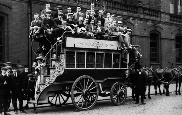
Figure 20. Photograph of a double-decker horse-drawn omnibus, produced as a postcard by Edward Ward.
_______________________________________________
Notes on some sources of microscope slides that were apparently in Edward Ward’s personal collection.
The following were noted as sources of slides shown in Figure 3.
Cognapier
Probably C.O.G. Napier (Charles Ottley Groom Napier), who wrote on microscopy and many other subjects during this era.
J. Ford
John Ford, amateur mounter and a member of the Postal Microscopical Society. Produced microscope slides between ca. 1870 and the 1890s. Lived in Stamford, Lincolnshire between ca. 1856 and ca. 1874, then in Tettenhall, Wolverhampton until ca. 1884, and Kew, Surrey after that.
J.C. Hutchinson
John C. Hutcheson. Advertised to exchange diatoms and other marine objects during the early 1870s. Lived at 9 Lansdowne Crescent, Glasgow.
A. Kempson
Augustus Kempson, bank manager. Was active in the Northampton Natural History
Society (president, 1882), and was noted as having exhibited microscopical
specimens in the Society’s reports. Lived at 6 Parade Square, Northampton.
F. Lazenby
Francis Lazenby of Basingstoke, Hampshire, who advertised microscope slide exchanges in the 1870s.
Col. Mason
Has not been identified. Supplied Ward with a louse from an “Indian secretary vulture”, in 1875. The identity of that bird is
not known. Sagittarius serpentarius
was formerly called the secretary vulture, and is now commonly called the
secretary bird, but is found only in Africa.
Mello
Reverend J. Magens Mello, president of the Derbyshire Microscopical Society. Wrote on the microscopical structure of rocks in 1875.
A.C. Rogers
Alfred C. Rogers. Advertised in Hardwicke’s
Science-Gossip, 1877: “A Twelve-inch
Plate Electrical Machine, with discharges, &c., complete in case, for
Microscopical Apparatus - Apply to A. C. Rogers, 132, High-street, Southampton.” Listed as a member of the Committee of
the Literary and Philosophical Society in the 1884 Stevens' Directory of Southampton, and Neighbourhood.
H.M.J. Underhill
Henry Michael John Underhill. Member of the Postal Microscopy Club and founding
member of the Oxford Natural History Society in 1889. One of the first to use a
microtome to make serial sections of insects, spiders, etc. Lived at 7 High Street, Oxford in 1889.
EW
Undoubtedly professional slide maker Edmund Wheeler. Wheeler’s papered slides bore his initials but often no other
identifying features. Ward
evidently exchanged for or purchased several of Wheeler’s mounts, then
re-wrapped them in his own distinctive green and orange papers.
Whyte – JB
A slide with both sources is shown in Figure 3. Whyte is
unknown. “JB” is probably a reference
to the monogram found on papered, professionally-prepared slides. Presumably, Whyte provided a “JB” slide to Ward. “JB” was most likely professional slide-maker John Barnett, who lived in Tottenham, Middlesex.
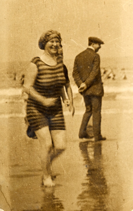
Figure 21. Undated seaside photograph by Edward Ward.
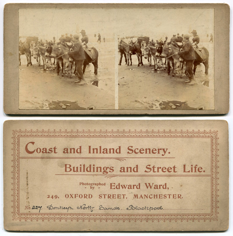
Figure 22. Undated photographic steroscope card by Edward Ward. Note that this is numbered 227, implying that Ward produced a substantial number of stereoviews.
Figure 23. A World War I era postcard, by Edward Vincent Ward, the microscopist’s son. He operated a photographic studio at the same address as had his father. The photographer died on November 2, 1921.
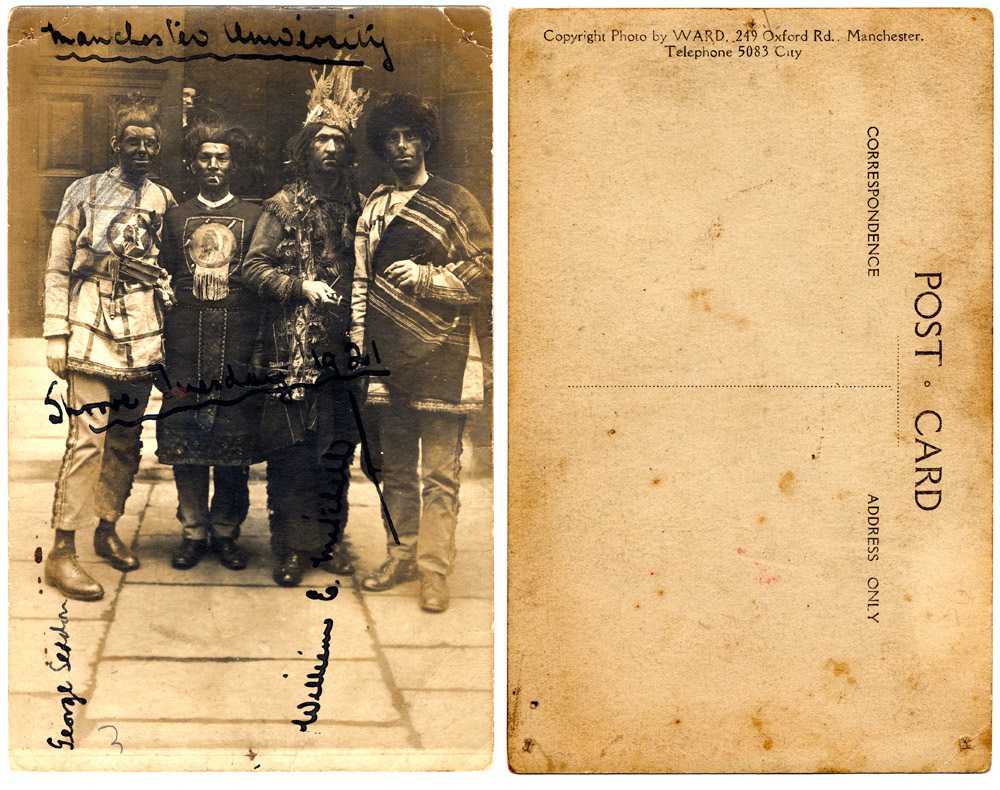
Figure 24. Front and rear views of a 1921 photograph of costumed University of Manchester students participating in Shrove Tuesday partying. Edward Vincent Ward took the photograph and produced this custom postcard.
Resources
The American Monthly Microscopical Journal (1883) Notes from abroad, Vol. 4, page 147
The American Monthly Microscopical Journal (1901) Edward Ward (obituary), Vol. 22, pages 282-283
Bracegirdle, Brian (1998) Microscopical Mounts and Mounters, Quekett Microscopical Society, London, pages 97, 178-181, and 212-213
Bracegirdle, Brian and James B. McCormick (1993) The Microscopic Photographs of J.B. Dancer, Science Heritage Ltd., Chicago, pages 45 and 2Ward
British Geological Survey GeoScenic Digital Asset P233272, “Pool Hall. Slickensided surface (and other views along the course of the Manchester Ship Canal)”, by Edward Ward, January 1, 1893. http://geoscenic.bgs.ac.uk/asset-bank/action/viewAsset?id=74671&index=75&total=76&categoryId=1048&categoryTypeId=1&collection=Cheshire&sortAttributeId=1019&sortDescending=false
The Charles Lamb Bulletin (1994) "On the back are . . presumably the photographer’s rubber stamp: E. Vincent Ward, 249 Oxford Road, Manchester"
Davis, George E. (1881) Photomicrography, “The most successful photographers of microscopic objects have been, Col. Woodward, Dr. Maddox, Mr. Wenham, Drs. Abercrombie and Wilson, Mr. Shadbolt, and the late Dr. Redmayne, of Bolton, some of whose pictures by the kindness of Mr. Ward, of Manchester, I am enabled to show you to-night.” Microscopical News and Northern Microscopist, Vol. 1, page 74
England census, birth, marriage and death records, accessed through ancestry.co.uk
Hardwicke’s Science Gossip (1870) Exchange offers from Edward Ward, Vol. 6, pages 192, 216, 240, and 264
Hardwicke’s Science Gossip (1871) Exchange offers from Edward Ward, Vol. 7, pages 48, 72, 96, 120, 168, and 284
Hardwicke’s Science Gossip (1871) Notices to correspondents: “E.W. – The ‘zoophyte’ is Diphasia roseacea, with female capsules”, Vol. 7, page 240
Hardwicke’s Science Gossip (1872) Exchange offers from Edward Ward, Vol. 8, pages 48 and 216
Hardwicke’s Science Gossip (1873) Exchange offer from Edward Ward, Vol. 9, page 24
Hardwicke’s Science Gossip (1874) Exchange offer from Francis Lazenby, Vol. 10, page 24
Hardwicke’s Science Gossip (1874) Exchange offers from Edward Ward, Vol. 10, page 240
Hardwicke’s Science Gossip (1881) Advertisements from Edward Ward, Vol. 17, pages x and lxxxvii
Hardwicke’s Science Gossip (1885) Advertisements from Edward Ward, Vol. 21, page cii
Hardwicke’s Science Gossip (1877) Exchange offer from A.C. Rogers, Vol. 13, page 120
Hardwicke’s Science Gossip (1879) Notices to correspondents: “Edward Ward. Your fresh-water algas are 1. Zygnema rizmlare, Hassall : 2. Zygnema [? sp.) ; 3. Z. nitidum ; 4. Vesiciilifera Candollii, Hassall.”, Vol. 15, page 144
The Journal of the Postal Microscopical Society (1882) Advertisement from Edward Ward, Vol. 2, inside cover
The Journal of the Postal Microscopical Society (1882) List of members: Ford, John, The Uplands, Tettenhall, Wolverhampton, Vol. 1, page 12
The Journal of the Postal Microscopical Society (1884) Advertisement from Edward Ward, September issue, inside cover
Journal of the Royal Microscopical Society (1880) Advertisement from Edward Ward, Vol. 3, advertising page 7
Journal of the Royal Microscopical Society (1881) New fellows, Series 2, Vol. 1, page 976
Mello, J. Magens (1875) On the microscopical structure of rocks, The Popular Science Review, Vol. 14, pages 1-19
Miles, J.L.W. (1905) The inception and formation of the Manchester Microscopical Society, Annual Report and Transactions, pages 39-40
Napier, C.O.G. (1869) Insects never grow, Hardwicke’s Science-Gossip, Vol. 4, 1869, page 23
Nature (1885) Advertisement from Edward Ward, Vol. 31, page xcviii
Nature (1886) Advertisement from Edward Ward, Vol. 35, page xxvi
The Northern Microscopist and Microscopical News (1882) News of the Northampton Natural History Society, A. Kempson, president, Vol. 2, page 161
The Owens College Calendar Advertiser (1880) Advertisement from Edward Ward, page 34
The Photographic Dealer (1901) Edward Ward (obituary), Vol. 11, page 53
Railway Signal (1915) "The pictures of Manchester railway men, which appeared in the January and February numbers of the Railway Signal, were reproduced from photographs by kind permission of Mr. EV Ward, Photographer, 249, Oxford Road, Manchester"
Research: the Monthly Illustrated Journal of Science (1890) Geological Sections on the Manchester Ship Canal, Vol. 2, page 232
Science-Gossip (1901) Edward Ward (obituary), New series, Vol. 8, page 147
Smith, Watson (1887) Report on section III of the Manchester Royal Jubilee Exhibition – the chemical and allied industries, Journal of the Society of Chemical Industry, Vol. 6, pages 624-643
(Stevens') Directory of Southampton, and Neighbourhood (1884) A.C. Rogers, Member of the Committee of the Literary and Philosophical Society, page 58
Thornton, Gail (accessed December, 2011) A pictorial archive of hose drawn vehicles: the omnibus (includes a photograph taken by Edward Ward), href="http://www.gail-thornton.co.uk/public-vehicles/omnibus.php
Ward, Edward (1882) Micro-crystallization, The Northern Microscopist and Microscopical News, Vol. 2, pages 25-33
Ward, Edward (1883) Mounts and mounting, abstracted, The American Monthly Microscopical Journal (1883) Vol. 4, pages 149-156
Wellcome Library no. 33165i (accessed January, 2012) A snake in the process of swallowing a small mammal, probably a mouse, viewed through x-ray. Photoprint from radiograph after Sir Arthur Schuster, 1896. href="http://catalogue.wellcome.ac.uk/record=b1190997~S8